Available at http://www.formulastudy.com
HFS
Original Article / 원저
防己茯苓湯이 백서에서 LPS로 유도된 우울증에서 면역 조직학적 변화에 미치는 효과
박성준, 이태희
*가천대학교 한의과대학 방제학교실
Effect of Banggibongnyeongtang on the immunohistological change in LPS-induced depression rats
Sung jun Park, Tae Hee Lee
*Department of Formulae Pharmacology, College of Korean Medicine, Gachon University
ABSTRACT
Objective : This study is accomplished in order to investigate the effect of banggibongnyeongtang on the immunohistological change in LPS-induced depression rats to confirm the histological result of the previous behavioral and biochemical effect.
Methods : LPS 5 ㎍ was injected to lateral ventricle and experimental groups were administered BBT intraperitoneally. The concentration of 5-HT in the Medial Prefrontal Cortex, Striatum, Hippocampus, Amygdala was measured by ELISA. IL-1β, TNF-α mRNA and BDNF mRNA expression in the hippocampus was examined by RT-PCR.
Result : BBT enhanced 5-HT concentration at all part of brain but no significantly difference at medial prefrontal cortex and striatum. LPS+BBT400 group increased 5-HT concentration significantly than LPS group at hippocampus and amygdala (p<0.05). BBT decreased IL-1β mRNA expression dose dependently but only with significantly decrease in LPS+BBT400 group than LPS group’s in Hippocampus (p<0.05). But BBT did not decrease TNF-α mRNA expression significantly in Hippocampus. BBT increased the expression of BDNF mRNA at hippocampus and LPS+BBT400 group significantly increased comparing with LPS group does (p<0.05).
ⓒ 2020 The Korean Medicine Society For The Herbal Formula Study
This paper is available at http://www.formulastudy.com which permits unrestricted non-commercial use, distribution, and reproduction in any medium, provided the original work is properly cited.
Conclusion : It is postulated that the anti-depressant effect of BBT can be validated through the anti-inflammatory effect, 5-HT concentration increase, and the neuro-protective effect mediated by BDNF by combining the results of the previous report about the behavioral and biochemical effect.
Key words : banggibongnyeongtang, LPS, antidepression, 5-HT, IL-1β, TNF-α, BDNF.
Ⅰ. 서론
a)우울증은 기분의 뚜렷한 변화를 필요로 한다. 슬픔 이나 초조함 그리고 적어도 몇 가지의 정신 생리적인 변화를 포함하는데, 수면, 식욕 혹은 성욕의 장애, 변비, 일이나 친구들과의 관계에서 즐거움 상실, 울 음, 자살 충동, 그리고 말과 행동의 둔마와 같은 것 을 특징으로 한다. 이러한 변화들은 반드시 적어도 2 주는 지속되어야 하고, 일과 가족관계를 방해해야 한 다1). 우울증은 자살과 밀접한 연관을 맺고 있으며, 현재 한국의 자살률은 2018년 현재 10만명 당 26.6 명으로 5위의 사망원인이다. 특히 10대에서 30대까지 자살은 가장 큰 사망 원인이다2).
우울증의 치료는 1960년대 소개된 1세대 항우울제 인 Tricyclic antidepressants (TCAs), inhibitor of monoamine oxidase (MAOI), 1990년대에 나온 2 세대 항우울제 selective serotonin reuptake inhibitor (SSRI), mixed serotonin/noradrenaline reuptake inhibitor (SNRI) 류가 사용되어 왔다. 1세대 항 우 울제(TCAs, MAOI)는 자율신경계와 심혈관계에 부작 용을 나타내고, 2세대 항 우울제(SSRI/SNRI)는 작용 까지 3~6주정도의 지연이 있고, 불면이나 구역, 성 기능 장애 등의 부작용을 나타낸다3). 최근 우울증을 초례할 수 있는 가능성 높은 가설로 염증 가설이 나 타났다. 이 가설에서 proinflammatory cytokine의 증가가 indolamine 2,3-dioxygeneas (IDO) 활성을 증가시킴으로써, 신경독성을 유발하는 N-methyl- D-aspartate receptor (NMDA R)의 작용이 활성화 되어서, neuron death가 유발되어 우울증이 될 수 있다고 설명 한다4). 박 등5)은 방기복령탕이 항 염증 효과를 통하여 항 우울 효과를 보고한 바 그 내용은 LPS를 투여하여 개방장 행동 실험(open field test) 과 강제수영부하실험(forced swimming test)을 통하
여 우울행동을 확인한 후 혈장에서 IL-1β, TNF-α 농도 그리고 CRH(cotritotropin relasing hormone), ACTH(adenocorticotropic hormone), Cort(corticosteron) 의 농도를 확인하였는데, 본 논문에서는 Medical Prefrontal Cortex, Hippocampus, Amygdala, Striatum 에서 5-hydroxytryptamine (5-HT)의 농도 변화, Hippocampus에서 Interleukin-1 beta (IL-1β), Tumor Necrosis Factor-alpha (TNF-α), Brain- Derived Neurotrophic Factor (BDNF) mRNA의 발 현을 면역조직학적으로 측정하여 이미 발표한 내용의 조직학적 소견에 대해 유의한 결과를 얻었기에 이를 보고하는 바이다.
Ⅱ. 실험 재료 및 방법
1. 실험재료 1) 실험 동물
실험 동물은 숫컷 흰쥐 成體로 Sprague-Dawley 계이고 Samtaco Animal Co. (Seoul, Korea)에서 구입하였다. 모든 실험은 동물이 실험실에 도착한 7 일 이후, 환경에 적응시켜 실험에 사용하였다. 주령 은 7주이고 체중은 210-230g이다. 이 흰쥐는 각 플 라스틱 사육 상자에 5 마리 내로 길렀다. 실험실 온 도는 22±2°C, 습도는 55±15%가 유지되도록 조절하 였고, 매일 12시간 동안 인공 햇빛을 쐬어 주었다.
흰쥐에게는 무제한으로 살균된 물과 고형 사료를 실 험기간 동안 공급하였다.
2) 약재
연구에 사용한 防己 (방기), 黃芪 (황기), 茯苓 (복 령), 桂枝 (계지), 甘草 (감초)는 표준품을 사용하였 으며, 처방은 상한론에 기재된 防己茯苓湯으로, 처방 구성 약제는 나눔 제약㈜(영천)에서 구입한 후 사용
*Corresponding author : Tae Hee Lee. Department of Formulae Pharmacology, College of Korean Medicine, Gachon University, 1342, Seongnam-daero, Sujeong-gu, Seongnam-si, Gyeonggi-do, 13120, Republic of Korea.
Tel: +82-31-750-5418, Fax: +82-31-750-5416, E-mail: [email protected]
∙Received : February 15, 2020 / Revised : February 21, 2020 / Accepted : February 24, 2020
하였다. 처방 내용은 다음과 같다.
Korean (Chinese) name
Pharmaceutical name Dose (g) 방기(防己) Stephaniae Tetrandrae Radix 36
황기(黃芪) Astragali Radix 36
복령(茯苓) Poria 72
계지(桂枝) Cinnamomi Ramulus 36 감초(甘草) Glycyrrhizae Radix 24
Total Amounts 204
防己茯苓湯(BBT) 204 g을 가루내어 섞어 정확히 200 g으로 평량한 후, 85% 메탄올을 사용하여 3 회 추출하였으며, 추출물을 감압 농축한 후, 그 추출물 을 동결 건조시켜 분말 건조하였다. 건조시켜 얻은 양은 25 g으로써, 수율은 12.5%였다.
2. 실험 1) 실험군
실험쥐는 무작위적로 몸무게에 따라 6마리씩으로 6 그룹으로 나누었다. LPS를 투여하지 않고 약재 대신 생리식염수를 주사한 군(0.9% NaCl, i.p., SAL group, n=6), LPS를 투여하고 매일 14일 동안 생리 식염수를 주사한 군(0.9% NaCl, i.p., LPS group, n=6), LPS를 투여하고 매일 14일 동안 100 mg/kg 의 防己茯苓湯 (BBT)을 주사한 그룹(i.p., LPS+BBT100 group, n=6), LPS를 투여하고 매일 14일동안 200
mg/kg의 防己茯苓湯 (BBT)을 주사한 그룹(i.p., LPS+BBT200 group, n=6), LPS를 투여하고 매일 14일 동안 400 mg/kg의 防己茯苓湯 (BBT)을 주사한 그룹(i.p., LPS+BBT400 group, n=6) 으로 나누었 다. 防己茯苓湯 (BBT)과 식염수는 14일간 복강 내로 주사하였다.
2) Lipopolysaccharide (LPS) 처리
염증에 의한 우울증 동물모델을 만들기 위해서, 쥐 에 sodium pentobarbital (50 mg/kg, i.p.)로 마취 시킨 후 stereotaxic technique를 이용하여 lateral ventricle (AP: -0.2, L: ±0.3, H: -6.2)위치에 LPS (Escherichia coli; 055:B5, Sigma)를 5 μg을 양쪽으로 주입하였다8,9). 미세주입은 1 ml gas-tight glass syringe (Hanilton, Reno, NV, USA)에 폴리 에틸렌 튜브 (polyethylene tubing)로 연결하여 관류 용 펌프(perfusion pump, Pump 22, Harvard Apparatus Inc., Holiston, MA, USA) 를 이용하여 2 μl/min 유속으로 주입 후에 5분간 방치한 다음 주사기(syringe)를 제거하였다. 각 손상군에 대한 약 물처치는 그 다음날부터 시행하였다. 모의 시술군은 손상 약물 대신 140 mM NaCl, 3.0 mM KCl, 2.5 mM CaCl2, 1.2 mM Na2HPO4로 만든 인공뇌척수액 (artificial CSF)을 같은 방식으로 주입하였다. 이 모 든 실험 과정은 다음과 같다. (Fig. 1)
Fig. 1. Experimental schedule for developing lipopolysaccharide-induced depression-like behaviors, and BBT treatment in rats. Separate groups of rats (n=6 animals per group) were used for all experiments.
3) 5-HT의 측정
실험동물 모두 행동 실험이 끝난 후 같은 날 decapitation 시켰다. 실험방법은 기존의 논문을 참 조하였다. 동일한 흰쥐의 뇌를 적출했다. 적출한 뇌 샘플에서 5-HT를 분석하기 위해서, medical prefrontal cortex, hippocampus, amygdala, striatum에서 분
리하여, 4개의 뇌 부위에서 5-HT를 측정하였다. 5-HT 는 rabbit polyclonal 5-HT antibody (Abcam) 프로 토콜 을 이 용하여 competitive enzyme-linked immunoassay (ELISA)로 측정하였다. 표본은 판에 옮겨져서 한 시간 동안 실온에서 공기에 노출시켜 배 양하였다. 표본을 buffers로 몇 번 씻어 내고 발색
시킨 후 450 nm에서 ELISA reader (MutiRead 400;
Authos Co., Vienna, Austria)로 optical density를 측정하였다6).
4) Total RNA 추출 및 Reverse transcription polymerase chain reaction (RT-PCR, 역전사중 합효소연쇄반응)
흰쥐의 적출한 뇌에서 trisol (Sigma)을 사용하여 total RNA를 분리하였다14). Reverse transcription 은 DEPC-DW로 최종부피를 50μl로 맞춘 후 total RNA에 RT-premix (Bioneer, Oakland, CA, USA) 혼합용액을 사용하여 cDNA를 합성하였다. RT 반응 은 65°C에서 10분 동안 RNA를 변성시키고, 42°C에 서 1시간 동안 역전사하여 cDNA를 합성하고, 95°C 에서 5분 동안 MuLV reverse transcriptase를 불 활성화시키는 과정으로 진행되었다. PCR 반응은 10 mM Tris-Hcl, 50 mM KCl, 1.5 mM MgCl2, 1 mM dNTP, 1 unit Taq polymerase, 10 pmol의 5‘과 3’의 BDNF (brain-derived neurotrophic factor) primer 그리고 RT product를 첨가하고 PCR을 수행 하였다. PCR 사이클은 94°C, 5 분 동안 denaturation, annealing 온도는 57°C에서 30 초, 72°C에서 30 초 동안 extention과정을 거쳐 PCR product를 얻었다.
GAPDH는 internal control로 수행하였다. 이렇게 얻어진 product는 1.2% agarose gel에서 전기영동하 여 UV (i-MaxTM, CoreBio System Co., Seoul, Korea)에서 관찰하였다. 또한 동일한 방법으로 IL-1 β와 TNF-α를 측정하였다.
3. 통계 처리
모든 측정값은 맹검하여 시행되었다. 모든 측정값 은 (평균값±표준오차)로 표시하였다. 각 실험군의 통 계학적 분석은 window용 SPSS 이용하고 사후 Tukey’s post-hoc test로 검정하였다. P값이 0.05 미만인 것을 통계적으로 유의한 것으로 인정하였다.
Ⅲ. 결과
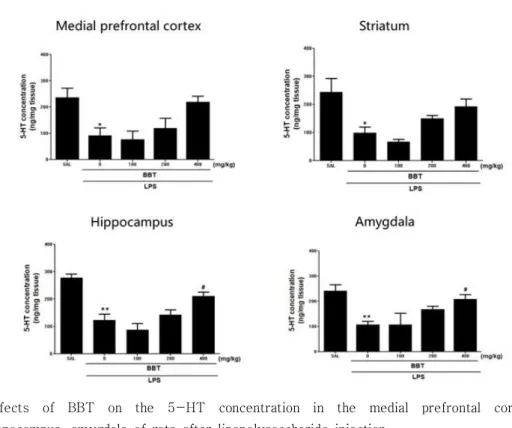
1. BBT의 Medial Prefrontal Cortex, Striatum, Hippocampus, Amygdala 에서 5-HT 농도에 대한 효과
LPS로 유도된 우울증 동물모델에서 BBT를 투여한 후 뇌의 Medial prefrontal cortex, Striatum, Hippocampus, Amygdala에서 5-HT의 농도를 측정 하였다. Medial prefrontal cortex에서 SAL group 에서 237.33±35.35 ng/mg, LPS group에서 92.00±29.87 ng/mg으로 5-HT의 농도가 유의하게 감소하였다(p<0.05). 그러나 LPS+BBT100 group에 서 78.00±30.62 ng/mg로 오히려 감소하였고, LPS+BBT200 group에서 120.67±37.78 ng/mg, LPS+BBT400 group에서는 220.00±22.12 ng/mg 로 5-HT 농도가 증가하였으나 유의하지는 않았다.
Striatum에서의 5-HT의 농도는 SAL group에서 244.67±48.75 ng/mg의 농도로 측정되고 LPS group에서 99.33±20.83 ng/mg로 유의하게 감 소하였다(p<0.05). 그러나 LPS+BBT100 group에서 67.33±9.33 ng/mg으로 오히려 감소 하였고 , LPS+BBT200 group에서 150.00±12.17 ng/mg, LPS+BBT400 group에서는 193.33±27.19 ng/mg로 증가하였지만 유의성은 없었다. Hippocampus에서 5-HT의 농도는 SAL group에서 277.33±13.33 ng/mg, LPS group에서는 123.33±22.19 ng/mg으 로 SAL group에 비해 유의하게 감소하였다(p<0.01).
LPS+BBT100 group에서 87.33±23.25 ng/mg로 오 히려 감소하였고, LPS+BBT200 group에서 142.67±17.52 ng/mg 로 증가하였으며, LPS+BBT400 group에서는 211.33±14.11 ng/mg으로 LPS group에 비하여 유의 하게 증가하며(p<0.05), 농도의존적으로 증가하는 경 향을 보였다. Amygdala에서의 SAL group에서 5-HT의 농도는 240.67±24.88 ng/mg였고, LPS group 에서는 106.67±13.87 ng/mg으로 유의하게 감소하였다(p<0.01). LPS+BBT100 group에서 107.33±44.56 ng/mg, LPS+BBT200 group에서 168.67±11.57 ng/mg, LPS+BBT400 group에서는 208.00±19.08 ng/mg으로 LPS group에 비하여 유의하게 증가하였 다(p<0.05). (Fig. 2)
Fig. 2. Effects of BBT on the 5-HT concentration in the medial prefrontal cortex, striatum, hippocampus, amygdala of rats after lipopolysaccharide injection.
LPS 5 ㎍ was injected to lateral ventricle for 15 days before the first behavior test.
Experimental groups were administered i.p. for 14 consecutive days prior to the test.
SAL group : Saline treated group for 14 consecutive days before OFT, FST.
LPS group : LPS and saline treated group for 14 consecutive days before OFT, FST.
LPS+BBT100 group : LPS and BBT (100 mg/kg) treated group for 14 consecutive days before OFT, FST.
LPS+BBT200 group : LPS and BBT (200 mg/kg) treated group for 14 cosnecutive days before OFT, FST
LPS+BBT400 group : LPS and BBT (400 mg/kg) treated group for 14 cosnecutive days before OFT, FST
*,** : Significant difference compared with the SAL group (p<0.05,p<0.01) # : Significant difference compared with the LPS group (p<0.05)
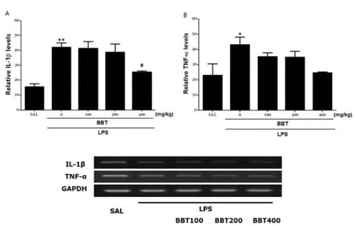
2. BBT의 Hippocampus 에서의 IL-1β, TNF-α mRNA 발현에 대한 효과
RT-PCR을 이용하여 상대적 IL-1β mRNA 발현 을 측정하였다. SAL group에서 15.86±1.88%, LPS group 에서 42.43±2.64%로 유의하게 증가하였다 (p<0.01). LPS+BBT100 group 에서 41.38±4.58 %, LPS+BBT200 group에서 38.89±5.41%로 발현이 감 소하였고, LPS+BBT400 group에서는 25.52±0.78%
로 발현되어 LPS group에 비하여 유의하게 감소하였 다(p<0.05). TNF-α mRNA 발현 비율을 측정한 바, SAL group에서 23.23±7.45%, LPS group에서 43.45±4.73%로 유의하게 증가하였다(p<0.05). LPS+BBT100 group에서 35.56±2.39%, LPS+BBT200 group에서 35.18±3.72%, LPS+BBT400 group에서 24.98±0.37%
로 LPS group 에 비하여 유의한 차이가 없었다.
(Fig. 3)
Fig. 3. Effects of BBT on Interleukin-1β (IL-1β) mRNA (A) and Tumor Necrosis Factor-α (TNF- α) mRNA (B) expression in the Hippocampus after lipopolysaccharide injection. PCR bands on an arose gel and their relative intensities.
LPS 5 ㎍ was injected to lateral ventricle for 15 days before the first behavior test.
Experimental groups were administered I.p. for 14 consecutive days prior to the test.
SAL group : Saline treated group for 14 consecutive days before OFT, FST.
LPS group : LPS and saline treated group for 14 consecutive days before OFT, FST.
LPS+BBT100 group : LPS and BBT (100 mg/kg) treated group for 14 consecutive days before OFT, FST.
LPS+BBT200 group : LPS and BBT (200 mg/kg) treated group for 14 cosnecutive days before OFT, FST
LPS+BBT400 group : LPS and BBT (400 mg/kg) treated group for 14 days before OFT, FST *,** : Significant difference compared with the SAL group (p<0.05,p<0.01)
# : Significant difference compared with the LPS group (p<0.05)
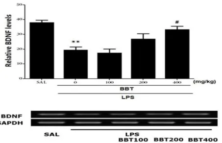
3. BBT의 RT-PCR을 이용한 Hippocampus에서의 BDNF mRNA의 발현에 대한 효과
PCR을 이용하여 Hippocampus에서 BDNF mRNA 발 현을 측정하였다. SAL group에서 38.07±1.47%, LPS group에서 19.45±2.03%에 비하여 유의하게 감 소하였다(p<0.01). LPS+BBT100 group에서 17.43±2.65 %
로 오히려 감소하였고, LPS+BBT200 group에서 27.03±3.39%, LPS+BBT400 group에서는 33.35±2.10%
가 발현하여 농도 의존적으로 증가하였고, LPS+BBT400group 에서 LPS group에 비하여 유의하게 증가하였다(p<0.05).
(Fig.4)
Fig. 4. Effects of BBT on BDNF mRNA expression in the hippocampus of rats with LPS-induced depression-like symptoms. PCR bands on an agarose gel and their relative intensities are shown. BDNF mRNAs levels were normalized to GAPDH levels as an internal control.
LPS 5 ㎍ was injected to lateral ventricle for 15 days before the first behavior test.
Experimental groups were administered i.p. for 14 consecutive days prior to the test.
SAL group : Saline treated group for 14 consecutive days before OFT, FST.
LPS group : LPS and saline treated group for 14 consecutive days before OFT, FST.
LPS+BBT100 group : LPS and BBT (100 mg/kg) treated group for 14 consecutive days before OFT, FST.
LPS+BBT200 group : LPS and BBT (200 mg/kg) treated group for 14 cosnecutive days before OFT, FST
LPS+BBT400 group : LPS and BBT (400 mg/kg) treated group for 14 cosnecutive days before OFT, FST
*,** : Significant difference compared with the SAL group (p<0.05,p<0.01) # : Significant difference compared with the LPS group (p<0.05)
Ⅳ. 고찰
우울증은 의욕 저하와 우울감을 특징으로 하며 다 양한 정신적, 신체적 증상을 보이는 질병 군 이다.
세계보건기구(WHO)의 International Classification of Disease, 10th edition(ICM-10)이나 Diagnostic and Statistical Manual for Mental Disorders, fourth edition(DSM-IV)에 의해 우울증을 진단 한 다7).
최근 염증과 우울증의 연관성에 대한 가설로서, Pro-inflammatory cytokine IL-1, IL-2 등이
5-HT의 전구물질인 트립토판(tryptophan)을 분해하 는 IDO의 활성을 증가시켜 5-HT의 합성을 저하시키 며, 신경 독성이 있는 NMDA R의 합성이 증가시켰 다. 그 결과 신경 독성이 증가함을 확인하였다4). 또 한 IL-1β 와 TNF-α 등의 pro-inflammatory cytokine은 norepinephrine, dopamine과 함께 CRF 분비를 촉진하며, 교감신경을 활성화시켜 염증이 증 폭되는데, 이 과정에서 ‘sickness behavior’ 를 유 발했다. sickness behavior는 주로 감염 상태에서 일 어나는 행동의 변화를 말하는데, 무력감, 우울, 불안, 과수면, 식욕저하, 집중력 저하 등의 양상의 행동 변
화를 가리킨다10). 우울증 환자의 혈중에 pro- inflammatory cytokines의 농도가 증가했고, 뇌척수 액 에서는 PGE2와 같은 염증 매개물질의 농도가 증가한 것을 확인하였다11).
염증반응은 면역계, 내분비계, 신경계가 복잡하게 엮여 cytokine, HPA axis, 신경전달 물질의 변화로 몸의 정상 상태를 되찾는 과정이 포함되어 있다. 그 래서 염증촉진 cytokine의 증가, HPA axis의 활성과 glucocorticoid 저항성의 증가, 5-HT, Norepinephrine, Dopamine의 합성과 제거, 신경세포의 사멸과 재생, 신경가소성에 모두 영향을 주어 우울증의 병태생리에 관여할 수 있다12).
실험에 사용된 防己茯苓湯은 『金櫃要略』에 나와 있는 처방이다. 처방을 구성하는 약물 중 방기는 mast cell에 작용하여 TNF-α의 유리를 억제한다고 밝혀져 있으며, PGE2생성을 억제하여 항 염증 효과 가 있다13). 계지는 5-HT1A 수용체의 결합력을 조절 하며, NMDA에 의한 세포 사멸을 억제하는 효과가 있으며13), LPS로 유도한 염증에서 TNF-α의 농도를 줄인다고 보고되었다14).
Pro-inflammatory cytokine은 5-HT의 합성을 억 제할 수 있다4). 뇌에서 5-HT의 농도는 쥐의 행동 변화와 관련이 깊다. 이를 확인하기 위하여 본 저자 는 뇌의 각 부위에서의 5-HT의 농도를 측정하였다.
Hippocampus, Medial Prefrontal Cortex, Striatum, Amygdala에서 5-HT의 농도는 우울증의 증상 중 무 게 감소와 무감각증, 운동행동, 학습된 절망 그리고 공포와 긴장과 관련이 깊다15). 또한, Hippocampus의 5-HT 농도가 증가하면 FST에서 immobility time이 감소되며, 5-HT와 norepinephrine (NE)의 농도가 증가하면 immobility time이 감소하여 항 우울 효능 을 나타내게 된다16). BBT에서 Hippocampus의 5-HT의 농도가 저농도에서 감소하였다가 증가되는 결과가 보였고, 고농도인 BBT400에서 유의하게 증가 하였다(p<0.05). 또한 Amygdala에서도 농도 의존적 으로 5-HT를 증가시켰고, 고농도군인 BBT400 group에서 유의한 증가를 보였다(P<0.05). 그러나 striatum과 medial prefrontal cortex에서는 BBT의 투약에 의해 5-HT농도가 저농도에서 감소하였다가 중농도와 고농도에서 증가하는 경향은 보였으나 유의 한 차이는 없었다.
Cytokine은 우울증의 병태생리와 가장 관련이 높
은 두 가지 조절 축, 시상하부-뇌하수체-부신피질 축(이하, HPA axis)과 카테콜아민/교감신경계 활성 과 관련이 있다17). IL-1β 와 TNF-α 등의 pro- inflammatory cytokine은 CRH, ACTH를 자극하여 HPA axis를 활성화 시킨다18, 19). 세로토닌의 전구물 질인 tryptophan에서 kyunurenine으로 대사시키는 indoleamine-2, 3-dioxygenase (IDO)를 활성화시 켜 뇌에서 5-HT 합성이 결핍 된다4). BBT를 투여한 group에서 IL-1β mRNA의 발현이 억제하는 효능이 나타났다. 이것은 농도와 비례하는 효과를 보였고, 고농도(BBT400)에서 mRNA의 발현이 유의한 차이를 보였다(p<0.05). 그러나, BBT를 투약한 group에서 TNF-α mRNA의 발현이 유의하게 감소하지 않았다.
BDNF는 뇌의 여러 단계에서 neurotrophin으로 행동 한다20). BDNF는 신경세포의 생존과 유지 그리고 성 장에 관여하는 것으로 보인다21). 사후 연구에 의하면 우울증 환자에서 hippocampus의 BDNF 수치가 감소 한 것으로 밝혀졌다22). 또한 MDD 환자에서 BDNF 농도가 비정상적으로 낮아져 있고, 항 우울 치료를 받은 환자에서 높아졌다23). BDNF는 5-HT 뉴런의 생존과 분화를 촉진하고, 5-HT과 함께 신경가소성을 제어 한다24). LPS는 뇌에서 pro-inflammatory cytokine을 이끌어내어 synapse의 기능을 악화 시킨 다25). BBT는 hippocampus에서 BDNF mRNA의 활 성을 증가 시키는 것을 확인할 수 있었는데, 저농도 에서는 감소하였지만 중농도와 고농도에서 유의한 증 가가 보였다(p<0.05).
서론에서 밝혔듯이 박 등5)이 밝힌 행동변화와 혈장 에서의 cytokine의 변화, 그리고 CRH, ACTH, Corticosterone의 변화의 결과와 같이 볼 때 BBT는 Hippocampus에서 IL-1β mRNA 발현을 억제하여 염증을 제어할 수 있고, Hippocampus, Amygdala에 서 5-HT의 농도를 증가시켜 긴장감과 무감각증, 행 동 둔마와 같은 우울증적 행동장애를 개선시킬 수 있 다. 그리고 신경 보호 효과를 지는 BDNF mRNA의 발현을 촉진시켜 염증으로 인한 신경세포의 파괴를 줄여 항 우울 효과를 낼 수 있을 것으로 보인다.
Ⅴ. 결론
흰쥐에 LPS를 주입하여 염증을 유발한 뒤, 防己茯 苓湯을 투약하여 뇌에서 5-HT 농도와 IL-1β, TNF-α, BDNF의 mRNA 발현을 측정한 결과를 통 해 LPS로 유발된 염증-우울증 모델에서 방기복령탕 에서 나타난 효과에 대해 다음과 같은 결론을 얻었다.
1. 뇌에서 5-HT의 농도에 대한 防己茯苓湯의 효과 는, Medial Prefrontal Cortex, Striatum 그리고 Hippocampus에서는 LPS group에 비해 저농도에 서 감소하였다가 중농도와 고농도에서 증가하는 경향은 보였고, Amygdala에서는 농도 의존적으로 증가하는 경향을 보이고, Hippocampus와 Amygdala의 LPS+BBT400 group에서 유의하게 증가하였다(p<0.05).
2. 防己茯苓湯은 모든 group에서 Hippocampus내의
IL-1β mRNA 발현을 억제하였으며,
LPS+BBT400 group에서 농도와 발현을 유의하 게 감소시켰다(p<0.05). 그러나 Hippocampus내 의 TNF-α mRNA 발현은 감소시키지 못하였다.
3. 防己茯苓湯이 Hippocampus내의 BDNF mRNA 발 현에 나타내는 효과는, LPS group에 비해 저농도 에서 감소하였다가 중농도, 고농도에서 증가하는 경향을 보이고, LPS+BBT400 group에서 유의하 게 증가하였다(p<0.05).
결론: LPS로 유발된 염증에 대한 우울증에 대하 여, 防己茯苓湯으로 뇌 조직 내에서 5-HT와 IL-1β, TNF-α 그리고 BDNF에 대한 효능을 확인한 결과 항 염증 효과 5-HT 농도 증가효과와 BDNF를 통한 신경세포 보호기능을 나타내며 선행 연구 결과를 같 이 고려할 때 방기복령탕이 항염증효과를 통한 항우 울 효과를 나타내는 것으로 사료된다.
References
1. Belmaker RH, Agam G. Major Depressive Disorder.
The new england journal of medicine. 2008;358(1):
55-68
2. Kim RH. 2018 Death Causes Statistics. 2019 sep 24. Availabel from; URL:http://kostat.go.kr/
portal/korea/kor_nw/1/6/2/index.board?bmode=re ad&bSeq=&aSeq=377606&pageNo=1&rowNum
=10&navCount=10&currPg=&searchInfo=&sTar get=title&sTxt=
3. Millan J, Mark. Multi-target strategies for the improved treatment of depressive states:
Conceptual foundations and neuronal substrates, drug discovery and therapeutic application. Pharmacology & Therapeutics.
2006;110(2):135-370
4. Jeon SW, Kim YK. Inflammation-induced depression: Its pathophysiology and therapeutic.
Journal of Neuroimmunology. 2017;313:92-98 5. SJ Park, TH Lee. Effect of banggibongnyeongtang on
LPS-induced Depression in rats. Herbal Formula Science. 2019;27(2):137-149.
6. 6.Lee BB, Sur BJ, Cho SG, Yeom MJ, Shim IS, Lee HJ, et al. Wogonin Attenuates Hippocampal Neuronal Loss and Cognitive Dysfunction in Trimethyltin-Intoxicated Rats.
Biomolecules & Therapeutics.2016;24(3):328-337 7. Lee JE, Kwon YJ, Cho SH. A Review of
Clinical Studies with Herbal Medicine for Depression - Based on Randomized Controlled Clinical Trial. The Journal of Oriental Neuropsychiatry. 2011;22(4):31-39
8. Hindmarch I. Beyond the monoamine hypothesis:
mechanisms, molecules and methods. European Psychiatry. 2002;17(3):294-299
9. Pigott HE, Leventhal AM, Alter GS, Boren JJ.
Efficacy and Effectiveness of Antidepressants: Current Status of Research. Psychother Psychosom.
2010;79(5):267-279
10. Raison CL, Capuron L, Miller AH. Cytokines sing the blues: inflammation and the pathogenesis of depression. TRENDS in Immunology. 2006;27(1):24-31
11. Miller AH, Maletic V, Raison CL. Inflammation and Its Discontents: The Role of Cytokines in the Pathophysiology of Major Depression.
Biological Psychiatry. 2009;65(9):732-741 12. Dooley LN, Kuhlman KR, Robles TF,
Eisenberger NI, Craske MG, et al. The role of inflammation in core features of depression:
Insights from paradigms using exogenously- induced inflammation. Neuroscience and Biobehavioral Reviews. 2018;94:219-237 13. Hanbangyangnihang gyojaepyeonchanwiwonhoe.
Hanbangyangnihak. 4thedition.Seoul:Sinilbukseu.
2015:193-8.
14. HongJW, YangGE, KimYB, EomSH, LewJH, Kangh. Anti-inflammatory activity of cinnamon water extract in vivo and in vitro LPS- induced models. ISCMR. 2012;12:237doi.org/
10.1186/1472-6882-12-237
15. Ressler KJ, Nemeroff CB. Role of serotonergic and noradrenergic systems in the pathophysiology of depression and anxiety disorders. Depress Anxiety. 2000;12Suppl1:2-19.
16. Sapolsky R, Rivier C, Yamamoto G, Plotsky P, Vale W. Interleukin-1 stimulates the secretion of hypothalamic corticotropin- releasing factor.
Science. 1987;238(4826): 522-4
17. Leonard BE. The HPA and immune axes in stress: the involvement of the serotonergic system. Eur Psychiatry, 200520(3),S302-S306.
18. Sapolsky R, Rivier C, Yamamoto G, Plotsky P, Vale W. Interleukin-1 stimulates the secretion of hypothalamic corticotropin- releasing factor.
Science. 1987;238(4826): 522-4
19. Berkenbosch F, van Oers J, del Rey A, Tilders F, Besedovsky H. Corticotropin- releasing factor-producing neurons in the rat activated by interleukin-1. Science.
1987;238(4826):524-6
20. Kruse N, Cetin S, Chan A, Gold R, Lühder F.
Differential expression of BDNF mRNA splice variants in mouse brain and immune cells.
Journal of Neuroimmunology. 2007Jan;
182(1-2):13-21
21. Wang Y, Zhang TY, Xin J, Li T, Yu H, Li N et al. Differential involvement of brain- derived neurotrophic factor in reconsolidation and consolidation of conditioned taste aversion memory. PLoS One. 2012;7(11):e49942. doi:
10.1371
22. Chen B, Dowlatshahi D, MacQueen GM, Wang JF, Young LT. Increased hippocampal BDNF immunoreactivity in subjects treated with antidepressant medication. Biol Psychiatry.
2001Aug15;50(4):260-5.
23. Sen S, Duman R, Sanacora G. Serum brain- derived neurotrophic factor, depression, and antidepressant medications: meta-analyses and implications. Biol Psychiatry. 2008Sep15;
64(6):527-32. doi: 10.1016
24. Martinowich K, Lu B. Interaction between BDNF and Serotonin: Role in mood disorders.
Neuropsychopharmacology. 2008Jan;33(1):73-83 25. Hritcu L, Gorgan LD. Intranigral lipopolysaccharide
induced anxiety and depression by altered BDNF mRNA expression in rat hippocampus.
Prog Neuropsychopharmacol Biol Psychiatry.
2014Jun3;51:126-32. doi: 10.1016