J Korean Surg Soc 2012;82:70-78
http://dx.doi.org/10.4174/jkss.2012.82.2.70
ORIGINAL ARTICLE
Journal of the Korean Surgical Society
JKSS
pISSN 2233-7903ㆍeISSN 2093-0488
Received July 5, 2011, Revised September 25, 2011, Accepted October 31, 2011 Correspondence to: Hyung-Ho Kim
Department of Surgery, Seoul National University Bundang Hospital, 300 Gumi-dong, Bundang-gu, Seongnam 463-802, Korea Tel: +82-31-787-7099, Fax: +82-31-787-4055, E-mail: [email protected]
cc Journal of the Korean Surgical Society is an Open Access Journal. All articles are distributed under the terms of the Creative Commons Attribution Non-Commercial License (http://creativecommons.org/licenses/by-nc/3.0/) which permits unrestricted non-commercial use, distribution, and reproduction in any medium, provided the original work is properly cited.
Local tissue reaction after injection of contrast media on gastric wall of mouse: experimental study for
application of contrast media to computed tomography lymphography
Sun-Hwi Hwang, Hyung-Ho Kim
1,2, Do Joong Park
2, Ye-Seob Jee
3, Kyoung Ho Lee
4, Young Hoon Kim
4, Hye Seung Lee
5, Hyuk-Joon Lee
1, Han-Kwang Yang
1Department of Surgery, Pusan National University Yangsan Hospital, Research Institute for Convergence of Biomedical Science and Technology, Yangsan, 1Department of Surgery, Seoul National University College of Medicine, Seoul,2Department of Surgery, Seoul National University Bundang Hospital, Seongnam, 3Department of Surgery, Dankook University Hospital, Cheonan, 4Department of Radiology, Seoul National University Bundang Hospital, Seoul National University College of Medicine, 5Department of Pathology, Seoul National University Bundang Hospital, Seongnam, Korea
Purpose: Computed tomography (CT) lymphography is a simple technique of sentinel node navigation but tissue reaction after injection of contrast media has not been reported yet. Methods: Ninety mice used in this study were divided into three groups: lipiodol, iopamidol, and normal saline. The test compounds were given by submucosal injection to the gastric wall of anesthetized mice. The specimens were subjected to histopathological examination. Results: The mean grades of acute in- flammatory response after iopamidol and lipiodol injection were significantly higher than control group. However, there was no significant difference between iopamidol and lipiodol injection. The mean grade of chronic inflammatory response and fibrosis showed no differences between groups. The presence or absence of fibrinoid necrosis and mesothelial hyper- plasia showed no statistical differences at each time point between groups. The foam cell, which is similar to human signet ring cell carcinoma, were not identified in normal saline and iopamidol group, but were detected by postoperative day 7 in lipiodol group. Conclusion: We conclude that iopamidol and lipiodol when used as a contrast media of CT lymphography is an available material for preoperative sentinel node navigation surgery for gastric cancer with an acceptable incidence of pathological alterations in a mouse model. Our results are potentially useful to clinical (human) application.
Key Words: Lymphography, Sentinel node, Tissue reaction, Contrast media
INTRODUCTION
The existence of sentinel lymph node (SLN) was dem- onstrated in penile cancer treatment by Cavanas from the
study of lymphangiography and surgical experience [1].
On the basis of the SLN concept, SLN mapping and biopsy are now becoming standard procedures for early stage breast cancer and malignant melanoma [2-4]. The sentinel
node concept also appears to be applicable to early gastric cancer, and SLN detection may contribute to minimally in- vasive surgery, selective lymphadenectomy, and accurate staging [5]. Therefore, SLN mapping and biopsy examina- tion can reduce operative morbidity, mortality, and com- plication, now has become standard practice for early stage breast cancer and malignant melanoma [3,6,7].
The general method of SLN mapping is a radiocolloid scintigraphic method with intra-operative gamma probe counting [8]. However, that method cannot be used to pre- dict the accurate anatomic location of primary SLNs pre- operatively because the resulting images have limited spa- tial resolution, which means that the detailed anatomy of surrounding structures cannot be sufficiently visualized [5,9-11]. Detection of high-uptake lymph nodes adjacent to the injection sites is difficult owing to the shine through phenomenon [5,9].
The concept of computed tomography (CT) closely matches that of sentinel node mapping. Small-sized con- trast media injected submucosally can reach lymphatic vessels owing to the increased permeability of the fenes- trated endothelial lining of distal capillaries [12]. Similarly to radio-isotopes, such agents then follow the lymphatic flow and progressively converge towards afferent nodes.
Wisner et al. [13] have assessed on CT imaging the behav- ior of locally administered iodinated contrast material whether injected in subcutaneous or within gastric mucosa.
CT lymphography is a safe technique with favorable re- sults that allows sentinel node navigation for some malig- nancies [14,15]. This modality is also easy and inex- pensive, requiring only a short time during routine CT to evaluate distant metastasis; thus, resulting in successful SLN navigation while saving time and cost. Iopamidol and lipiodol are possible agents to visualize the lymphatic pathway during CT lymphography, but injection of con- trast media into organs or tissues can cause inflammation and a tissue reaction. Frozen sections during a gas- trectomy are important for treating gastric cancer, because they reduce the risk of tumor recurrence. Thus, injection of contrast media can create confusion when examining fro- zen sections. These difficulties interpreting a frozen sec- tion are an obstacle when performing a gastrectomy.
The literature remains limited to direct visualization of sentinel node using contrast media in animal studies, and tissue reaction after injection of contrast media still have to be conducted to apply the CT lymphography on clinical practices.
METHODS
All procedures were performed under a protocol ap- proved by the Guidelines for Animal Experimentation of Seoul National University Bundang Hospital.
Materials
Studies were conducted in adult (8-week-old, 18 to 20 g) female C57BL/6NCrj mice (Charles River Japan Inc., Yokohama, Japan). The mice were maintained in a light- and-temperature-controlled environment (14-h light, 10-h dark cycle, 22 to 25oC) and allowed a 2-week period of ac- climation to the vivarium before any procedure was performed. Total ninety mice are used. The mice are div- ided into 3 groups. The animals fasted at least before and 12 hours after surgery. Two main groups of 30 mice re- ceived contrast agents. One group of 30 mice received iso- tonic saline. Two contrast agents and normal saline for negative controls were tested:
1) Group 1: normal saline with a amount of 0.1 mL 2) Group 2: iopamidol with a amount of 0.1 mL 3) Group 3: lipiodol with a amount of 0.1 mL
Methods
The mice are anesthetized with ketamine (60 mg/kg) and xylazine (8 mg/kg) by subcutaneous injection. The stomach is exposed by a 1 cm upper midline incision. A 30 gauge syringe is inserted into the submucosa, lesser curva- ture of antrum under microscopy. After injection of con- trast agents or normal saline, upper midline wounds are closed by 4-0 prolene. Dissections are performed in 5 ani- mals in each contrast media group postoperative day (POD) 1, 3, 7, 14, 28, 56 after injection. No antibiotics were administered during the study. Of each animal gastric specimen for histology were obtained.
Microscopic sections are taken from the harvested
Fig. 1. (A-D) Grade of acute inflammation. (E, F) Grade of chronic inflammation. (G-J) Grade of fibrosis. (K) Presence of foam cell. (L) Presence of fibrinoid necrosis. (M) Presence of mesothelial hyperplasia. A, D, E, G-J, L (H&E, ×40), B, C, F, K, M (H&E, ×100).
stomach. The sections are hematoxylin-eosin stained.
Histologic reactions are evaluated according to type and severity of edematous or inflammatory reaction. Acute and chronic inflammatory reactions were graded (0 to 3) for leukocyte and lymphocyte infiltrations, respectively [16].
For grading of histologic reaction the system was used (Fig. 1):
1) Grade 0: normal or no significant reaction
2) Grade 1: mild reaction (a few foci more than 5 cells) 3) Grade 2: moderate reaction (a few foci more than 20 cells or one confluent focus)
4) Grade 3: severe reaction (diffuse and dense in- flammation)
Also, grading systems of fibrosis are categorized as fol- lowing:
1) Grade 0: no fibrosis
2) Grade 1: mild fibrosis(a few mild patchy fibrotic foci) 3) Grade 2: moderate fibrosis (continuous fibrotic foci) 4) Grade 3: severe fibrosis (dense and diffuse fibrosis) The presence or absence of fibrinoid necrosis, meso- thelial hyperplasia, foreign body reaction to injection ma- terial, and uptake of contrast medium on high resolution X-ray examination using mammography were also eval- uated to distinguish the difference between injection materials.
Fig. 2. Comparisons of acute inflammatory response between groups.
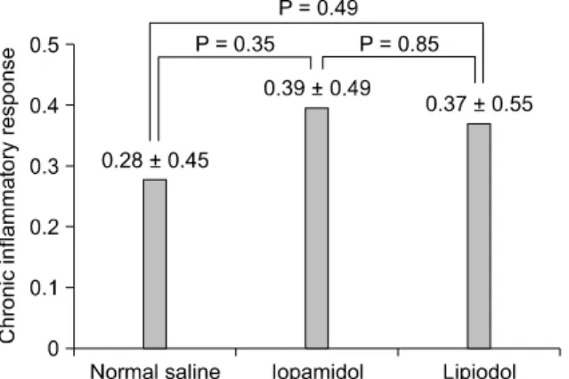
Fig. 3. Comparisons of chronic inflammatory response between groups.
Fig. 4. Comparisons of fibrosis between groups.
Statistical analysis
The SPSS ver. 11.0 (SPSS Inc., Chicago, IL, USA) was used for statistical analysis. Comparisons were made us- ing the one-way analysis of variance (ANOVA) with Tukey post hoc analysis for multiple comparisons, in- dependent t-test and chi-square test at each time point.
Statistical significance was defined as P < 0.05.
RESULTS
There were 3 mortalities after operation. One mouse died of unknown cause after injection of normal saline at POD 54, and another 2 mice in iopamidol group were kil- led by cannibalism at POD 20 and 47, respectively.
Gross observations
Adhesions were either viscera-to-viscera, viscera-to- solid organ, or viscera-to-omentum. There was no gross evidence of peritonitis in any of the groups, although mac- roscopic abscess formation was seen in two rats, one with a lipiodol-injected and one control animal, at 1 week and 2 weeks, respectively.
Microscopic observations Acute inflammatory reaction
A comparison of mean grade of acute inflammatory re- action of harvested stomach after injection is provided in Fig. 2. No significant difference of mean grade was ob- served between iopamidol and lipiodol group. However,
the mean grade after injection of lipiodol and iopamidol was significantly higher than control group (iopamidol vs.
normal saline; 1.29 ± 0.76 vs. 0.86 ± 0.74, P = 0.03, lipiodol vs. normal saline; 1.60 ± 0.93 vs. 0.86 ± 0.74, P = 0.001).
Chronic inflammatory response
The mean grade of chronic inflammatory response caused by injection of normal saline, iopamidol, and lip- iodol was the similar for all the time period (0.28 ± 0.45, 0.39 ± 0.49, 0.37 ± 0.55, P = 0.65) (Fig. 3). There were no sig- nificant difference between groups using independent t-test (normal saline vs. iopamidol, 0.28 ± 0.45 vs. 0.39 ± 0.49, P = 0.35; normal saline vs. lipiodol, 0.28 ± 0.45 vs. 0.37
± 0.55, P = 0.49; iopamidol vs. lipiodol, 0.39 ± 0.49 vs. 0.37 ± 0.55, P = 0.85).
Fibrosis
There was no significant difference between groups in
POD 1 POD 3 POD 7 POD 14 POD 28 POD 56 P-value Acute inflammatory response
Normal salinea) 1.00 ± 0.00 1.40 ± 0.54 1.00 ± 1.00 0.40 ± 0.89 0.80 ± 0.83 0.50 ± 0.57 0.32 Iopamidolb) 2.00 ± 0.70 1.40 ± 0.54 1.20 ± 0.44 1.00 ± 1.22 1.25 ± 0.50 0.75 ± 0.50 0.19 Lipiodolc) 2.00 ± 0.70 1.80 ± 0.83 1.80 ± 0.83 2.40 ± 0.54 0.80 ± 0.83 0.80 ± 0.83 0.01 Chronic inflammatory response
Normal salined) 0.00 ± 0.00 0.00 ± 0.00 0.60 ± 0.54 0.40 ± 0.54 0.60 ± 0.54 0.00 ± 0.00 0.04 Iopamidole) 0.00 ± 0.00 0.20 ± 0.44 0.60 ± 0.54 0.60 ± 0.54 0.50 ± 0.57 0.50 ± 0.57 0.32 Lipiodolf) 0.00 ± 0.00 0.00 ± 0.00 0.40 ± 0.54 0.00 ± 0.00 1.00 ± 0.00 0.60 ± 0.54 <0.01 Fibrosis
Normal salineg) 0.00 ± 0.00 0.00 ± 0.00 1.00 ± 0.70 2.20 ± 0.83 1.40 ± 1.14 1.25 ± 1.25 <0.01 Iopamidolh) 0.00 ± 0.00 1.20 ± 0.44 1.60 ± 0.89 1.00 ± 1.22 1.50 ± 0.57 1.00 ± 0.81 0.04 Lipiodoli) 0.20 ± 0.44 1.00 ± 0.70 1.20 ± 1.30 1.20 ± 0.44 1.40 ± 0.54 1.60 ± 0.54 0.08 POD, postoperative day.
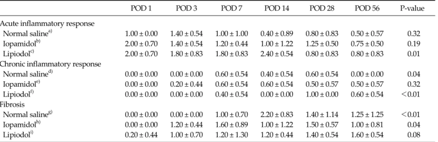
a)There are no differences between all periods according to analysis of variance (ANOVA) and Tukey post hoc analysis. b)There are no differences between all periods according to ANOVA and Tukey post hoc analysis. c)Acute inflammatory response of lipiodol injection after POD 14 was significantly higher than POD 28 and POD 56 in ANOVA and Tukey post hoc analysis. d)There are nodifferences between all periods according to ANOVA and Tukey post hoc analysis. e)There are no differences between all periods according to ANOVA and Tukey post hoc analysis. f)Chronic inflammatory response of lipiodol injection after POD 28 was significantly higher than POD 1, POD 3, POD 7 and POD 14 in ANOVA and Tukey post hoc analysis. g)Fibrosis of normal saline injection after POD 14 was significantly higher than POD 1 and POD 3 in ANOVA and Tukey post hoc analysis. h)Fibrosis of iopamidol injection after POD 7 was significantly higher than POD 1 in ANOVA and Tukey post hoc analysis. i)There are no differences between all periods according to ANOVA and Tukey post hoc analysis.
Table 1. Comparison of acute inflammatory response, chronic inflammatory response, and fibrosis between 3 groups according to injection time
Normal saline Iopamidol Lipiodol
P-valuea)
Yes No Yes No Yes No
POD 1 2 3 2 3 4 1 0.53
POD 3 2 3 1 4 3 2 0.80
POD 7 1 4 0 5 1 4 1.0
POD 14 0 5 1 4 0 5 1.0
POD 28 0 5 1 4 0 5 1.0
POD 56 0 4 0 4 0 5 N/A
Values are presented as number.
POD, postoperative day; NA, not available.
a)Chi-square test.
Table 2.Comparison of fibrinoid necrosis between groups at each time point
mean grade of fibrosis (0.97 ± 1.08, 1.04 ± 0.88, 1.10 ± 0.80, P = 0.85) (Fig. 4). No significant difference was observed between groups (normal saline vs. iopamidol, 0.97 ± 1.08 vs. 1.04 ± 0.88, P = 0.79; normal saline vs. lipiodol, 0.97 ± 1.08 vs. 1.10 ± 0.80, P = 0.59; iopamidol vs. lipiodol, 1.04 ± 0.88 vs. 1.10 ± 0.80, P = 0.77).
Changes of acute inflammation, chronic inflammation, and fibrosis at each time point
Table 1 summarizes the results of acute, chronic in- flammatory response, and fibrosis at POD 1, 3, 7, 14, 28, 56 after injection of contrast agent and normal saline. There are no significant differences at each point after injection of normal saline and iopamidol. However, acute in- flammatory response of POD 14 in lipiodol group was sig- nificantly higher than POD 28 and POD 56 using ANOVA and Tukey post hoc analysis. Similarly, in lipiodol group, chronic inflammatory response at POD 28 was sig- nificantly higher than POD 1, POD 3, POD 7 and POD 14 in ANOVA and Tukey post hoc analysis. Fibrosis of normal saline injection after POD 14 and iopamidol injection after POD 7 was significantly higher than POD 1and POD 3 in
normal saline group and POD 1 in iopamidol group using ANOVA and Tukey post hoc analysis. However, there are no differences between all periods in lipiodol group using ANOVA and Tukey post hoc analysis.
Fibrinoid necrosis
The presence of acute fibrinoid necrosis was observed in each group on POD 1, 3, 7, but did not show the difference between groups (Table 2).
Normal saline Iopamidol Lipiodol
P-valuea)
Yes No Yes No Yes No
POD 1 0 5 0 5 0 5 N/A
POD 3 0 4 0 5 4 1 0.011
POD 7 0 5 0 5 2 3 0.286
POD 14 0 5 0 5 0 5 N/A
POD 28 0 5 0 4 0 5 N/A
POD 56 0 4 0 4 0 5 N/A
POD, postoperative day; N/A, not available.
a)Chi-square test.
Table 4. Foreign body reaction (presence of foam cell)
Fig. 5. (A) Large histiocytic cells showing foamy cytoplasm in murine gastric mucosa after injection of lipiodol (post-operative 3 days). (B) Human signet ring cell carcinoma (A, H&E, ×200, B, H&E, ×400).
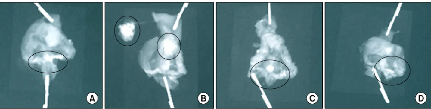
Fig. 6. Uptake of contrast agent on radiologic examination. The circled part of each figure shows the uptake of contrast agent. The uptake of iopamidol at postoperative day (POD) 1 is identified in Fig. 6A. Fig. 6B-D also show lipiodol uptake at POD 1, 3, and 7, respectively.
Normal saline Iopamidol Lipiodol
P-valuea)
Yes No Yes No Yes No
POD 1 0 5 2 3 0 5 0.286
POD 3 3 2 5 0 3 2 0.451
POD 7 3 2 2 3 4 1 0.800
POD 14 0 5 2 3 3 2 0.251
POD 28 1 4 2 2 1 4 0.600
POD 56 0 4 0 4 1 4 1.0
Values are presented as number.
POD, postoperative day.
a)Chi-square test.
Table 3. Comparison of mesothelial hyperplasia between groups at each time point
Mesothelial hyperplasia
There was no significant difference in mesothelial hy- perplasia at each time points between groups (Table 3).
Foreign body reaction
Especially, foam cell resulting from foreign body re-
action was observed in lipiodol group, and statistically significant differences was identified in POD 3 (Table 4, Fig. 5) (P = 0.011).
High resolution X- ray examination (using mammography) Uptake of iopamidol was identified on radiologic ex- amination on POD 1, but since then, no more uptake was
Iopamidol uptake Lipiodol uptake
P-valuea)
Yes No Yes No
POD 1 2 3 5 0 0.167
POD 3 0 5 2 3 0.444
POD 7 0 5 1 4 1.0
POD 14 0 5 0 5 N/A
POD 28 0 4 0 5 N/A
POD 56 0 4 0 5 N/A
Values are presented as number.
POD, postoperative day; NA, not available.
a)Chi-square test.
Table 5. X-ray examination (presence of contrast uptake)
found in iopamidol group. However, the uptake of lip- iodol was present until POD 7 (Table 5, Fig. 6).
DISCUSSION
Surgeons frequently express concerns about complica- tions from gastric cancer surgery in patients receiving large area lymph node dissection. Location of lymph node metastasis from gastric cancer is reported to be distributed widely as a result of the complicated perigastric lymphatic network. Generally, when the depth of invasion confined to mucosa in early gastric cancer, the rate of lymph node metastasis have been reported as 1 to 3%, and submucosa as 11 to 20% [17-19]. However, because preoperative diag- nostic techniques, including CT and endoscopic ultra- sonography, do not provide an accurate prediction of metastasis in the regional lymph nodes, gastrectomy with extensive lymphadenectomy (D2 or D2 + a) is still consid- ered as a standard surgical treatment for early gastric can- cer in some centers [20]. In addition to prolonged oper- ation time and hospital stay, the operative complications accompanying extended lymphadenectomy, such as leak- age of anastomosis, bleeding, pancreatitis, intra-abdomi- nal abscess, leakage of lymphatics cannot be trivialized [21,22]. Therefore, limited surgery such as laparoscopic wedge resection with limited regional lymph node dis- section was attempted by Ohgami et al. [23] to overcome the complication and improve the quality of life in patients with early gastric cancer.
There are several methods of detecting the sentinel
node. However, it is more difficult to detect the sentinel node in gastric cancer, because the lymphatic drainage of the stomach is considerably more complex than that of ec- todermal organs like breast and skin due to the complex embryological development. Although it has been consid- erable debate on the advantages and disadvantages of dif- ferent detection methods, a growing number of inves- tigators used radiocolloid or a combination of both meth- ods in more recent studies [24,25]. However, this method has the disadvantage of impossibility of predicting the ac- curate locations of primary sentinel nodes preoperatively because of the limited spatial images and the lack of dis- tinct anatomy of the surrounding structures. Another problem which may occur are that use of radioisotope needs a special facilities, detection of correct sentinel node needs an experience and a technical learning curve, and the detection of radioactive lymph nodes adjacent to the injection site is difficult because of the shine-through effect.
Although there is insufficient evidence for applying CT lymphography to gastric cancer, recent studies have shown that CT lymphography can be an alternative SLN navigation surgery method for esophageal cancer [26]. In that study, they identified SLNs with 100% sensitivity by CT lymphography. However, another study of CT lym- phography for gastric cancer reported only a 30% de- tection rate. Not only the technical aspects of CT lymphog- raphy, but also the tumor characteristics are important fac- tors for achieving a higher rate of SLN detection in gastro- intestinal cancers.
A technique using interstitial CT lymphography with the widely available iodine contrast medium such as iopa- midol or lipiodol is an alternative method for preoperative sentinel node mapping and biopsy examination with a scintigraphic method [14,27,28]. Suga et al. [28] showed an interstitial CT lymphography with endoscopic mucosal injection of iopamidol was applicable for sentinel node navigation of superficial esophageal cancer. In this man- ner, it may be useful for planning the operative field and limited lymph node dissection, and for avoiding un- necessary extended lymph node dissection if surgeons identify the preoperative visualization of lymphatic spread and sentinel node draining from the primary can-
cer on CT lymphography. However, there is a limitation of application of this method to clinical practice, because tis- sue reactions after injection of iodinated contrast media have not been reported recently.
Our experimental investigation was performed to eval- uate the response of gastric wall histology to direct effects of iopamidol or lipiodol. The degree of safety in using a contrast media needs to be determined before intelligent contrast media choices can be made. Our study demon- strated a lesser pathological response with iopamidol than lipiodol. We presume that this may be due to the wa- ter-soluble characteristics and relatively rapid wash-out nature of iopamidol. However, we did confirm that ap- pearance of foreign body reaction, such as foam cell, after injection of lipiodol can make confuse the diagnosis of sig- net ring cell carcinoma.
A frozen section examination of the proximal cut-end adjacent to the lesion is an important step during gastrectomy. Previous reports have demonstrated that a positive margin is associated with a worse outcome [29-31]. Injection of contrast media into specimens for CT lymphography can cause a tissue reaction, including acute and chronic inflammation and a foreign body reaction.
This process can create confusion when performing frozen and routine pathological examinations. These findings prompted us to investigate the histological reactions after injection of contrast media during CT lymphography.
Another purpose of this study was to determine how long contrast media can remain in tissue. We examined high-resolution X-rays using mammography after inject- ing contrast media. As a result, we found that lipiodol re- mained in the tissue much longer than iopamidol.
Because we only focused on the feasibility of using con- trast media during CT lymphography, we cannot validate the clinical feasibility of CT lymphography. We only found potential difficulties with contrast media during CT lymphography. Further study including SLN mapping techniques is needed to show clinical feasibility and to provide the clinical significance of SLN detection using CT lymphography.
In conclusion, we conclude that iopamidol and lipiodol when used as a contrast media of CT lymphography is an available material of preoperative sentinel node navi-
gation surgery for gastric cancer with a acceptable in- cidence of pathological alterations in an mouse model, and our results are potentially useful to clinical (human) application.
However, these agents can produce an acute inflam- matory reaction within 7 days after injection. In particular, there is a possibility of causing a foreign body reaction af- ter lipiodol injection from POD 3.
CONFLICTS OF INTEREST
No potential conflict of interest relevant to this article was reported.
ACKNOWLEDGEMENTS
This study was supported by a grant of the Korea Healthcare Technology R&D Project, Ministry of Health &
Welfare, Republic of Korea (A060151).
REFERENCES
1. Cabanas RM. An approach for the treatment of penile carcinoma. Cancer 1977;39:456-66.
2. Giuliano AE, Jones RC, Brennan M, Statman R. Sentinel lymphadenectomy in breast cancer. J Clin Oncol 1997;15:
2345-50.
3. Veronesi U, Paganelli G, Galimberti V, Viale G, Zurrida S, Bedoni M, et al. Sentinel-node biopsy to avoid axillary dis- section in breast cancer with clinically negative lymph- nodes. Lancet 1997;349:1864-7.
4. Bass SS, Cox CE, Ku NN, Berman C, Reintgen DS. The role of sentinel lymph node biopsy in breast cancer. J Am Coll Surg 1999;189:183-94.
5. Kitagawa Y, Kitajima M. Gastrointestinal cancer and senti- nel node navigation surgery. J Surg Oncol 2002;79:188-93.
6. Schwartz GF, Giuliano AE, Veronesi U; Consensus Confer- ence Committee. Proceedings of the consensus conference on the role of sentinel lymph node biopsy in carcinoma of the breast, April 19-22, 2001, Philadelphia, Pennsylvania.
Cancer 2002;94:2542-51.
7. Morton DL, Wen DR, Wong JH, Economou JS, Cagle LA, Storm FK, et al. Technical details of intraoperative lym- phatic mapping for early stage melanoma. Arch Surg 1992;127:392-9.
8. Borgstein PJ, Pijpers R, Comans EF, van Diest PJ, Boom RP,
Meijer S. Sentinel lymph node biopsy in breast cancer:
guidelines and pitfalls of lymphoscintigraphy and gamma probe detection. J Am Coll Surg 1998;186:275-83.
9. Kitagawa Y, Fujii H, Mukai M, Kubota T, Otani Y, Kitajima M. Radio-guided sentinel node detection for gastric cancer.
Br J Surg 2002;89:604-8.
10. Méndez J, Wallace AM, Hoh CK, Vera DR. Detection of gastric and colonic sentinel nodes through endoscopic ad- ministration of 99mTc-DTPA-mannosyl-dextran in pigs. J Nucl Med 2003;44:1677-81.
11. Yasuda S, Shimada H, Chino O, Tanaka H, Kenmochi T, Takechi M, et al. Sentinel lymph node detection with Tc-99m tin colloids in patients with esophagogastric cancer. Jpn J Clin Oncol 2003;33:68-72.
12. Moghimi SM, Bonnemain B. Subcutaneous and intra- venous delivery of diagnostic agents to the lymphatic sys- tem: applications in lymphoscintigraphy and indirect lymphography. Adv Drug Deliv Rev 1999;37:295-312.
13. Wisner ER, Katzberg RW, Koblik PD, McGahan JP, Griffey SM, Drake CM, et al. Indirect computed tomography lym- phography of subdiaphragmatic lymph nodes using iodi- nated nanoparticles in normal dogs. Acad Radiol 1995;2:
405-12.
14. Hayashi H, Tangoku A, Suga K, Shimizu K, Ueda K, Yoshino S, et al. CT lymphography-navigated sentinel lymph node biopsy in patients with superficial esophageal cancer. Surgery 2006;139:224-35.
15. Ueda K, Suga K, Kaneda Y, Li TS, Ueda K, Hamano K.
Preoperative imaging of the lung sentinel lymphatic basin with computed tomographic lymphography: a prelimi- nary study. Ann Thorac Surg 2004;77:1033-7.
16. Kumar V. Acute and chronic inflammation. In: Kumar V, Abbas AK, Fausto N, Robbins SL, Cotran RS, editors.
Robbins and Cotran pathologic basis of disease. 7th ed.
Philadelphia: Elsevier Saunders; 2005. p.47-86.
17. Adachi Y, Shiraishi N, Kitano S. Modern treatment of early gastric cancer: review of the Japanese experience. Dig Surg 2002;19:333-9.
18. Borie F, Millat B, Fingerhut A, Hay JM, Fagniez PL, De Saxce B. Lymphatic involvement in early gastric cancer:
prevalence and prognosis in France. Arch Surg 2000;135:
1218-23.
19. Roviello F, Rossi S, Marrelli D, Pedrazzani C, Corso G, Vindigni C, et al. Number of lymph node metastases and its prognostic significance in early gastric cancer: a multi- center Italian study. J Surg Oncol 2006;94:275-80.
20. Hayes N, Karat D, Scott DJ, Raimes SA, Griffin SM. Radical
lymphadenectomy in the management of early gastric cancer. Br J Surg 1996;83:1421-3.
21. Yonemura Y, Wu CC, Fukushima N, Honda I, Bandou E, Kawamura T, et al. Operative morbidity and mortality af- ter D2 and D4 extended dissection for advanced gastric cancer: a prospective randomized trial conducted by Asian surgeons. Hepatogastroenterology 2006;53:389-94.
22. Smith JW, Shiu MH, Kelsey L, Brennan MF. Morbidity of radical lymphadenectomy in the curative resection of gas- tric carcinoma. Arch Surg 1991;126:1469-73.
23. Ohgami M, Otani Y, Kumai K, Kubota T, Kim YI, Kitajima M. Curative laparoscopic surgery for early gastric cancer:
five years experience. World J Surg 1999;23:187-92.
24. Nakahara T, Kitagawa Y, Yakeuchi H, Fujii H, Suzuki T, Mukai M, et al. Preoperative lymphoscintigraphy for de- tection of sentinel lymph node in patients with gastric can- cer--initial experience. Ann Surg Oncol 2008;15:1447-53.
25. Kitagawa Y, Saha S, Kubo A, Kitajima M. Sentinel node for gastrointestinal malignancies. Surg Oncol Clin N Am 2007;16:71-80.
26. Yuasa Y, Seike J, Yoshida T, Takechi H, Yamai H, Yamamoto Y, et al. Sentinel lymph node biopsy using intraoperative indocyanine green fluorescence imaging navigated with preoperative CT lymphography for superficial esophageal cancer. Ann Surg Oncol 2011 Jul 27. [Epub]. DOI:10.1245/
s10434-011-1922-x.
27. Tangoku A, Seike J, Nakano K, Nagao T, Honda J, Yoshida T, et al. Current status of sentinel lymph node navigation surgery in breast and gastrointestinal tract. J Med Invest 2007;54:1-18.
28. Suga K, Shimizu K, Kawakami Y, Tangoku A, Zaki M, Matsunaga N, et al. Lymphatic drainage from esoph- agogastric tract: feasibility of endoscopic CT lymphog- raphy for direct visualization of pathways. Radiology 2005;237:952-60.
29. Songun I, Bonenkamp JJ, Hermans J, van Krieken JH, van de Velde CJ. Prognostic value of resection-line involvement in patients undergoing curative resections for gastric cancer. Eur J Cancer 1996;32A:433-7.
30. Kakeji Y, Tsujitani S, Baba H, Moriguchi S, Mori M, Maehara Y, et al. Clinicopathologic features and prognostic significance of duodenal invasion in patients with distal gastric carcinoma. Cancer 1991;68:380-4.
31. Papachristou DN, Agnanti N, D'Agostino H, Fortner JG.
Histologically positive esophageal margin in the surgical treatment of gastric cancer. Am J Surg 1980;139:711-3.