http://dx.doi.org/10.4174/astr.2015.89.2.102 Annals of Surgical Treatment and Research
Necrotizing fasciitis of the breast in a pregnant woman successfully treated using negative-pressure wound therapy
Jina Lee, Kwan Ju Lee, Woo Young Sun
Department of Surgery, Daejeon St. Mary’s Hospital, The Catholic University of Korea College of Medicine, Daejeon, Korea
INTRODUCTION
Necrotising fasciitis (NF) is the most aggressive form of soft tissue infection which involves necrosis of subcutaneous fat and spreads along the fascial planes. NF can occur in any part of the body with or without injury. In this report, we present the first case of NF of the breast in a pregnant woman successfully treated using negative-pressure wound therapy.
CASE REPORT
A 31-year-old woman in 33 weeks of pregnancy presented to the Emergency Department with a 2-day history of pain in the left breast and fever. The patient had previously given birth vaginally to a single child with no complications. On physical examination, the patient had a fever (temperature, 39.0oC), tachypnea (32 breaths/min), tachycardia (heart rate, 132 beats/
min), and a blood pressure of 90/50 mmHg. The whole left
breast was swollen, and the skin over the left breast, especially the outer-central area, was reddish. There was no fluctuation, but the breast was extremely painful. No palpable masses could be felt in the left breast, and the axillary lymph nodes were not palpable. On initial admission, laboratory evaluation revealed leukocytosis (18.4 × 10³/L), an increased CRP level (41.1 mg/
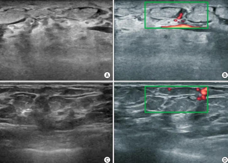
L), and an increased ESR (108 mg/L). Ultrasound of the breasts showed marked subcutaneous edema, diffuse edema of the glandular tissue, intraglandular fluid collection, bright echo, and decreased superficial blood flow (Fig. 1).
The initial impression was acute mastitis. The patient was resuscitated with intravenous fluids, and empiric treatment was begun with cefazolin 1.0 g and clindamycin 600 mg every 8 hours intravenously. Physical examination one day later revealed skin discoloration (Fig. 2A), and the patient described unbearable pain radiating to the left flank. Hence, necrotizing fasciitis (NF) was suspected, and emergency operation (excision of necrotic skin and debridement with sparing of the nipple- Necrotizing fasciitis (NF) is a rare and rapidly progressive disease involving the skin, subcutaneous tissue, and deep soft tissue. Although NF can occur any part of the body, the breast is an uncommon primary site for NF, and its occurrence in the breast during pregnancy has never previously been reported. Here, we report the case of a healthy 31-year- old pregnant woman who presented with NF of the left breast that was successfully treated with breast-conserving debridement and secondary wound closure using negative-pressure wound therapy.
[Ann Surg Treat Res 2015;89(2):102-106]
Key Words: Necrotizing fasciitis, Breast, Pregnancy, Negative-pressure wound therapy
Reviewed January February March April May June July August September October November December
Received February 9, 2015, Revised April 28, 2015, Accepted May 12, 2015
Corresponding Author: Woo Young Sun
Department of Surgery, Daejeon St. Mary’s Hospital, The Catholic University of Korea College of Medicine, 64 Daeheung-ro, Jung-gu, Daejeon 301-723, Korea
Tel: +82-42-220-9268, Fax: +82-42-220-9565 E-mail: [email protected]
Copyright ⓒ 2015, the Korean Surgical Society
cc Annals of Surgical Treatment and Research is an Open Access Journal. All articles are distributed under the terms of the Creative Commons Attribution Non- Commercial License (http://creativecommons.org/licenses/by-nc/4.0/) which permits unrestricted non-commercial use, distribution, and reproduction in any medium, provided the original work is properly cited.
areolar complex as it was nonnecrotic) was performed. The intraoperative findings were highly suggestive of NF because the deep and superficial fat layers showed liquefied necrosis, and there was loss of normal resistance of the fascia to finger dissection (positive “finger test”).
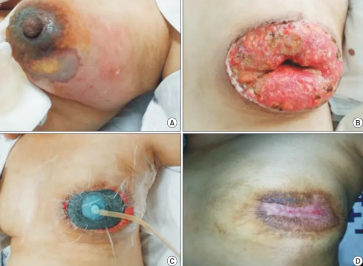
Microscopic examination of the surgical specimen revealed coagulative and liquefactive necrosis involving the skin, subcutaneous fat, and breast tissue, and the patient was ultimately diagnosed with NF. There was no growth in blood cultures, and Streptococcus pyogenes grew in the necrotic tissue culture. Seven days after the initial operation, the nipple-areolar complex turned necrotic and was subsequently excised under local anesthesia. Primary closure of the wound was not possible due to the large skin defect (Fig. 2B). On postoperative day 6, the wound defect was covered with negative-pressure wound therapy (NPWT; CuraVac, Daewoong Pharmaceutical Co., Seoul, Korea) system that provided a continuous vacuum force (120 mmHg) across a closed wound dressing (Fig. 2C). The NPWT dressing was changed two or three times a week for 1 month.
The patient delivered a healthy baby in her 39th week, while still undergoing NPWT treatment. After the NPWT dressings had been applied for 4 weeks, the open wound had shrunk in size significantly. Then, a silicon adhesive foam dressing was applied twice a week. At the follow-up visit 2 months after her initial presentation with symptoms, the patient had no local or systemic signs of infection, and her healed state was demonstrated by progressive re-epithelialization (Fig. 2D).
This study was approved by Institutional Review Board of the Daejeon St. Mary’s Hospital, The Catholic University College of Medicine (IRB No. DC15ZISE0004).
DISCUSSION
NF is a rare disease but is associated with systemic toxicity and a high death rate despite aggressive treatment. Although there have been significant advances in our understanding of this disease and great improvements in treatment, the mortality rate has not changed in the last 30 years and remains 25% to
A B
C D
Fig. 1. Ultrasound image of necrotizing fasciitis. Ultrasound revealing marked subcutaneous and glandular edema, intraglandular fluid collection, brighter echo (A), and decreased superficial blood flow in Doppler sonogram (B) compared to that in the contralateral normal breast (C, D).
35%; the rate is directly proportional to the time to intervention [1]. Suspecting and diagnosing potential cases of NF as early as possible are the most important tactics in the approach to this disease, since aggressive surgery and appropriate antibiotics are required. In the treatment of NF in the breast, mastectomy has been reported to be the main treatment in over 90% of cases in the published literature [2]. Most patients with NF initially experience severe pain and tenderness with respect to clinical findings and fever. However, the skin might have normal appearance in the early stages of the disease because the infection tracks the subcutaneous tissue. Skin changes will become evident only with resulting skin ischemia, usually a late feature of the disease. In our case, acute mastitis was first suspected due to the presence of no signs of skin discoloration, and the rapidly exacerbating pain and bluish discoloration subsequently led us to perform an emergency operation under the diagnosis of NF. Gram staining and culture may be helpful, but these procedures can delay appropriate therapy.
Standard NF risk factors include: chronic debilitating comor- bidities (diabetes mellitus, peripheral vascular disease, smoking, alcohol abuse, liver disease, obesity, and immunosuppression) and conditions compromising skin integrity (surgery, trauma, burn, intravenous drug use, biopsy, pressure ulcers, and chronic skin disease) [3,4]. Breast cancer, operation, wound dehiscence, and previous biopsies are other possible risk factors that could facilitate the development of NF in breast [2,5]. Though pregnancy can induce a mildly immunosuppressive state, there had been no previous reported cases of NF during pregnancy. In our case, the patient was previously healthy and had no other risk factors for NF.
Depending on the pathogen, NF may be categorized into three types. Type 1 infections are the most common form of disease and are polymicrobial in nature. Type 2 comprises monomicrobial infections caused by group A beta-hemolytic streptococci. Type 3 NF is less common and involves skin wounds with infections caused by marine insects hosting Vibrio
A B
C D
Fig. 2. (A) Necrotizing fasciitis of the left breast demonstrating edema, inflammation, and an area of necrosis. (B) Postoperative photograph demonstrating open wound after removal of the nipple-areolar complex. (C) Wound defect closed with negative- pressure wound therapy (NPWT) on postoperative day 6. (D) Healed wound after 4 weeks of application of NPWT on postoperative day 62.
vulnificus [3]. Type 1 NF occurs most frequently following abdominal and perineal surgeries in patients with predisposing factors such as diabetes mellitus, obesity, high comorbid index scores, and atherosclerosis, as well as in patients who are immunocompromised, while type 2 NF has been associated with minor injuries and breaks in the skin and usually occurs in individuals without underlying disease [6]. We dealt with a type 2 infection without any underlying disease, although type 1 infection has been reported in over half of the cases in breast [2].
The Laboratory Risk Indicator for Necrotizing Fasciitis score can be utilized to stratify patients presenting with signs of mastitis to determine the likelihood of NF. Wong et al.
[7] proposed a practical scoring system that uses six easily obtainable blood tests (CRP, total white cell count, hemoglobin, sodium, creatinine, and glucose). In their system, a score >6 of 13 indicates that NF should be seriously considered, and they also showed that the positive predictive value for NF increases as the score rises above 7. In the case described here, a score of 8 was obtained. However, currently, the Laboratory Risk Indicator for Necrotizing Fasciitis score remains unvalidated in larger, prospective studies.
Yen et al. [8] have described the sonographic findings of NF as diffuse thickening of the subcutaneous tissue accompanied by a layer of fluid accumulation more than 4 mm in depth along the deep fascial layer, when compared with the contralateral position on the corresponding normal limb. Hanif and Bradley [9] reported that focal bright echoes can be seen due to microbubbles of gas in advanced cases. In our case, a diffuse brighter echo was observed in the affected breast compared to that in the contralateral healthy breast on the initial admission.
Limited data on the diffuse brighter echo exist. However, providers should include NF in the differential diagnosis, and consider the possible consequence of necrosis, when ultrasound shows a brighter echo in the diseased breast. As a different radiological method to diagnose NF, MRI has a high sensitivity of 80%–100% but a low specificity of 46%–86% [9]. MRI may not be readily available in an emergent setting and can be time- consuming.
Intraoperatively, an incision should be made down the pec-
toralis muscle over the area of maximal tenderness and most obvious skin involvement. Few data exist on the optimal method of wound management following debridement. As with all infected wounds, the wound should be left open and treated with wet-to-dry dressings initially. NPWT has become a common method of treating large wounds once infection is controlled, although there have been no well-designed studies evaluating the role of NPWT in patients with large wounds following infection [1]. NPWT increases local oxygenation by enhancement of dermal perfusion and accelerates formation of granulation tissue by stimulating fibroblasts [10]. NPWT has been used for lesions in the breast such as chest wall defects following bilateral NF and salvage of infected breast implants [2].
There was a case report describing NF of the breast following a core needle biopsy, in which surgical debridement and NPWT allowed breast conservation after split thickness skin graft [5].
Skin graft requires considerable skill and donor-site surgery.
Furthermore, the results are usually less cosmetically attractive and graft surgery still carries a risk of infection. In our case, split thickness skin graft was also considered, but we decided to allow the wound to heal by secondary closure to avoid a second operation because the patient was pregnant. About 2 months were required for full recovery of the open wound, which might be comparable to the length of time required for healing following a second graft operation.
To the best of our knowledge, this is the first case of NF of the breast in a pregnant woman, whose wound defect was successfully managed by secondary closure using NPWT. It is reasonable to assume that this case demonstrates the benefits of secondary wound closure using NPWT. These benefits are enhanced by the fact that providers can avoid a second skin graft operation, including donor-site surgery, for wound closure.
Secondary wound closure using NPWT can be a viable option in the management of huge wound defects.
CONFLICTS OF INTEREST
No potential conflict of interest relevant to this article was reported.
1. Sarani B, Strong M, Pascual J, Schwab CW.
Necrotizing fasciitis: current concepts and review of the literature. J Am Coll Surg 2009;208:279-88.
2. Angarita FA, Acuna SA, Torregrosa L,
Tawil M, Sanchez EF, Heilbron O, et al.
Bilateral necrotizing fasciitis of the breast following quadrantectomy. Breast Cancer 2014;21:108-14.
3. Salcido RS. Necrotizing fasciitis: reviewing
the causes and treatment strategies. Adv Skin Wound Care 2007;20:288-93.
4. Cho YS, Yang HT, Yim H, Park JM, Kim D, Hur J, et al. Necrotizing fasciitis follow- ing a small burn. J Korean Surg Soc 2010;
REFERENCES
Low CO. The LRINEC (Laboratory Risk Indicator for Necrotizing Fasciitis) score:
a tool for distinguishing necrotizing fas- ciitis from other soft tissue infections.
Crit Care Med 2004;32:1535-41.
8. Yen ZS, Wang HP, Ma HM, Chen SC, Chen WJ. Ultrasonographic screening of cli- nically-suspected necrotizing fasciitis.
Acad Emerg Med 2002;9:1448-51.
79:71-4.
5. Flandrin A, Rouleau C, Azar CC, Dubon O, Giacalone PL. First report of a necrotising fasciitis of the breast following a core needle biopsy. Breast J 2009;15:199-201.
6. Elliott D, Kufera JA, Myers RA. The mi- crobiology of necrotizing soft tissue infec- tions. Am J Surg 2000;179:361-6.
7. Wong CH, Khin LW, Heng KS, Tan KC,
9. Hanif MA, Bradley MJ. Sonographic find- ings of necrotizing fasciitis in the breast. J Clin Ultrasound 2008;36:517-9.
10. Liao EC, Breuing KH. Breast mound sal- vage using vacuum-assisted closure de vice as bridge to reconstruction with infe- rolateral AlloDerm hammock. Ann Plast Surg 2007;59:218-24.