• 검색 결과가 없습니다.

회전근개 파열의 수술적 치료 후 회전근개 초음파 소견

N/A
N/A
Protected

Academic year: 2021

Share "회전근개 파열의 수술적 치료 후 회전근개 초음파 소견"

Copied!
6
0
0

로드 중.... (전체 텍스트 보기)

전체 글

(1)

https://doi.org/10.35827/cp.2020.19.2.64

접수일 : 2020 년 11 월 22 일 , 게재승인일 : 2020 년 12 월 11 일 책임저자 : 권동락 , 대구시 남구 두류공원로 17 길 33

42472, 대구가톨릭대학교 의과대학 재활의학교실

Tel: 053-650-3627, Fax: 053-622-4687 E-mail: [email protected]

회전근개 파열의 수술적 치료 후 회전근개 초음파 소견

대구가톨릭대학교 의과대학 재활의학교실

권 동 락

Postoperative Ultrasound Findings of the Rotator Cuff Tendon after Arthroscopic Repair of a Rotator Cuff Tear

Dong Rak Kwon, M.D., Ph.D.

Department of Rehabilitation Medicine, Catholic University of Daegu School of Medicine, Daegu, Korea

Ultrasound (US) imaging is an efficient, easy to use, rapid, dynamic, noninvasive, with rare side-effects and inexpensive tool allowing for facilitated diagnosis and management of the painful shoulder. It also has advantages over other imaging modalities in the evaluation of the postoperative shoulder for rotator cuff integrity and correct anchor and suture placement, as well as rotator cuff analysis following repair surgery. Early postoperative tendons frequently had a hypo- echoic echo texture and the absence of a fibrillar pattern, which might be misinterpreted as recurrent tears. however, these features often normalized into tendons with an increased echo texture and the reappearance of a fibrillar pattern at 6 months. Based on these sequential findings, the US findings within 3 months after surgery should be interpreted with caution to accurately understand and monitor the repaired tendon status. (Clinical Pain 2020;19:64-69)

Key Words: Rotator cuff, Tear, Musculoskeletal ultrasound, Repair

서 론

회전근개는 극상근, 극하근, 소원근, 견갑하근의 네 개의 근육으로 구성되어 있으며, 모두 견갑골에서 시작하여 상 완골의 대결절과 소결절에 부착하며, 상완골두와 견봉 사 이에 위치한다. 회전근개의 주요 기능은 견관절이 운동 시 상완골두의 아래로 잡아당기는 근육으로 작용하여 상완골 두가 관절와의 중심으로 향하도록 압박하여 상완관절의 안 정 기전에 능동적으로 기여하며, 상완골의 회전에도 참여 한다.

회전근개 힘줄파열은 매년 25만 건 이상의 회전근개 봉 합술을 하는 미국에서 만성 어깨통증의 가장 흔한 원인 중 하나로 보고되고 있다.

1

사체연구에서 60세 미만의 경우 전 층회전근개 파열의 발병률이 6%인 반면, 60세 이상의 경우 는 발병률이 30%인 것으로 보고되었다.

2

회전근개 파열은 많은 환자들에게 상당한 장애를 초래하고 삶의 질을 떨어 뜨린다.

3

회전근개 파열의 치료는 운동, 전기 치료, 침, 도수치료, 테이핑, 스테로이드 및 혈소판이 풍부 혈장치료, 줄기세포 치료, 폴리디옥시리보뉴클레오티드, 콜라겐 주사 등 보존 적치료법과 수술적 치료가 있다.

4-9

회전근개 수술은 정형외 과영역에서 가장 흔히 시행되는 수술 중 하나이다. 이에 본 종설에서는 회전근개 수술(봉합술) 후 적절한 힘줄의 치유 과정에서 보이는 초음파 소견에 대해 알아보고, 이를 바탕 으로 임상에서 초음파의 효용성에 대해 기술하고자 한다.

본 론 1. 회전근개 파열의 분류

신뢰할 수 있는 분류법을 사용한 회전근개 파열의 분류 는 회전근개 질환의 치료에 매우 중요하다. 이러한 분류는 각 회전근개 파열 유형별로 최상의 결과를 얻기 위한 적절 한 치료법을 결정하는 데 사용되었다.

DeOrio와 Cofield

10

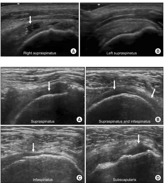
는 회전근개 전층파열에 대한 분류 시스템을 개발했는데, 수술시 상완골두에서 떼어낸 힘줄의 앞쪽-뒤쪽 길이 측정이 포함되었다. 이들의 시스템은 힘줄 파열 크기가 1 cm 미만일 경우 작은(small), 1∼3 cm일 경 우 중간(medium), 3∼5 cm일 경우 큰(large), 5 cm 이상일 경우 광범위(massive) 등으로 분류했다(Fig. 1).

Southern California Orthopaedic Institute는 이와 유사한

(2)

Fig. 1. Longitudinal view of supra- spinatus tendon (A) supraspinatus tendon full thickness tear (white arrow, 2.3 cm, medium): “sagging peribursal fat sign” and cortical irregularities. (B) Normal supra- spinatus tendon.

Fig. 2. Representative image of massive full thickness rotator cuff tendon tear. (A) Longitudinal view of supraspinatus tendon full thick- ness tear (white arrow): “sagging peribursal fat sign” and cortical irregularities. (B) Transverse view of supraspinatus and infraspinatus tendon full thickness tear: naked tuberosity (white arrows). (C) Longi- tudinal view of infraspinatus ten- don full thickness tear (white arrow).

(D) Longitudinal view of sub- scapularis tendon full thickness tear (white arrow).

분류의 작은(pinhole), 중간(< 2 cm, 수축(retraction)이 없 는 하나의 힘줄만 포함), 큰(수축이 있는 전체 힘줄), 광범 위(수축이 있는 두 개 이상의 힘줄)로 정의한다(Fig. 2).

11

2. 조기수술 대상 및 목적

조기 수술적 치료가 고려되는 경우는 급성 파열의 경우, 그리고 65세 미만의 환자의 경우, 만성적인 근육의 큰 변화 (근 위축, 지방 침윤, 그리고 봉합부위의 생체역학적 특성 의 손상) 없이 1 cm 이상의 회복 가능한 퍄열과 파열 진행 의 위험성과 보존적 치료의 실패율

12

이 높은 큰 윤활낭 파 열(힘줄두께의 6 mm 이상 또는 50% 이상)에서도 고려할 수 있다.

회전근개 수술의 목적은 첫째, 통증을 없애는 것이고, 둘 째, 회전근개 기능 및 근력을 회복시키는 것이다. 회전근개 손상의 크기에 따라 차이가 있으나 회전근개 봉합술 후 힘

줄 회복 실패율은 약 20∼90% 정도로 보고되고 있다.

13

3. 회전근개 파열 봉합술 후 영상학적 검사 회전근개 봉합의 온전함(integrity) 및 해부학적 결과를 평가하려면 봉합된 힘줄의 영상 평가가 중요하다. 자기공 명영상(MRI)은 진단 정확도가 매우 높아 수술 전 회전근개 를 평가하기 위한 가장 좋은 영상촬영 형식이지만, 수술 후 에는 수술 시 사용한 봉합사 및 봉합 나사, 수술로 인한 연 부조직 및 골 조직의 변화, 높은 비용 등으로 인한 수술 후 회전근개 평가에는 한계가 있는 것으로 알려져 있다.

14-16

초음파는 봉합 재료가 있는 상태에서 회전근개를 평가하

기 위한 대안적 영상 촬영 양식으로 제안되어 역동적, 기능

적, 해부학적 평가가 가능하다.

14,17

또한 필요할 경우 수술

후 다양한 시간에 반복적으로 초음파를 수행할 수 있다. 그

러나 수술 후 힘줄이 봉합물, 수술 후 염증, 과립조직 등

(3)

Fig. 3. Images from a 55-year-old man with an initial supraspinatus tendon tear. Longitudinal view at 6 weeks after surgery (A) repaired cuff with sufficient thickness (> 2 mm) associated with partial hy- po-echogenicity (B) shows no in- creased vascularity.

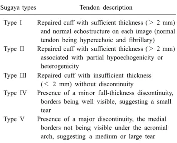
Table 1. Sugaya Classification for Tendon Healing

Sugaya types Tendon description

Type I Repaired cuff with sufficient thickness (> 2 mm) and normal echostructure on each image (normal tendon being hyperechoic and fibrillary) Type II Repaired cuff with sufficient thickness (> 2 mm)

associated with partial hypoechogenicity or heterogenicity

Type III Repaired cuff with insufficient thickness (< 2 mm) without discontinuity

Type IV Presence of a minor full-thickness discontinuity, borders being well visible, suggesting a small tear

Type V Presence of a major discontinuity, the medial borders not being visible under the acromial arch, suggesting a medium or large tear 다양한 수술 후 변화로 인해 변형될 수 있기 때문에 초음파 로 힘줄의 수술 후 외관을 해석하는 데 어려움이 지속되고 있다. 수술 후 정상적인 외관, 초음파를 사용한 수술 후 봉 합된 힘줄의 시간적 변화 또는 도플러 분석을 통해 봉합된 회전근개 힘줄의 혈관 반응 등에 대한 연구 보고가 있 다.

14,18-23

이전 연구에서 회전근개 질환에 대한 수술적 치료 후 통증 을 호소하는 환자에 대해 수술 후 회전근개의 상태를 평가하 는 데 있어 적용할 수 있으며 재수술 시 시행되는 관절경 소 견과 비교해 볼 때 초음파는 민감도가 91%, 특이도가 86%, 수 술 후 회전근개 상태 평가 정확도가 89%로 보고하였다.

24

4. 회전근개 파열 봉합술 후 초음파소견

초음파검사는 특히 회전근개 봉합 수술 후 어깨를 분석 하는 데 유용한 영상검사이다. 회전근개 파열의 봉합 수술 후 봉합 온전함의 초음파 평가는 수술 후 어깨 기능의 예측 에 도움이 되는 것으로 보고하였다.

25

연구에서는 봉합 수 술 후 봉합 온전함을 Sugaya 초음파 분류법(Table 1) 5단계

(I-V)로 분류하였고, 어깨 기능평가는 세 가지 등급 시스템 (Constant 점수, UCLA 및 어깨 주관척도)으로 평가하였다.

결과는 I 유형 봉합이 다른 모든 유형보다 기능적 결과가 유의하게 높은 것으로 나타났으며, V 유형 봉합(68.7)이 II, III 및 IV 유형 봉합(각각 80.2, 71.1, 71.0)보다 기능 결과가 좋지 않았다. 봉합 유형과 가장 잘 상관된 기능평가 등급 시스템은 Constant 점수였다.

수술 3개월 후 봉합된 힘줄의 초음파 평가가 중요할 수 있는데, 이때의 임상 검사는 움직이지 않고 통증이 있어 제 한적이고, 대부분의 재발된 파열은 수술 후 처음 3개월 이 내에 발생하기 때문이다. 또한, 수술 후 힘줄은 정상적인 섬유질 패턴이 없는 저에코형태, 힘줄주변부 혈관성(힘줄 은 상대적 무혈관성)을 분석하는 것으로 설명되어 왔으며, 이 세 가지 특성은 6개월에 걸쳐 정상화가 된다고 알려져 있다.

19,26

Fealy 등

20

는 또한 수술 후 초기에는 초음파에서 전층 파 열을 발견하는 것이 드문 일이 아니라고 보고했다. 그러나 그들은 시간이 지남에 따라 점차 좋아지기 때문에 이러한 결함은 진정한 재파열이라기 보다는 재생성 흉터라고 가정 했다. 이전 연구는 수술 후 4∼6주 사이에, 뼈와 힘줄 경계 면에서 붕괴된(disorganized) 콜라젠과 신생혈관이 있는 콜 라젠 다발 사이에 불규칙한 부종이 뼈-힘줄 경계면 바로 근 위부에 같이 있음을 보고하여 이러한 결과를 뒷받침하였

다.

27,28

경계면 조직은 힘줄과 함께 점진적으로 더욱 조직화

되었고, 힘줄 섬유모세포는 힘줄을 따라 점점 방향을 잡고 있었다.

회전근개파열의 관절경 봉합 후 회전근개 힘줄의 수술

후 형태에 대한 연속적인 초음파 연구에서 5주와 3개월의

봉합된 힘줄에서 에코 질감 저하, 섬유패턴 부재, 표면 불규

칙성 등 붕괴된 형태가 있었으나, 수술 후 6개월이 지나도

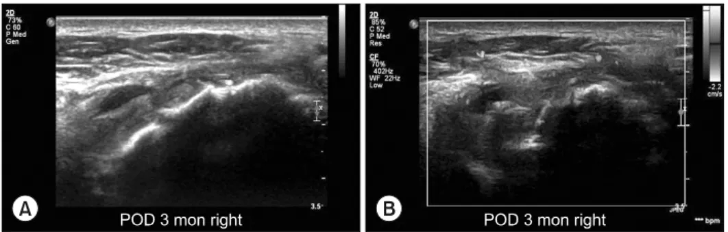
록 재형성을 통해 정상화됨을 보고하였다(Fig. 3∼5).

26

속적인 초음파 검사 내내 봉홥된 힘줄의 연속성, 두께 및

형태적 특징을 평가하였다. 주관적인 점수 체계는 힘줄 에

코결, 힘줄 섬유성, 표면 불규칙성, 혈관성, 견봉밑-삼각근

밑 윤활낭염 등 형태론적 힘줄 특성을 평가하기 위해 사용

(4)

Fig. 4. Longitudinal view at 3 months after surgery (A) repaired tendon show a localized fluid col- lection within the subacromial- subdeltoid bursa (B) color Doppler image shows the vascularity in the peritendinous and intratendinous areas.

Fig. 5. Longitudinal view at 6 months after surgery (A) repaired tendon has normalization of the morphologic appearance, including a hyperechoic echo texture, re- storation of the fibrillar pattern, and a clear surface margin (B) col- or Doppler image shows no vascu- larity in the peritendinous and in- tratendinous areas.

되었다. 연구에서 평가한 항목들의 구체적인 정의는 다음 과 같다.

1) 힘줄 온전함 (tendon integrity): 상완골두의 대결절에 부 착된 연속적인 힘줄 섬유가 가시화되는 것이었다. 확실한 전층 파열을 시각화하면 힘줄이 파열된 것으로 고려되었 다.

18

2) 힘줄 두께 (tendon thickness): 힘줄 두께는 봉합된 힘줄 의 종축(longitudinal) 스캔에서 측정하였다. 상완골두의 대 결절과 상완골두의 관절연골이 만나는 접합부에서 힘줄 섬 유와 평행한 선에 직각의 선을 긋고, 이 선을 따라 봉합된 힘줄의 최대 두께를 측정하였다.

3) 힘줄 에코결 : 힘줄 에코결은 봉합 재료들로 인해 생성 되는 허상들(artifacts)을 제외하고 저에코 영역의 범위를 기준으로 힘줄 에코결을 평가하였다. 힘줄 에코결은 봉합 된 힘줄 부위의 50% 이상에서 저에코 부위가 확인되는 경 우 저에코성 부위로, 저에코 부위가 봉합된 힘줄 부위의 50%를 초과하지 않는 경우 고에코성 부위로 간주되었다.

4) 근섬유 패턴과 표면 불규칙성 : 봉합한 힘줄의 근섬유 패 턴과 표면 불규칙성은 유 또는 무로 분류되었다. 근섬유 패 턴은 종축 스캔에 힘줄 내 선형 고에코 밴드가 있는 것이 특징이다.

5) 혈관성 : 봉합된 회전근개 힘줄은 저유량 민감도 설정 컬러 도플러 영상에서 국소 혈류 패턴을 다음의 3가지 영역 으로 (1) 힘줄 내, (2) 대결절 내의 봉합 고정기 부위, (3) 힘줄주위로 정의하였다.

6) 견봉밑 - 삼각근밑 윤활낭염 : 견봉밑-삼각근밑 윤활낭염은 다음 3등급 중 하나의 등급이 부여되었다. (1) 없음, (2) 윤 활낭이 액체 없이 두께가 2 mm 이상 측정된 경우 윤활낭두 께화 또는 (3) 견봉밑-삼각근밑 윤활낭의 액체확장성이 있 는 견봉밑-삼각근밑 윤활낭염.

29

결 론

회전근개 파열 수술 후 3개월 이내에 재파열이 대부분 발생하고, 봉합된 힘줄의 두께와 형태적 모양이 시간이 지 남에 따라 개선되며 봉합술 후 6개월 이내에 거의 정상화되 는 경과를 잘 이해하고 시간 경과에 따른 초음파 소견을 숙지한다면 환자의 진단, 치료방법 결정 및 예후 예측에 도 움이 될 것으로 판단된다.

ACKNOWLEDGEMENT

This research was supported by Basic Science Research Program through the National Research Foundation of Korea (NRF) funded by the Ministry of Education (NRF- 2016R1D1A1B01014260).

REFERENCES

1. Vitale MA, Vitale MG, Zivin JG, Braman JP, Bigliani LU,

(5)

and cost-effectiveness. J Shoulder Elbow Surg 2007; 16:

181-187

2. Lehman C, Cuomo F, Kummer FJ, Zuckerman JD. The in- cidence of full thickness rotator cuff tears in a large cadav- eric population. Bull Hosp Jt Dis 1995; 54: 30-31 3. Feeley BT, Gallo RA, Craig EV. Cuff tear arthrop-

athy:current trends in diagnosis and surgical management.

J Shoulder Elbow Surg 2009; 18: 484-494

4. Berger MJ, Adams SD, Tigges BM, Sprague SL, Wang XJ, Collins DP, et al. Differentiation of umbilical cord blood- derived multilineage progenitor cells into respiratory epi- thelial cells. Cytotherapy 2006; 8: 480-487

5. Park GY, Kwon DR, Lee SC. Regeneration of full-thick- ness rotator cuff tendon tear after ultrasound-guided in- jection with umbilical cord blood-derived mesenchymal stem cells in a rabbit model. Stem Cells Transl Med 2015;

4: 1344-1351

6. Kwon DR, Park GY, Lee SC. Treatment of full-thickness rotator cuff tendon tear using umbilical cord blood-derived mesenchymal stem cells and polydeoxyribonucleotides in a rabbit model. Stem Cells Int 2018; 7146384

7. Kwon DR, Park GY, Moon YS, Lee SC. Therapeutic ef- fects of umbilical cord blood-derived mesenchymal stem cells combined with polydeoxyribonucleotides on full-thickness rotator cuff tendon tear in a rabbit model.

Cell Transplantation 2018; 27: 1613-1622

8. Kwon DR, Park GY. Adult mesenchymal stem cells for the treatment in patients with rotator cuff disease: present and future direction. Ann Transl Med 2018; 6: 432

9. Jo CH, Chai JW, Jeong EC, Oh S, Kim PS, Yoon JY, et al. Intratendinous injection of autologous adipose tissue-de- rived mesenchymal stem cells for the treatment of rotator cuff disease: a first-in-human trial. Stem Cells 2018; 36:

1441-1550

10. DeOrio JK, Cofield RH. Results of a second attempt at sur- gical repair of a failed initial rotator cuff repair. J Bone Joint Surg Am 1984: 66: 563-567

11. Hinsley H, Nicholls A, Daines M, Wallace G, Arden N, Carr A. Classification of rotator cuff tendinopathy using high definition ultrasound. Muscles Ligaments Tendons J 2014; 4: 391-397

12. Tashjian RZ. Epidemiology, natural history, and indications for treatment of rotator cuff tears. Clin Sports Med 2012;

31: 589-604.

13. Castricini R, Longo UG, De Benedetto M, Panfoli N, Pirani P, Zini R, et al. Platelet rich plasma augmentation for arthroscopic rotator cuff repair: a randomized controlled trial. Am J Sports Med 2011; 39: 258-265

14. Lasbleiz J, Benkalfate T, Morelli JN, Jan J. Sonographic

thickness matter? Open J Clin Diagn 2013; 3: 78-84 15. de Jesus JO, Parker L, Frangos AJ, Nazarian LN. Accuracy

of MRI, MR arthrography, and ultrasound in the diagnosis of rotator cuff tears: a metaanalysis. AJR Am J Roentgenol 2009; 192: 1701-1707

16. Zlatkin MB. MRI of the postoperative shoulder. Skeletal Radiol 2002; 31: 63-80

17. Sipola P, Niemitukia L, Kröger H, Höfling I, Väätäinen U.

Detection and quantification of rotator cuff tears with ultra- sonography and magnetic resonance imaging: a prospective study in 77 consecutive patients with a surgical reference.

Ultrasound Med Biol 2010; 36: 1981-1989

18. Miller BS, Downie BK, Kohen RB, Kijek T, Lesniak B, Jacobson JA, et al. When do rotator cuff repairs fail? Serial ultrasound examination after arthroscopic repair of large and massive rotator cuff tears. Am J Sports Med 2011; 39:

2064-2070

19. Cadet ER, Adler RS, Gallo RA, Gamradt SC, Warren RF, Cordasco FA, et al. Contrast-enhanced ultrasound charac- terization of the vascularity of the repaired rotator cuff ten- don: short-term and intermediate-term follow-up. J Shoulder Elbow Surg 2012; 21: 597-603

20. Fealy S, Adler RS, Drakos MC, Kelly AM, Allen AA, Cordasco FA, et al. Patterns of vascular and anatomical re- sponse after rotator cuff repair. Am J Sports Med 2006; 34:

120-127

21. Tham ER, Briggs L, Murrell GA. Ultrasound changes after rotator cuff repair: is supraspinatus tendon thickness related to pain? J Shoulder Elbow Surg 2013; 22: e8-e15 22. Adler RS, Johnson KM, Fealy S, Maderazo A, Gallo RA,

Gamradt SC, et al. Contrast-enhanced sonographic charac- terization of the vascularity of the repaired rotator cuff:

utility of maximum intensity projection imaging. J Ultrasound Med 2011; 30: 1103-1109

23. Gamradt SC, Gallo RA, Adler RS, Maderazo A, Altchek DW, Warren RF, et al. Vascularity of the supraspinatus ten- don three months after repair: characterization using con- trast enhanced ultrasound. J Shoulder Elbow Surg 2010;

19: 73-80

24. Prickett WD, Teefey SA, Galatz LM, Calfee RP, Middleton WD, Yamaguchi K. Accuracy of ultrasound imaging of the rotator cuff in shoulders that are painful postoperatively. J Bone Joint Surg Am 2003; 8: 1084-1089

25. Barth J, Fotiadis E, Barthelemy R, Genna S, Saffarini M.

Ultrasonic evaluation of the repair integrity can predict functional outcomes after arthroscopic double-row rotator cuff repair. Knee Surg Sports Traumatol Arthrosc 2015; 23:

376-385

26. Yoo HJ, Choi JY, Hong SH, Kang Y, Park J, Kim SH, et

(6)

al. Assessment of the post-operative appearance of the rota- tor cuff tendon using serial sonography after arthroscopic repair of a rotator cuff tear. J Ultrasound Med 2015; 34:

1183-1190

27. St Pierre P, Olson EJ, Elliott JJ, O’Hair KC, McKinney LA, Ryan J. Tendon-healing to cortical bone compared with healing to a cancellous trough: a biomechanical and histo-

logical evaluation in goats. J Bone Joint Surg Am 1995;

77: 1858-1866

28. Cohen DB, Kawamura S, Ehteshami JR, Rodeo SA.

Indomethacin and celecoxib impair rotator cuff ten- don-to-bone healing. Am J Sports Med 2006; 34: 362-369 29. Jacobson JA. Ultrasound in sports medicine. Radiol Clin

North Am 2002; 40: 363-386

수치

Fig. 1. Longitudinal view of supra- supra-spinatus tendon (A) suprasupra-spinatus  tendon full thickness tear (white  arrow, 2.3 cm, medium): “sagging peribursal fat sign” and cortical  irregularities
Fig. 3. Images from a 55-year-old  man with an initial supraspinatus  tendon tear. Longitudinal view at 6  weeks after surgery (A) repaired  cuff with sufficient thickness (> 2  mm) associated with partial  hy-po-echogenicity (B) shows no  in-creased vascu
Fig. 4. Longitudinal view at 3  months after surgery (A) repaired  tendon show a localized fluid  col-lection within the subacromial-  subdeltoid bursa (B) color Doppler  image shows the vascularity in the  peritendinous and intratendinous  areas.

참조

관련 문서

– Embryonic stem cell : Pluripotent cells that can give rise to all tissue types. – Adult stem cell : Multipotent cells

국내 초음파 기술의 경우 치료 초음파보다 진단을 위한 초음파 영상 시스템 분야에 치우쳐 있고 국내 대학연구소 연구로는 제주대학교의 ‘체외 고강도 집속 초음파 치료

Approved clinical use of bone marrow stem cells for myocardial infarction treatment... Cardiac

• Generation of different specialized kinds of cells from zygote (fertilized egg) or other precursor cells.. – Generate blood cells, muscle

Graph showing improving Constant Shoulder Score in both groups but more improvement in autologous bone marrow injection group which is statistically

Results: The results demonstrated a significant positive correlation between the muscle atrophy and the tendon tear.(p<0.05) And there was statistically

Sequencing results of groEL gene of Anaplasma phagocytophilum detected in blood, kidney and spleen of wild rodents captured in Jeollanam-do area using a

This research is a research to examine change in physical strengh, thickness of subcutaneous fat, and blood lipid after twelve week execution of