INTRODUCTION
It has been reported that the incidence of thyroid nodule in Graves’ disease patients varies from 10% to 35% (1-9). It is possible that high-resolution ultrasonography during ini- tial diagnosis allowed identification of small thyroid nodules in Graves’ disease patients. However, the prevalence of dif- ferentiated thyroid cancer (DTC) in Graves’ disease patients remains as a matter of debate, with previous reports show- ing the prevalence was 0.3-16.6% (1-14).
The biologic behaviors and optimal management of DTC with concurrent Graves’ disease are still controversial. Some investigators have reported that patients of DTC associated with Graves’ disease experienced a more aggressive course than euthyroid patients (15, 16). Pellegriti et al. (17) suggested that the extent of surgery in such patients must be extensive, involving at least a total thyroidectomy and lymph node dis- section followed by 131radioactive iodine therapy (RI). In con- trast, others reported that the aggressiveness and the treat- ment outcomes of DTC in Graves’ disease patients are simi- lar to those of euthyroid patients (6, 18). Thus, there remains controversy as to the optimal extent of surgery in these cases.
The aims of the present study were to identify clinicopatho- logic features, treatment outcome, and indicators for pre- dicting locoregional recurrences and to suggest the optimal extent of surgery in DTC patients with concurrent Graves’
disease.
MATERIALS AND METHODS Study patients
During the 20-yr period from 1986 to 2005, 779 patients with Graves’ disease underwent surgery at our hospital. The diagnosis of Graves’ disease was based on the history and signs of thyrotoxicosis, increased levels of serum triiodothy- ronine (T3) and thyroxine (T4), low levels of thyroid-stimu- lating hormone (TSH), the presence of autoantibodies (thy- roid stimulating antibodies [TSAbs]and/or antiperoxidase antibodies), increased radionuclide uptake at scintiscan, and the histological features of diffuse hyperplasia of the thyroid gland. Patients were treated preoperatively with propylth- iouracil, cabimazole, or methimazole. They also received Lugol
796
Jandee Lee, Kee Hyun Nam*, Woung Youn Chung*, Euy-Young Soh, and Cheong Soo Park*
Department of Surgery, Ajou University College of Medicine, Suwon; Department of Surgery*, Yonsei University College of Medicine, Seoul, Korea
Address for correspondence Cheong Soo Park, M.D.
Department of Surgery, Yonsei University College of Medicine, 134 Sinchon-dong, Seodaemun-gu, Seoul 120-752, Korea
Tel : +82.2-2228-2100, Fax : +82.2-313-8289 E-mail : [email protected]
DOI: 10.3346/jkms.2008.23.5.796
Clinicopathologic Features and Treatment Outcomes in Differentiated Thyroid Carcinoma Patients with Concurrent Graves' Disease
The clinical behaviors and treatment outcomes of thyroid carcinomas in patients with Graves’ disease is a matter of controversy. This study aimed to identify the clinicopathologic features, treatment outcome, and the indicators for predicting recur- rence, and to suggest the optimal extent of surgery in these patients. We retrospec- tively analyzed data of 58 patients who underwent surgical treatment for differenti- ated thyroid cancer and concurrent Graves’ disease. The follow-up period ranged from 23 to 260 months (mean±standard devuation, 116.8±54.0). In our series, the mean age was 40.8±12.7 yr (range, 15-70), with a male-to-female ratio of 1:
6.25. The mean tumor size was 13±9 mm (range, 3-62). The surgical methods included 19 cases of total thyroidectomy, 38 cases of subtotal thyroidectomy, and 1 case of completion total thyroidectomy. Locoregional recurrence occurred in four patients (6.9%). The 10-yr overall survival and disease-free survival of patients were 95.8% and 91.1%, respectively. Age over 45 yr (p=0.031), tumor size over 10 mm (p=0.049), multiplicity (p=0.007), extracapsular invasion (p=0.021), and clinical can- cer (p=0.035) were significantly more prevalent in patients with locoregional recur- rence than in those without recurrence. We recommend that Graves’ disease patients should undergo regular ultrasonography screening for early detection of thyroid carcinoma. We also suggest that the choice of extent of surgery should depend on the diagnostic timing (clinical or incidental) and factors for predicting recurrence.
Key Words : Graves Disease; Differentiated Thyroid Cancer; Incidental Carcinoma; Clinical Carcinoma; Extent of Surgery
Received : 27 March 2007 Accepted : 25 January 2008
solution (8 drops per 6 hr) for 7 to 10 days before surgery.
The indications for surgery were the presence of a large com- pressive goiter, recurrent thyrotoxicosis, side effects of anti- thyroid medications, unlikelihood or inability of taking medi- cine, presence of cold nodules on thyroid scanning, and sus- picion of malignancy on fine needle aspiration biopsy.
Diagnostic timing
Study patients were classified as clinically overt carcino- ma or incidental carcinoma. In clinically overt carcinoma, a patient with Graves’ disease was found to have a specific symptom or sign and thyroid carcinoma was diagnosed pre- operatively. However, in incidental carcinoma, Graves’ dis- ease was the only clinical diagnosis before operation and thy- roid carcinoma was identified during surgery or following histological examination of permanent sections.
Surgical method
Patients with incidental carcinoma underwent a bilateral subtotal thyroidectomy or a Hartley-Dunhill operation (16) (a unilateral lobectomy with a contralateral subtotal lobec- tomy, leaving 4-6 g of thyroid tissue). Intraoperative frozen sections were carried out in all the suspicious lesions. In case of clinical carcinoma, we performed the operative methods according to the TNM classification system (T, tumor size;
N, involvement of lymph nodes; M, presence of distant metas- tasis) (19).
Postoperative follow-up
All patients were postoperatively followed up every 3-6 months. Patients were evaluated for locoregional or distant metastasis by the following diagnostic methods: serum thy- roglobulin/anti-thyroglobulin antibody level, ultrasonogra- phy, computed tomography, radioiodine scintigraphy.
Statistical analysis
Comparisons between groups were performed using Mann- Whitney U test and Fisher’s exact test. Estimated survival rates were calculated by the Kaplan-Meier method. Statisti- cal significance was set at p<0.05.
RESULTS Clinical characteristics
Fifty-eight cases of DTC (7.4%) were diagnosed among 779 Graves’ disease patients who underwent thyroidectomy between 1986 and 2005. The study enrolled 8 male and 50 female patients with a mean age of 40.8±12.7 yr (range, 15-70). No patients had previous history of radiation expo- sure or radioactive iodine therapy. Twenty-six cases of clini- cally overt thyroid carcinomas were diagnosed among 779 Graves’ disease patients (3.3%). The prevalence of inciden- tal carcinoma in the Graves’ disease patients was calculated as 4.2% (32 patients with occult thyroid carcinoma out of 753 patients who underwent operation represented only part of Graves’ disease). Comparison of clinical findings of inci- dental and clinical carcinoma patients revealed no difference between the two groups in terms of duration of thyrotoxic symptoms, T3/T4 serum levels before the administration of antithyroid medication, and postoperative 6-week serum concentration of thyroglobulin/TSH (Table 1).
Surgical treatment and histopathologic findings
Of the 26 patients with clinical carcinoma, 14 underwent total thyroidectomy and 12 underwent subtotal thyroidec- tomy. Of the 32 patients with incidental carcinoma, 5 under- went total thyroidectomy and 26 subtotal thyroidectomy (Table 2). There was one patient of completion thyroidecto-
Characteristics Total (n=58) Clinical cancer (n=26) Incidental cancer (n=32) p value
Gender
Male 8 4 4
Female 50 22 28 0.751*
Mean age (yr) 40.84±12.69 42.50±14.87 39.50±10.67 0.376�
Tumor size (mm) 13.14±9.78 19.52±14.43 7.32±6.76 0.017�
Duration of thyrotoxic 27.62±26.40 29.54±27.90 26.06±25.46 0.622�
symptoms (month)
Serum T4 (mM/L)� 212.43±13.64 226.32±31.46 204.19±19.76 0.196�
Serum T3 (mM/L)� 7.98±0.76 8.02±0.99 7.42±0.85 0.241�
Thyroglobulin (μg/L)� 13.41±17.35 11.540±12.64 15.13±10.04 0.482�
Serum TSH (mIU/L)� 19.32±21.08 22.06±20.19 17.78±24.64 0.186�
Table 1. Clinical characteristics of clinical and incidental cancers
Mean±SD: median, with the range presented in parenthesis.
*, Fisher’s exact test; �, Mann-Whitney test; �, before antithyroid medication; �, six-week postoperative serum level.
T4, thyroxine; T3, triiodothyronine; TSH, thyroid-stimulating hormone.
my, who had initially underwent a subtotal thyroidectomy for Graves’ disease; the subsequent histopathology analysis, however, showed a widely invasive follicular carcinoma. Of the total 58 carcinomas, 57 (98.3%) were papillary thyroid carcinoma and 1 (1.7%) was a follicular thyroid carcinoma.
The mean tumor size was 13±9 mm in diameter (range, 3-62). Extracapsular invasion and invasion to adjacent struc- ture were found in 23 (39.7%) and 3 cases (5.2%), respec- tively. Multiple lesions were noted in 18 cases (31.0%). Cen- tral compartment node metastasis and lateral cervical lymph node metastasis were shown in 18 (31.0%) and 5 (8.6%) cases, respectively.
Postoperative adjuvant therapy
Sixteen patients treated with total thyroidectomy under- went postoperative adjuvant RI. Eleven of these patients
TT, total thyroidectomy; MRND, modified radical neck dissection; CCND, central compartment neck dissection.
Characteristics Total
(n=58) (%)
Clinical cancer (n=26) (%)
Incidental cancer (n=32) (%)
Bilateral subtotal 22 (37.9) 22 (68.8)
thyroidectomy
Dunhill procedure 4 (6.9) 4 (12.5)
TT with CCND 14 (24.2) 9 (34.6) 5 (15.6) Ipsilateral total and 12 (20.7) 12 (46.2)
contralateral subtotal thyroidectomy with CCND
Total thyroidectomy with 5 (8.6) 5 (19.2) MRND
Completion thyroidectomy 1 (1.7) 1 (3.1)
Table 2. Surgical methods
Overall survival (%)
100
80
60
40
20
0
0 30 60 90 120 150
Months
10-yr overall survival: 95.8%
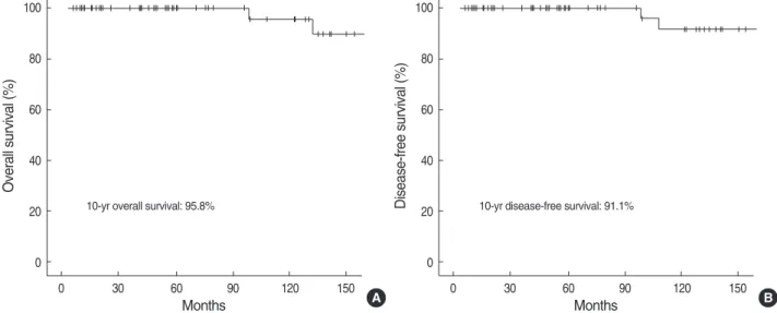
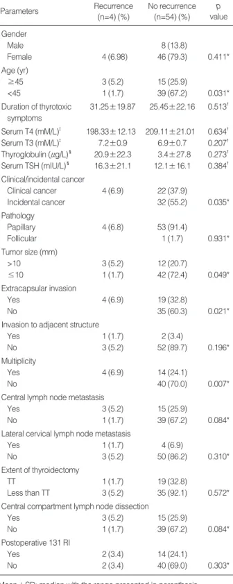
Fig. 1. The overall (A) and disease-free (B) survival of patients who underwent surgical treatment for differentiated thyroid carcinoma and concurrent Graves’ disease.
A
Disease-free survival (%)
100
80
60
40
20
0
0 30 60 90 120 150
Months
10-yr disease-free survival: 91.1%
B
Case 1 Case 2 Case 3 Case 4
Gender F F F F
Age (yr) 48 21 61 46
Duration of recurrence (months) 153 91 167 107
Tumor size (mm) 17 9 24 16
Clinical/incidental Clinical Clinical Clinical Clinical
TNM T3N1aM0 T3N1aM0 T3N0M0 T3N1bM0
Stage Stage III Stage I Stage III Stage IVa
1st operation TT /c CCND TT /c CCND TT /c CCND TT /c MRND
Site of LR Strap muscle Thyroid bed Thyroid bed Thyroid bed
LCLN LCLN
Treatment of LR Reoperation+H-RI No treatment Reoperation+H-RI No treatment
Treatment outcome Remission Death Remission Death
Cause of death Ovary cancer /c SM LR
Table 3. Disease patterns in four locoregional recurrence patients
Clinical/incidental, clinical cancer or incidental cancer; TT, total thyroidectomy; /c,with; CCND, central compartment node dissection; MRND, modified radical neck dissection; LR, local recurrence; LCLN, lateral cervical lymph node; H-RI, high-dose radioactive iodine threapy; SM, systemic metastasis.
received low-dose (30-60 mCi) therapy for ablation of resid- ual thyroid tissue. Four patients in which lateral cervical lymph node metastasis was present received high-dose (150- 250 mCi) therapy.
Treatment outcome and Indicators for predicting recur- rence
The mean follow-up period was 116.8±54.0 months (range, 23-260). For all the patients, the 10-yr overall sur- vival and disease-free survival rates were calculated to be 95.8% and 91.1%, respectively (Fig. 1). Local recurrence was identified in 4 patients (6.9%), all of whom belonged to the clinical carcinoma group. The mean duration of recur- rence was 129±36 months (range, 91-167), with no patients diagnosed with distant metastases during the follow-up.
Of the 4 loco-regional recurrence patients, 2 patients under- went re-operation and high-dose RI. Neither of these patients has shown any evidence of recurrence up to now (77 months and 54 months, respectively) since the second operation. For the other 2 patients, local recurrence was noted in the thy- roid bed and lateral cervical lymph nodes, respectively (Table 3). These patients were not treated for this local recurrence since both refused to undergo treatment: one died of aggres- siveness of local recurrence 132 months after the initial thy- roidectomy and the other of combined ovary cancer with systemic metastasis 98 months after the surgery.
The patient age of over 45 yr, tumor size, multiplicity, extracapsular invasion, and clinical cancer were significant factors in predicting the locoregional recurrence (Table 4).
DISCUSSION
The prevalence of thyroid carcinoma in Graves’ disease has been reported to be from 0.3% to 16.6% (1-14). It is sug- gested that part of the explanation for the wide variation in incidence could be due to differences in the extent of resec- tion as well as in the number of histologic sections examined per specimen (20). Moreover, the increasing use of high res- olution ultrasonography has revealed greater numbers of thy- roid nodules, and the routine use of such devices has allowed diagnosis of small thyroid carcinoma in Graves’ disease pati- ents. The 7.4% prevalence found in our work seems to be relevant to that found in other reports.
Controversy remains regarding the pathogenic relation- ship between thyroid carcinoma and Graves’ disease. It is well known that the binding of TSH to TSH receptors might promote the growth of cancer cells in euthyroid patients with thyroid carcinoma. In Graves’ disease in which serum TSH is suppressed, TSAbs rather than TSH are reported to acti- vate the TSH receptor (16, 21, 22). Some studies showed that autoimmune responses in Graves’ disease including TSAbs are closely linked to angiogenesis, which plays a crucial role
Parameters Recurrence
(n=4) (%)
No recurrence (n=54) (%)
p value Gender
Male 8 (13.8)
Female 4 (6.98) 46 (79.3) 0.411*
Age (yr)
≥45 3 (5.2) 15 (25.9)
<45 1 (1.7) 39 (67.2) 0.031*
Duration of thyrotoxic 31.25±19.87 25.45±22.16 0.513� symptoms
Serum T4 (mM/L)� 198.33±12.13 209.11±21.01 0.634� Serum T3 (mM/L)� 7.2±0.9 6.9±0.7 0.207� Thyroglobulin (μg/L)� 20.9±22.3 3.4±27.8 0.273� Serum TSH (mIU/L)� 16.3±21.1 12.1±16.1 0.384� Clinical/incidental cancer
Clinical cancer 4 (6.9) 22 (37.9)
Incidental cancer 32 (55.2) 0.035*
Pathology
Papillary 4 (6.8) 53 (91.4)
Follicular 1 (1.7) 0.931*
Tumor size (mm)
>10 3 (5.2) 12 (20.7)
≤10 1 (1.7) 42 (72.4) 0.049*
Extracapsular invasion
Yes 4 (6.9) 19 (32.8)
No 35 (60.3) 0.021*
Invasion to adjacent structure
Yes 1 (1.7) 2 (3.4)
No 3 (5.2) 52 (89.7) 0.196*
Multiplicity
Yes 4 (6.9) 14 (24.1)
No 40 (70.0) 0.007*
Central lymph node metastasis
Yes 3 (5.2) 15 (25.9)
No 1 (1.7) 39 (67.2) 0.084*
Lateral cervical lymph node metastasis
Yes 1 (1.7) 4 (6.9)
No 3 (5.2) 50 (86.2) 0.310*
Extent of thyroidectomy
TT 1 (1.7) 19 (32.8)
Less than TT 3 (5.2) 35 (92.1) 0.572*
Central compartment lymph node dissection
Yes 3 (5.2) 15 (25.9)
No 1 (1.7) 39 (67.2) 0.084*
Postoperative 131 RI
Yes 2 (3.4) 14 (24.1)
No 2 (3.4) 40 (69.0) 0.303*
Table 4. Parameters predicting recurrence of differentiated thy- roid cancer associated with Graves’ disease
Mean±SD: median with the range presented in parenthesis.
*, Fishers exact test; �, Mann-Whitney U test; �, measured before antithy- roid treatment; �, measured after postoperative six week.
T4, thyroxine; T3, triiodothyronine; TSH, thyroid-stimulating hormone;
TT, total thyroidectomy; RI, radioactive iodine therapy.
in tumor growth and development (15, 21, 23). Therefore, patients of Graves’ disease with DTC has been found to have a worse clinical outcome than euthyroid patients with DTC (4, 17, 18, 24, 25). In contrast, other investigators suggest- ed that Graves’ disease was not related to the aggressiveness of coexisting thyroid carcinoma (6, 9, 26). These studies iden- tified that the severity of thyrotoxicosis was not related to the prognosis of DTC patients with concurrent Graves’ dis- ease (27-29). In the present series of our work, the 10-yr over- all and disease-free survival rates were 95.8% and 91.1%, respectively, showing favorable treatment outcomes in these patients, and the severity of thyrotoxicosis and serum thy- roid hormone levels did not affect the prognosis.
A few studies examined recurrence-predicting indicators in patients of DTC with concurrent Graves’ disease. Although some authors suggested patient age, tumor size and preop- erative T3 levels are significant factors to predict metastasis (6, 30), recent studies indicate that the most crucial prog- nostic indicator is diagnostic timing (clinical or incidental) (26, 31). In the present investigation, patient age over 45 yr, the presence of extracapsular invasion, multiplicity, and clinical cancer, but not severity of thyrotoxicosis, were cor- related with a high recurrence rate.
Debate continues regarding the proper extent of surgery for Graves’ disease patients with DTC. There has been a pref- erence for radical surgery in patients with DTC with concur- rent Graves’ disease, involving at least total thyroidectomy and lymph node dissection followed by RI (16, 17). How- ever, recent approaches have suggested that the choice of surgical strategies depend on the diagnostic timing and stage of tumor (26-31). This indicates that the extent of surgery should be similar for thyroid cancer in euthyroid patients, and that subtotal thyroidectomy is sufficient, whereas addi- tional completion total thyroidectomy is not necessary in incidental cases. In the present investigation, neither the extent of thyroidectomy nor the presence of lymph node dis- section was found to be correlated with the recurrence rate.
Therefore, complete thyroidectomy might be helpful only in cases in which indicators predicting recurrence are present.
In this study, two of the four patients who had local recur- rence underwent second operation and survived with no fur- ther episodes. The other two patients refused further treat- ment and died. Even though the present study involved only a small number of patients, the findings suggest that early detection and active treatment lead to a good prognosis even in cases of local recurrence.
Nevertheless, we encountered with several limitations when we tried to investigate the treatment outcome and signifi- cant factors to predict recurrence in thyroid carcinoma pati- ents with concurrent Graves’ disease. First, there were only a limited number of patients. Second, the natural course of DTC is characterized by very slow progression, therefore, our observation period might not have been enough to detect the evolution from silent disease to overt clinical metastasis.
In conclusion, we recommend that patients of Graves’ dis- ease should undergo regular screening both meticulous phys- ical examination and ultrasonography for early detection of thyroid carcinoma. Our present results show that predicting factors for recurrence in DTC patients with concurrent Graves’
disease were age over 45 yr, tumor size over 10 mm, multi- plicity, extracapsular invasion, and clinical carcinoma. We also suggest that the choice of extent of surgery in these pati- ents should depend on the diagnostic timing (clinical or inci- dental) and factors for predicting recurrences, and that a subto- tal thyroidectomy would be sufficient in incidental carcino- mas without factors predicting recurrence. On the other hand, in clinical cancers or in cases where recurrence-predicting factors are present, at least a total thyroidectomy with lymph node dissection might be helpful.
REFERENCES
1. Dobyns BM, Sheline GE, Workman JB, Tompkins EA, McCona- hey WM, Becker DV. Malignant and benign neoplasms of the thy- roid in patients treated for hyperthyroidism: a report of the cooper- ative thyrotoxicosis therapy follow-up study. J Clin Endocrinol Metab 1974; 38: 976-98.
2. Pacini F, Elisei R, Di Coscio GC, Anelli S, Macchia E, Concetti R, Miccoli P, Arganini M, Pinchera A. Thyroid carcinoma in thyroto- sic patients treated by surgery. J Endocrinol Invest 1988; 11: 107-12.
3. Ruggieri M, Scocchera F, Genderini M, Mascaro A, Luongo B, Paolini A. Hyperthyroidism and concurrent thyroid carcinoma. Eur Rev Med Pharmacol Sci 1999; 6: 265-8.
4. Belfiore A, Garofalo MR, Giuffrida D, Runello F, Filetti S, Fiumara A, Ippolito O, Vigneri R. Increased aggressiveness of thyroid can- cer in patients with Graves’ disease. J Clin Endocrinol Metab 1990;
70: 830-5.
5. Carnell NE, Valente WA. Thyroid nodules in Graves’ disease or thyrotoxicosis affect the prognosis of thyroid cancer. Thyroid 1998;
8: 571-6.
6. Hales IB, McElduff A, Crummer P, Clifton-Bligh P, Delbridge L, Hoschl R, Poole A, Reeve TS, Wilmshurst E, Wiseman J. Does Graves’ disease or thyrotoxicosis affect the prognosis of thyroid can- cer. J Clin Endocrinol Metab 1992; 75: 886-9.
7. Prades JM, Dumollard JM, Timoshenko A, Chelikh L, Michel F, Estour B, Martin C. Multinodular goiter: surgical management and histopathological findings. Eur Arch Otorhinolaryngol. 2002; 259:
217-21.
8. Belfiore A, Russo D, Vigneri R, Filetti S. Graves’ disease, thyroid nodules and thyroid cancer. Clin Endocrinol (Oxf) 2001; 55: 711-8.
9. Gerenova J, Buysschaert M, de Burbure CY, Daumerie C. Prevalence of thyroid cancer in Graves’ disease: a retrospective study of a cohort of 103 patients treated surgically. Eur J Med 2003; 14: 321-5.
10. Kim WB, Han SM, Kim TY, Nam-Goong IS, Gong G, Lee HK, Hoong SJ, Shong YK. Ultrasonographic screening for detection of thyroid cancer in patients with Graves’ disease. Clin Endocrinol (Oxf) 2004; 60: 719-25.
11. Olen E, Klinck GH. Hyperthyroidism and thyroid cancer. Arch Pathol 1966; 81: 531-5.
12. Nicolosi A, Addis E, Calo PG, Tarquini A. Hyperthyroidism and cancer of the thyroid. Minerva Chir 1994; 49: 491-5.
13. Ragni F, Pinelli D, Facchini M, Ghedi M, Piccini I, Pasini M, Ron- cali S, Pezzola D, Braga M. Thyroid carcinoma in hyperthyroid syndromes. G Chir 1996; 17: 158-65.
14. Farbota LM, Calandra DB, Lawrence AM, Paloyan E. Thyroid car- cinoma in Graves’ disease. Surgery 1985; 98: 1148-53.
15. Sato K, Yamazaki K, Shizume K, Kanaji Y, Obara T, Ohsumi K, Demura H, Yamaguchi S, Shibuya M. Stimulation by thyroid-stim- ulating hormone and Graves’ immunoglobulin G of vascular endothe- lial growth factor mRNA expression in human thyroid follicles in vitro and flt mRNA expression in the rat thyroid in vivo. J Clin Invest 1995; 96: 1295-302.
16. Kashima K, Yokoyama S, Daa T, Takahashi K, Nakayama I, Noguchi S. C-myc expression is associated with increased proliferation activ- ity in thyroid follicle cells of Graves’ disease as stimulated by autoan- tibodies. Eur J Endocrinol 1996; 135: 69-76 .
17. Pellegriti G, Belfiore A, Giuffrida D, Lupo L, Vigneri R. Outcome of differentiated thyroid cancer in Graves’ patients. J Clin Endocrinol Metab 1998; 83: 2805-9 .
18. Kikuchi S, Noguchi S, Yamashita H, Uchino S, Kawamoto H. Prog- nosis of small thyroid cancer in patients with Graves’ disease. Br J Surg 2006; 93: 434-9.
19. Greene FL, Page DL, Fleming ID, Fritz A, Balch CM. AJCC Can- cer Staging Manual, 6th edn. Chicago: Springer Verlag, 2003.
20. Behar R, Arganini M, Wu TC, McCormick M, Straus FH 2nd, Deg- root LJ, Kaplan EL. Graves’ disease and thyroid cancer. Surgery 1986; 100: 1121-7.
21. Filetti S, Belfiore A, Amir SM, Daniels GH, Ippolito O, Vigneri R, Ingbar SH. The role of thyroid-stimulating antibodies of Graves’
disease in differentiated thyroid cancer. N Engl J Med 1988; 318:
753-9.
22. Van Sande J, Lejeune C, Ludgate M, Munro DS, Vassart G, Dumont
JE, Mockel J. Thyroid stimulating immunoglobulins, like thyrotropin activate both the cyclic AMP and the PIP2 cascades in CHO cells expressing the TSH receptor. Mol Cell Endocrinol 1992; 88: R1-5.
23. Viglietto G, Romano A, Manzo G, Chiappetta G, Paoletti I, Califano D, Galati MG, Mauriello V, Bruni P, Lago CT, Fusco A, Persico MG. Upregulation of the angiogenic factors PIGF, VEGF and their receptors (Flt-1, Flk-1/KDR) by TSH in cultured thyrocytes and in the thyroid gland of thiouracil-fed rats suggest a TSH-dependent paracrine mechanism for goiter hypervascularization. Oncogene 1997; 15: 2678-98.
24. Stocker DJ, Burch HB. Thyroid cancer yield in patients with Graves’
disease. Minerva Endocrinol 2003; 28: 205-12.
25. Cappelli C, Braga M, De Martino E, Castellano M, Gandossi E, Agosti B, Cumetti D, Pirola I, Mattanza C, Cherubini L, Rosei EA.
Outcome of patinets surgically treated for various forms of hyper- thyroidism with differentiated thyroid cancer: Experience at an endocrine center in Italy. Surg Today 2006; 36: 125-30.
26. Duh QY. Thyroid cancer in Graves disease: Incidental cancer ver- sus clinical cancer. Ann Surg Oncol 2004; 11: 356-7.
27. Miki H, Oshimo K, Inoue H, Kawano M, Tanaka K, Komaki K, Uyama T, Monden Y. Diagnosis and surgical treatment of small papillary carcinoma of the thyroid gland. J Surg Oncol 1993; 54:
78-80.
28. Soh EY, Jung WH, Park CS. Thyroid carcinoma in Graves’ disease:
Clinical features and diagnostic approach. J. Korean Med Assoc 1991; 34: 1229-35.
29. Yeo PP, Wang KW, Sinniah R, Aw TC, Chang CH, Sethi VK, Tan BC, Lim P. Thyrotoxicosis and thyroid cancer. Aust N Z J Med 1982; 12: 589-93.
30. Chao TC, Lin JD, Chen MF. Surgical treatment of thyroid cancers with concurrent Graves disease. Ann Surg Oncol 2004; 11: 407-12.
31. Yano Y, Shibuya H, Kitagawa W, Nagahama M, Sugino K, Ito K, Ito K. Recent outcome of Graves’ disease patients with papillary thyroid cancer. Eur J Endocrinol 2007; 157: 325-9.