INTRODUCTION
Surgeons are increasingly challenged by opportunistic fun- gal infections. During the last four decades, the incidence of invasive opportunistic fungal infections has risen dramati- cally (1). Aspergillus species are ubiquitous molds that are easily isolated from air, soil, decaying vegetation, and dust.
These species are the second-most common cause of oppor- tunistic fungal infections, surpassed only by Candida species (2). Cutaneous aspergillosis is a rare form of a locally inva- sive disease. It generally enters through breaks in the skin, colonizing burns, surgical wounds, or intravenous catheter sites, and subsequently invades viable tissue. Human beings are constantly exposed to this organism, which results in frequent colonization. Cutaneous aspergillosis is most com- monly seen in immunocompromised hosts; however, only rarely does Aspergillus behave as a pathogen in an immuno- competent host (2, 3).
Despite the advent of efficacious antimicrobial therapy, combined surgical therapy is still advocated. There could be various surgical therapeutic methods: incision and drainage of abscesses, fistulotomy, sinus tract excision, and more exten- sive debulking of infected tissue with skin graft or flap cov- erage are recommended. To the best of our knowledge, this is the first report of flap reconstruction of cutaneous aspergillo- sis. Therefore, we report our clinical experience with a review of the relevant literature (1, 4).
CASE REPORT
A 45-yr-old man presented with a painful ulcerative lesion on his right elbow. The patient had been treated for herpes zoster 4 yr before and erythematous 1-cm sized painful nod- ule was noted on his upper arm in the following years. His past medical history was not specific and he had worked as a carpenter for 20 yr. The nodular lesion developed into ulcer- ative lesion pregressively and finally became a dry, black, escharous lesion with a maximal diameter of 6 cm (Fig. 1).
The surrounding skin was erythematous. Neither purulence nor odor was noted, and no localized axillary or generalized lymphadenopathy was observed. Laboratory studies revealed unspecific results, except a mildly elevated white blood cell count. The patient did not exhibit any sensory or motor deficits in his upper extremities, although movement resulted in inter- mittent pain.
Three-dimensional (3-D) upper extremity computed tomo- graphic (CT) angiography of the lesion revealed a skin defect at the posterolateral aspect of the distal humerus and a low attenuated lesion 3 cm in size that was located intramuscu- larly and showed rim enhancement. A candidate recipient artery, a 1-2 mm-sized branch of the brachial artery, was marked. Magnetic resonance imaging (MRI) examination showed a chronic inflammatory change of the skin at the subcutaneous fat defect portion and infectious myositis at the triceps muscle (Fig. 2). The possibility of squamous cell
920
Chan Yeong Heo, Seok Chan Eun, Rong Min Baek, and Kyung Won Minn
Department of Plastic and Reconstructive Surgery, Seoul National University College of Medicine, Seoul, Korea
Address for correspondence Seok Chan Eun, M.D.
Department of Plastic and Reconstructive Surgery, Seoul National University College of Medicine, Seoul National University Bundang Hospital, 300 Gumi-dong, Bundang-gu, Seongnam 463-707, Korea
Tel : +82.31-787-7223, Fax : +82.31-787-4055 E-mail : [email protected]
J Korean Med Sci 2008; 23: 920-3 ISSN 1011-8934
DOI: 10.3346/jkms.2008.23.5.920
Copyright � The Korean Academy of Medical Sciences
Free Flap Coverage of Extensive Soft Tissue Defect in Cutaneous Aspergillosis: A Case Report
Isolated fungal soft-tissue infections are uncommon, but may cause severe morbid- ity or mortality. Aspergillosis infection is rare, but the frequency in increasing over the last two decades. Here, we present a patient with cutaneous aspergillosis of his right elbow with unusual clinical and radiological features suggestive of a malig- nant disease, which remained undiagnosed for an extended period of time. The patient presented with necrotic, black-colored skin ulcerations. We completely removed the skin ulcer with the surrounding erythematous skin lesion, and then we reconstructed the area with thoracodorsal perforator free flap. The biopsy speci- men contained septate hyphae with dichotomous branching, which is morphologi- cally consistent with a finding of Aspergillus. After surgery, we initiated antifungal medication therapy with amphotericin B and itraconazole. At the time of follow-up, the elbow with the reconstructed flap had fully healed, and no recurrent disease was found.
Key Words : Aspergillosis; Flap
Received : 30 May 2007 Accepted : 23 December 2007
Free Flap Reconstruction in Cutaneous Aspergillosis 921
carcinoma could not be ruled out. Diagnostic skin punch biopsy demonstrated chronic active inflammation with ulcer- ation and necrosis with regenerating epithelia, but failed to show any microorganism on direct microscopy or in culture.
We planned surgical resection of the necrotic skin ulcer and free flap coverage. The necrotic and infected central tissue extending into the subcutaneous fat was removed, and a 2-3 cm rim of non-necrotic, erythematous skin was also removed
as the surgical margin (Fig. 3). The lateral side of the triceps muscle and periosteum was also partially excised. No bony involvement was seen on the gross view but elbow joint exter- nal capsule was exposed. The wound bed was severely scarred owing to chronic inflammation. We dissected the profunda brachii artery and the vein between the biceps and triceps brachii muscles as a recipient vessel. After identification of
Fig. 1. Primary cutaneous aspergillosis of the elbow. Note the open weeping ulcers, black necrotic eschars, and diffuse erythe- matous skin changes.
Fig. 2. Magnetic resonance imaging (fat saturated T1-enhancing) examination showed a soft tissue defect and severe muscular inflammatory infiltration around bone.
Fig. 3. The surrounding 2-3 cm margin of non-necrotic, erythe- matous skin was removed with central ulceration.
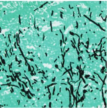
Fig. 4. A periodic acid-Schiff stained section of necrotic lesion.
Septate hyphae are surrounded by dermal necrosis and acute inflammation (original magnification, ×40).
922 C.Y. Heo, S.C. Eun, R.M. Baek, et al.
the perforator using Doppler sound, a 13×8 cm-sized ipsi- lateral thoracodorsal perforator flap was designed and elevat- ed. After flap transfer, we performed arterial microanastomo- sis first between the radial collateral artery and thoracodor- sal artery. Finally, we did venous anastomosis between each of vena commitantes.
One week postoperatively, permanent pathology with Gomori methenamine silver (GMS) and periodic acid-Schiff (PAS) staining confirmed chronic active inflammation and extensive necrosis with numerous fungal hyphae showing septation and branching consistent with Aspergillus species (Fig. 4, 5). Tissue cultures grew A. fumigatus. After surgery, the patient was started on amphotericin B (20 mg/day) IV for 5 days and switched to oral itraconazole (200 mg q 12 hr) medication. The wound went on to heal satisfactorily, and there has been no evidence of recurrent disease at 2 yr of fol- low-up (Fig. 6).
DISCUSSION
All Aspergillus species are molds, which together with yeasts are classified as fungi. Aspergillosis is the second-most frequent opportunistic infection, surpassed only by candidi- asis (1, 3, 4). There are more than 1,000 species of Aspergillus, but A. fumigatus is the most common cause of colonization and invasive aspergillosis (2, 3). A. fumigatus rarely behaves as a pathogen in an immunocompetent host; however, in an immunocompromised host, it may be invasive and may take a fulminant course. Cutaneous aspergillosis may be primary or
secondary to systemic dissemination. It presents as erythema- tous papules or plaques that evolve into necrotic skin lesions, often at sites of skin trauma associated with intravenous cathe- ters and placement of adhesive tape or monitor leads, or at sites of surgical or other traumas (5, 6). If hyphae are some- how able to develop, functioning neutrophils remove them efficiently. If these host defenses fail, infection may develop.
The prompt recognition and appropriate treatment of cuta- neous fungal disease is critical to the prevention of adverse outcomes, but aspergillosis poses some unique diagnostic and therapeutic challenges. Skeletal aspergillosis usually con- flicts with neoplastic or inflammatory diseases. Radiograph- ic studies may be helpful, but not confirmatory. Aspergillo- sis may initially present to the clinician as a painful and locally destructive lesion. The typical lesion is an erythematous (or violaceous), edematous, indurated papule or nodule that pro- gresses to a blue-black necrotic ulcer with a black eschar due to regular invasion into blood vessels, causing local throm- bosis and hemorrhage (2-5). Tissue biopsy with special stains and tissue culture are the preferred methods for the diagno- sis of aspergillosis. Fungal hyphae can be seen in hematoxylin- eosin-stained sections, but staining with periodic acid-Schiff or Gomori methenamine silver highlights their morpholog- ical characteristics, as in our case (2-4).
Treatment of cutaneous aspergillosis includes a combina- tion of surgical debridement and multi-drug antifungal chemotherapy. Amphotericin B, often in combination with flucytosine, is considered the first-line therapy. Then a switch to itraconazole (or voriconazole) oral medication for several months is a recent trend of drug therapy. Itraconazole has some disadvantage of gastrointestinal trouble and unpredic-
Fig. 5. Gomori methenamine silver stain (GMS) of the lesion reveals numerous spores and hyphae with a morphology consistent with aspergillosis (original magnification, ×40).
Fig. 6. A postoperative 2 yr view shows a completely healed flap coverage area.
Free Flap Reconstruction in Cutaneous Aspergillosis 923
table outcome. If patients do not respond to oral itracona- zole therapy, serum itraconazole levels must be checked to ensure therapeutic dosages, and cultures checked for sensi- tivity to itraconazole (2-5).
Despite the advent of efficacious antimicrobial therapy, combined surgical therapy is still advocated (5-7). Surgical treatment ranges from simple excision to radical debridr- ment. Simple, reliable coverage should be the goal for debili- tated patients with multisystem dysfunction (coagulation factors, steroids, immunosuppression, anemia, malnutrition, etc.). Skin grafting, healing by secondary intention, and local flaps are the safest, most reliable choices in this compromised population (8-10). Our patient was relatively healthy, and the wound demanded free flap coverage as a treatment choice in the aspects of defect size and depth. Our case is unique in the cutaneous fungal infections, and suggests that free flap coverage could be a useful addition to the therapeutic arse- nal in select cases of extensive cutaneous fungal infections.
REFERENCES
1. Heinz T, Perfect J, Schell W, Ritter E, Ruff G, Serafin D. Soft-tissue fungal infections. Surgical management of 12 Immunocompromised
patients. Plast Reconstr Surg 1996; 97: 1391-9.
2. Murakawa GJ, Harvell JD, Lubitz P, Schnoll S, Lee S, Berger T.
Cutaneous aspergillosis and acquired immunodeficiency syndrome.
Arch Dermatol 2000; 136: 365-9.
3. Gupta M, Weinberger B, Whitley-Williams PN. Cutaneous aspergillo- sis in a neonate. Pediatr Infect Dis J 1996; 15: 464-5.
4. Smolinski KN, Shah SS, Honig PJ, Yan AC. Neonatal cutaneous fungal infections. Curr Opin Pediatr 2005; 17: 486-93.
5. Herron MD, Vanderhooft SL, Byington C, King JD. Aspergillosis in a 24-week newborn: a case report. J Perinatol 2003; 23: 256-9.
6. Goel R, Wallace ML. Pseudoepitheliomatous hyperplasia secondary to cutaneous Aspergillus. Am J Dermatopathol 2001; 23: 224-6.
7. Roilides E, Farmaki E. Human immunodeficiency virus Infection and cutaneous aspergillosis. Arch Dermatol 2000; 136: 412-4.
8. Colwell AS, Mentzer SJ, Vargas SO, Orgill DP. The role of muscle flaps in pulmonary aspergillosis. Plast Reconstr Surg 2003; 111:
1147-50.
9. Salerno CT, Ouyang DW, Pederson TS, Larson DM, Shake JP, John- son EM, Maddaus MA. Surgical therapy for pulmonary aspergillo- sis in immunocompromised patients. Ann Thorac Surg 1998; 65:
1415-9.
10. Anderson LL, Giandoni MB, Keller RA, Grabski WJ. Surgical wound healing complicated by aspergillus infection in a nonimmunocom- promised host. Dermatol Surg 1995; 21: 799-801.