INTRODUCTION
Placement of endosseous implants has become a predictable option in comprehensive periodontal treat- ment planning for both fully and partially edentulous patients. The initial stability of an implant is a crit- ical factor for the achievement of osseointegration1). But, it is often difficult to obtain proper implant sta- bility in soft bone. The lack of initial stability in soft bone can lead to lower success rates, which can vary from 50% to 94%2). Occasionally, the placement of im- plant in the posterior maxilla is limited by insufficient bone volume. However, it can be solved by sinus aug-
mentation using various surgical procedures3-5). Indeed, when the width and height of residual alveolar ridges were significantly modified after tooth ex- traction, it may jeopardize the correct implant place- ment and stability. To effect more ideal implant placement or allow the fabrication of better restora- tions, the application of the principle of guided bone regeneration (GBR) has become a predictable treatment option in implant dentistry6-7).
Since the 1980s, it has been tried to overcome the high failure rate of machined surface implants and gain adequate primary stability in sites with poor bone quality and quantity. Firstly, the evolution of implant design has been proposed. Many manufactures developed more variable implants using an increase in implant diameter, double–spiraled thread or root shape anatomy. Secondly,bone condensation using osteotomes was proposed by Summers8). This is an useful and a predictable procedure for implant placement in soft maxillary bone. Finally, the development of new sur-
A comparative clinical study on oxidized titanium implants and sandblasted large-grit acid etched implants in soft bone
Jun-Young Lee, Ji-Eun Song, Ui-Won Jung, Chang-Sung Kim, Seong-Ho Choi, Kyoo-Sung Cho
*Department of Periodontology, Research Institute for Periodontal Regeneration, College of Dentistry, Yonsei University
ABSTRACT
Purpose: The aim of this retrospective study was to compare the survival rate of oxidized titanium implants and sandblasted large-grit acid etched implants in soft bone.
Methods: 201 oxidized titanium implants were inserted in 84 patients between May 1999 and May 2004. 120 sandblasted large-grit acid etched implants were inserted in 74 patients between December 2000 and May 2004. The patients were followed-up 0~5 years in ITI group or 0~6 years in BRA group, respectively. The following information was collected from the patient records: age, gender, systemic disease, implant type, number, length and diameter of the implants, their location in the jaws, bone quantity, the number of failed implants, the causes of failure, and advanced surgery for bone augmentation.
Results: In the oxidized titanium implants, 8 implants showed early failure, and 1 implant showed late failure, respectively.
The cumulative survival rate was 95.48%. In the sandblasted large-grit acid etched implants, 1 implant showed late failure and cumulative survival rate was 99.10%. The cumulative survival rate and the survival rates in the case of the advanced procedure during the implant placement were not significantly different in both groups.
Conclusions: Oxidized titanium implants and sandblasted large-grit acid etched implants can be used successfully in soft bone regardless of the surgical methods used during the implant placement. (J Korean Acad Periodontol 2009;39:205-212) KEY WORDS: dental implants; survival rate; retrospective study.
Correspondence: Dr. Kyoo-Sung Cho
Department of Periodontology, Research Institute for Periodontal Regeneration, College of Dentistry, Yonsei University, 134 Shinchon- Dong, Seodaemun-gu, Seoul, 120-752, Korea.
E-mail: [email protected], Tel: 82-2-2228-3188, Fax: 82-2-392-0398 Received: May 7, 2009; Accepted: Jun 18, 2009
This study was partially supported by research grant of Research institute for Periodontal Regeneration, College of Dentistry, Yonsei University.
face textures has been studied widely with the aim of improving the initial implant stability and bone healing. There are many implants of new surface, but we were interested in two typical implant surfaces.
One is a novel titanium porous oxide implant surface (Ti-UniteTM) which has been introduced by the Nobel Biocare AB (Gothenburg, Sweden) since 2000. The highly porous titanium oxide layer is thickened toward the apex of the threaded root-form oral implant. The other is a sandblasted large-grit acid etched implant surface (SLA) which has been proposed by the Straumann Institute since the early 1990s. The tita- nium surface is first sandblasted with large particles causing a grossly rough surface which is secondarily acid-etched, forming a finely rough surface.
Recently, a few studies have compared Br 。anemark System◯R implants with ITI System◯R implants. SLA ITI implants (98%) have a significant higher survival rate than machine-surfaced Br 。anemark implants (81%) in autogenous grafted maxillary bone9). In a 3-year fol- low-up of a randomized study, there was a high sur- vival rate (97.3%) and low marginal bone loss for both ITI (TPS surface) implants and Br 。anemark (turned surface) implants in the treatment of a partially edentulous maxilla10). The simultaneous placement of the Brånemark Ti-UniteTM and ITI SLA implants with BAOSFE procedure showed a predictable clinical re- sults during the observation periods of 12 months. The Br 。anemark Ti-UniteTM implants showed 100% survival rate and the ITI SLA implants showed 94.4% survival rate11). In the atrophic posterior maxilla with sinus floor elevation procedure, the survival rate was 96.4%
in Br 。anemark Ti-UniteTM implants and 98.8% in ITI SLA implants12).
However, there have been few studies that have compared the survival rate between the Br 。anemark Ti-UniteTM implants and the ITI SLA implants in soft bone. The aim of this study was to compare the sur- vival rate of Br 。anemark Ti-UniteTM implants and ITI SLA implants in soft bone.
MATERIALS AND METHODS
1. Patients and Implants
In the Br 。anemark Ti-UniteTM (Nobel Biocare AB, Gothenburg, Sweden) group, 84 patients (39 men and 45 women, mean age of 54 years, age range of 21 to 75 years) were treated with 201 Br 。anemark Ti-UniteTM (BRA) MK Ⅲ or MK Ⅳ implants between May 1999 and May 2004. In the ITI SLA (Straumann Institute, Basel, Switzerland) group, 74 patients (44 men and 30 wom- en, mean age of 57 years, age range of 21 to 81 years) were treated with 120 ITI SLA (ITI) implants between December 2000 and May 2004. The patients were fol- lowed-up 0~5 years in ITI group or 0~6 years in BRA group, respectively. The patients were mainly healthy or had well-controlled systemic disease. All implants were placed in soft bone at the Department of Periodontology, College of Dentistry, Yonsei University.
2. Implant distribution
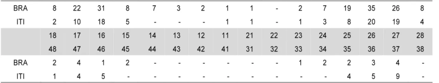
In both groups, the implants were mainly placed in the posterior maxilla (Table 1) or the type C bone (Table 2) in accordance to the Lekholm and Zarb in- dex13). As shown in Table 3, MK Ⅳ implants were mostly installed in the BRA group (81.1% ), and ITI solid screw implants were mostly installed in the ITI group (75.8%), respectively.
3. Study design
This study was carried out retrospectively using the patients’chart. The following information was col- lected from the patient records: age, gender, systemic disease, the type, number, length and diameter of the implants, their location in the jaws, bone quantity, the number of failed implants, the causes of failure, and advanced surgery for bone augmentation [Osteotome Sinus Floor Elevation (OSFE), Bone Added Osteotome Sinus Floor Elevation (BAOSFE), Sinus graft (1-stage), Sinus graft (2-stage), and GBR].
4. Survival criteria
The survival rates were calculated according to the method reported by Buser et al14) as follows:
1. The absence of persistent subjective complaints, such as pain, foreign body sensation, and/or dysesthesia 2. The absence of recurrent peri-implant infections
with suppuration 3. The absence of mobility
4. The absence of continuous radiolucency around the implant
5. The possibility for restoration
5. Statistical analysis
The results were evaluated using the life table anal- ysis described by Cutler & Ederer.15) The differences in the survival rates between the implant types were ex- amined using a Mantel-Haenszel chi-square, and the differences among the advanced surgical techniques were examined using the Fisherʼs exact test.
Table 1. Implant Distribution according to the Location (WHO Site Classification)
BRA 8 22 31 8 7 3 2 1 1 - 2 7 19 35 26 8
ITI 2 10 18 5 - - - 1 1 - 1 3 8 20 19 4
18 17 16 15 14 13 12 11 21 22 23 24 25 26 27 28
48 47 46 45 44 43 42 41 31 32 33 34 35 36 37 38
BRA 2 4 1 2 - - - - - - 1 2 2 3 4 -
ITI 1 4 5 - - - - - - - - - 4 5 9 -
BRA: Brånemark Ti-UniteTM implants, ITI: ITI SLA implants.
Table 2. Implant Distribution according to the Bone Quantity
Quantity (%) B C D Total
BRA 55 (27.4%) 119 (59.2%) 27 (13.4%) 201
ITI 35 (29.2%) 48 (40.0%) 37 (30.8%) 120
BRA: Brånemark Ti-UniteTM, implants, ITI: ITI SLA implants.
Table 3. Implant Distribution according to the Diameter and Length
Brånemark Ti-UniteTM ITI SLA
MK Ⅲ MK Ⅳ Solid screw Esthetic plus TETM
Length\
Diameter Ø 3.75 Ø 4 Ø 5 Ø 4 Ø 5 Ø 4.1 /
4.8
Ø 4.8 / 4.8
Ø 4.8 / 6.5
Ø 4.1 / 4.8
Ø 4.8 / 4.8
Ø 4.1 / 4.8
Ø 4.8 / 6.5
7 ㎜ 3
8 ㎜ 2 2 1
8.5 ㎜ 7 2 11
10 ㎜ 2 10 11 21 27 13 12 4 13
11.5 ㎜ 4 2 21 23
12 ㎜ 12 9 6 1 1 4 5
13 ㎜ 1 8 3 49 15 2
14 ㎜ 6
15 ㎜ 1 7
Total 1 15 22 93 70 47 26 18 5 1 4 19
RESULTS
1. Cumulative survival rate
In the BRA group, 2 submerged implants were lost before healing abutment connection following flap de- hiscence with suppuration, 5 submerged implants were lost at the time of abutment connection and 1non- submerged implant was l ost 5 weeks postoperatively following healing abutment loosening and fixture mobility. Of the failed implants, one upper anterior implant (MK Ⅳ Ø 4×15 mm, # 11 area) was installed 7 weeks after removal of MK Ⅱ Ø 3.75 ×18 mm. The previous MK Ⅱ implant was installed with GBR tech- nique because of labial bone penetration, but it was lost 10 months postoperatively due to repeated pus discharge. One lower posterior implant (MK Ⅲ Ø 3.75
×13 mm, #45 area) was failed at the time of healing abutment connection. The six upper posterior failed implants (MK Ⅲ Ø 5×8.5 mm, # 26, 27 area MK Ⅳ Ø 4×13 mm, # 25 area; MK Ⅳ Ø 5×8.5 mm, # 16, 26 area; MK Ⅳ Ø 5×11.5 mm, # 25 area) were re- lated to sinus augmentation. One MK Ⅲ Ø 5 ×8.5 mm fixture on # 26 area was installed with sinus membrane perforation at the time of OSFE technique.
Two patients (3 implants) had smoking habit and one patient (1 implant) had bruxism, and one patient (1 implant) had a stable angina pectoris. A total of 8 implants failed early, resulting in a 96.02% survival rate. After loading, one implant (MK Ⅳ Ø 4 ×13 mm,
# 24 area) was lost at the 7th month after using an overdenture due to overloading, resulting in a cumu- lative survival rate of 95.48%. In the ITI group, no implant was removed but one implant (ITI TETM Ø 4.1 / 4.8×12 mm, # 27 area) showed repeated suppu- ration after installation of the permanent prosthesis.
After being treated with antibiotics, chlorhexidine ir- rigation, and curettage, the peri-implantitis was controlled. The implant was left in place but a suppu- rative peri-implant infection was found at the last annual examination. This implant was considered to
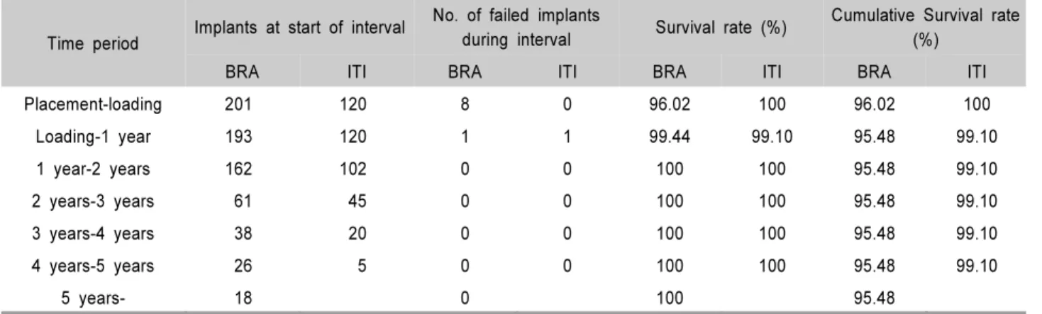
be a failure, resulting in a cumulative survival rate of 99.10% (Table 4, Fig. 1). Therefore, there were 1of 120 failure in the ITI SLA implants and 9 of 201 failures in the Br 。anemark Ti-UniteTM implants, respectively.
However, there was no significant difference between both groups (Mantel -Haenszel =0.138).
2. Survival rate for each surgical method The surgical methods used at the time of implant placement are described below (Table 5). In the case of OSFE, or 1-stage sinus graft, or 2-stage sinus graft, respectively, there was higher percentage of BRA cases than ITI cases. Figure 2 shows the survival rate according to the additional surgical procedures and implant type. In all cases, the survival rate was not significantly different in the two implant types according to Fisherʼs exact test (p>0.05).
DISCUSSION
Many studies have demonstrated that a lack of ini- tial stability in soft bone, particularly in the posterior maxilla, leads to lower success rates than in other lo- cations and bone qualities16-18). In order to overcome the high failure rate of implants in soft bone, a mod- ification of the surgical methods during implant placement has been suggested that bone condensation with osteotomes, minimal or no countersinking, not to drill to the total implant length, and light forces dur- ing implant insertion. In addition, wide diameter im- plant, wide collar, and the implant design for in- creasing the surface of bone to implant contact are recommended. Finally, the implant surface texture have been modified to enhance the cellular activity and primary stability. Rough surfaces of implant are advocated not only to increase primary stability but mainly to improve bone healing2). To improve the ini- tial implant stability, high removal torques and max-
imize the quality of the bone-implant interface, a novel titanium porous oxide implant surface or a sandblasted large-grit acid etched implant surface are studied respectively19-22). However, there have been few studies that have compared the survival rate be- tween both implants in soft bone.
In this study, 201 (BRA) and 120 (ITI) implants were placed in soft bone. Among the 8 early failed implants
(BRA), 6 implants were related to the sinus augmen- tation procedure in the posterior maxilla, and 2 im- plants were rotated at the time of healing abutment connection because of osseointegration failure. There was only 1 implant failure within 1 year after loading in each group, and no implant failed in both group after 1 year. Therefore, the cumulative survival rate was 95.48% in BRA group, and 99.10% in ITI group, Table 4. Life Table Analyses
Time period Implants at start of interval No. of failed implants
during interval Survival rate (%) Cumulative Survival rate (%)
BRA ITI BRA ITI BRA ITI BRA ITI
Placement-loading 201 120 8 0 96.02 100 96.02 100
Loading-1 year 193 120 1 1 99.44 99.10 95.48 99.10
1 year-2 years 162 102 0 0 100 100 95.48 99.10
2 years-3 years 61 45 0 0 100 100 95.48 99.10
3 years-4 years 38 20 0 0 100 100 95.48 99.10
4 years-5 years 26 5 0 0 100 100 95.48 99.10
5 years- 18 0 100 95.48
BRA: Brånemark Ti-UniteTM, implants, ITI: ITI SLA implants.
Table 5. Implant Distribution according to the Additional Surgical Procedures and Implant Group
None OSFE BAOSFE Sinus graft (1-stage) Sinus graft (2-stage) GBR BAOSFE+GBR BRA (%) 46 (22.9%) 55 (27.4%) 25 (12.4%) 28 (13.9%) 40 (19.9%) 5 (2.5%) 2 (1%)
ITI (%) 61 (50.8%) 10 (8.3%) 35 (29.2%) 4 (3.3%) 8 (6.7%) 2 (1.7%) 0 (0%)
BRA: Brånemark Ti-UniteTM implants ITI: ITI SLA implants
None: No additional surgery OSFE: Osteotome Sinus Floor Elevation BAOSFE: Bone Added Osteotome Sinus Floor Elevation GBR : Guided Bone Regeneration.
BRA I
Figure 1. Cumulative survival rates in relation to the im- plant type (BRA: Brånemark Ti-UniteTM implants, ITI: ITI SLA implants).
Figure 2. Implant survival rate according to the additional surgical procedures and implant type (BRA: Brånemark Ti-UniteTM implants, ITI: ITI SLA implants).
respectively. At the time of implant placement, none or the BAOSFE method were more frequently used in the ITI group, while other procedures were more fre- quently used in the BRA group. The survival rates in the BRA group (97.5%) and ITI group (87.5%) were significantly different in the case of sinus graft (2-stage). However, there was no overall significant difference between the two groups because the number of implant placement in the ITI group (8) was sig- nificantly lower than in the BRA group (40). The cu- mulative survival rate and overall survival rate for each surgical method was similar in the two groups (Mantel-Haenszel=0.138 and Fisherʼs exact test>0.05, respectively), and this results were comparable to previous study23).
High survival and success rates (90.7-100%) for the two systems have been individually reported in many earlier studies24-37). Regarding the Br 。anemark Ti-UniteTM implant placement in type 4, Glauser et al24) demon- strated a 97.1% success rate after 4 years of pros- thetic loading in soft bone. In addition, Friberg et al25) reported a 96.2% survival rate over a follow-up period of 1 year. Likewise, Pinholt9) reported a 98% overall survival rate of ITI SLA surface implants in the hu- man bone-grafted maxilla, bone quality 4, over a fol- low-up period of 20-67 months. Stricker et al26) dem- onstrated a 99.5% survival rate after 15-40 months of implant placement during maxillary sinus augmenta- tion with autogenous bone grafts. Therefore, the sur- vival rate in the BRA group (95.48%) and ITI group (99.10%) in this study is comparable to other studies.
In this retrospective article, most cases had been ap- plied a delayed loading after the placement of implant. Further studies will be needed to evaluate the radiographic changes over a long follow-up period in each implant system and to study the survival rate after immediate loading.
In conclusion, the survival rates of the oxidized ti- tanium implants and the sandblasted large-grit acid etched implants were similarly high in soft bone. Both implants can be used successfully in soft bone regard-
less of the surgical methods used at the time of im- plant placement.
REFERENCES
1. Albrektsson T, Brånemark PI, Hansson HA, Lindstrom J.
Titanium implants. Requirements for ensuring a long-lasting direct bone anchorage in man. Acta Orthop Scand 1981;
52:155-170.
2. Martinez H, Davarpanah M, Missika P, Celletti R, Lazzara R. Optimal implant stabilization in low density bone. Clin Oral Implants Res 2001;12:423-432.
3. Boyne PJ, James RA. Grafting of the maxillary sinus floor with autogenous marrow and bone. J Oral Surg 1980;38:
613-616.
4. Tatum OH. Maxillary and sinus implant reconstruction.
Dent Clin North Am 1986;30:207-229.
5. Summers RB. The osteotome technique: Part Ⅲ: Less in- vasive methods of elevating the sinus floor. Compendium 1994;15:698,700,702-704.
6. Buser D, Dula K, Lang NP, Nyman S. Long-term stability of osseointegrated implants in bone regenerated with the membrane technique. Five-year results of a prostective study with 12 implants. Clin Oral Implants Res 1996;7:
175-183.
7. Fugazzotto PA. Success and failure rates of osseointegrated implants in function in regenerated bone for 72 to 133 months. Int J Oral Maxillofac Implants 2005;20:77-83.
8. Summers RB. A new concept in maxillary implant surgery:
The osteotome technique. Compendium 1994;15:152,154- 156, 158.
9. Pinholt EM. Brånemark and ITI dental implants in the hu- man bone grafted maxilla: a comparative evaluation. Clin Oral Implants Res 2003;14:584-592.
10. Åstrand P, Engquist B, Anzén B et al. A three-year fol- low-up report of a comparative study of ITI dental im- plants® and Brånemark System® implants in the treatment of the partially edentulous maxilla. Clin Implant Dent Relat Res 2004;6:130-141.
11. Kang NW, Jung UW, Choi SH et al. Bone added osteo- tome sinus floor elevation with simultaneous placement of Br 。anamark Ti-Unite and ITI SLA implants. J Korean Acad
Periodontol 2005;35:609-621.
12. Hong SB, Chai GJ, Jung UW et al. Clinical evaluation of Brånemark Ti-Unite implant and ITI SLA implant in the post maxillary area with sinus elevation technique. J Korean Acad Periodontol 2005;35:813-822.
13. Lekholm U, Zarb GA. Patient selection and preparations.
In: Brånemark PI, Zarb GA, Albretksson T, eds. Tissue-in- tegrated prostheses: Osseointegration in clinical dentistry.
1st ed. Chicago: Quintessence Publishing Co Inc; 1985:
199-220.
14. Buser D, Weber HP, Lang NP. Tissue integration of non-submerged implants: 1-year results of a prospective study with 100 ITI hollow-cylinder and hollow-screw implants. Clin Oral Implants Res 1990;1:33-40.
15. Cutler SJ, Ederer F. Maximum utilization of the life table method in analyzing survival. J Chronic Dis 1958;6:699-712.
16. Engquist B, Bergendal T, Kallus T, Linden U. A retro- spective multicenter evaluation of osseointegrated implants supporting overdentures. Int J Oral Maxillofac Implants 1988;3:129-134.
17. Friberg B, Jemt T, Lekholm U. Early failures in 4,641 con- secutively placed Brånemark dental implants: a study from stage I surgery to the connection of completed prostheses.
Int J Oral Maxillofac Implants 1988;3:129-134.
18. Jaffin R, Berman C. The excessive loss of Brånemark fix- tures in type Ⅳ bone: a 5-year analysis. J Periodontol 1991;62:2-4.
19. Wilke HJ, Claes L, Stenemann S. Clinical implant material.
In: Heimke G, Soltesz U, Lee AJC, eds. Advances in biomaterials. Vol. 9. Amsterdam: Elsevier Science Publishing Co Inc; 1990:390.
20. Buser D, Nydegger T, Hirt HP, Cochran DL, Nolte LP.
Removal torque values of titanium implants in the maxilla of miniature pigs. Int J Oral Maxillofac Implants 1998;13:
611-619.
21. Henry P, Tan A, Allan B, Hall J, Johansson C. Removal torque comparison of TiUnite and turned implants in the Greyhound dog mandible. Appl Osseointegration Res 2000;
1:15-17.
22. Glauser R, Portmann M, Ruhstaller P et al. Stability meas- urements of immediately loaded machined and oxidized im- plants in the posterior maxilla. A comparative clinical study using resonance frequency analysis. Appl Osseointegration Res 2001;2:27-29.
23. Lee ES, Ahn YB, Lee WJ, Kim HS. Survival rate of im- plant placement in the maxilla treated with sinus elevation by the lateral approach: A retrospective study. J Korean Acad Periodontol 2008;38:589-594.
24. Glauser R, Ruhstaller P, Windisch S et al. Immediate oc- clusal loading of Brånemark TiUniteTM implants placed pre- dominantly in soft bone: 4-year results of a prospective clinical study. Clin Implant Dent Relat Res 2005;7:s52-s59.
25. Friberg B, Dahlin C, Widmark G, Östman PO, Billström C.
One-year results of a prospective multicenter study on Brånemark System® implants with a TiUniteTM surface.
Clin Implant Dent Relat Res 2005;7:s70-75.
26. Stricker A, Voss PJ, Gutwald R, Schramm A, Schmelzeisen R. Maxillary sinus floor augmentation with autogenous bone grafts to enable placement of SLA-surfaced implants:
preliminary results after 15-40 months. Clin Oral Implants Res 2003;14:207-212.
27. Glauser R, Gottlow J, Lundgren AK et al. Immediate oc- clusal loading of Brånemark Mk Ⅳ TiUniteTM implants placed in bone quality type 4. Appl Osseointegration Res 2002;3:22-24.
28. Vanden Bogaerde L, Pedretti G, Dellacasa P et al. Early function of splinted implants in maxillas and posterior mandibles, using Brånemark System® TiUniteTM implants:
An 18-month prospective clinical multicenter study. Clin Implant Dent Relat Res 2004;6:121-129.
29. Rocci A, Martignoni M, Gottlow J, Rangert B. Immediate function of single and partial reconstructions in the maxilla using Mk Ⅳ fixtures: A retrospective analysis. Appl Osseointegration Res 2001;2:22-26.
30. Bornstein MM, Schmid B, Belser UC, Lussi A, Buser D.
Early loading of nonsubmerged titanium implants with a sandblasted and acid-etched (SLA) surface: 5-year results of a prospective study in partially edentulous patients. Clin Oral Implants Res 2005;16:631-638.
31. Ferrigno N, Laureti M. Surgical advantages with ITI TE® implants placement in conjunction with split crest techni- que: 18-month results of an ongoing prospective study.
Clin Oral Implants Res 2005;16:147-155.
32. Fugazzotto PA, Vlassis J, Butler B. ITI implant use in pri- vate practice: Clinical results with 5,526 implants followed up to 72+ months in function. Int J Oral Maxillofac Implants 2004;19:408-412.
33. Luongo G, Raimondo R, Filippini P, Gualini F, Paoleschi
C. Early loading of sandblasted, acid-etched implants in the posterior maxilla and mandible: A 1-year follow-up report from a multicenter 3-year prospective study. Int J Oral Maxillofac Implants 2005;20:84-91.
34. Nedir R, Bischof M, Briaux JM et al. A 7-year life table analysis from a prospective study on ITI implants with spe- cial emphasis on the use of short implants: Results from a private practice. Clin Oral Implants Res 2004;15:150-157.
35. Nordin T, Nilsson R, Frykholm A, Hallman M. A 3-arm study of early loading of rough-surfaced implants in the completely edentulous maxilla and in the edentulous poste-
rior maxilla and mandible: results after 1 year of loading.
Int J Oral Maxillofac Implants 2004;19:880-886.
36. Salvi GE, Gallini G, Lang NP. Early loading (2 or 6 weeks) of sandblasted and acid-etched (SLA) ITI® implants in the posterior mandible: A 1-year randomized controlled clinical trial. Clin Oral Implants Res 2004;15:142-149.
37. Vanden Bogaerde L, Rangert B, Wendelhag I. Immediate / early function of Brånemark System® TiUniteTM implants in fresh extraction sockets in maxillae and posterior man- dibles: An 18-month prospective clinical study. Clin Implant Dent Relat Res 2005;7:s121-s130.