대한소화기학회지 2005;46:388-395
서 론
Tamoxifen은 유방암 치료에 가장 효과적이고 널리 쓰이는 약물이다. Tamoxifen은 경쟁적으로 수용체에 작용하여 에스
트로젠 작용을 억제함으로써 항암효과를 보이지만, 약물의 작용기전은 매우 다양하다. 일부에서 세포 내 protein kinase C를 억제하여 세포 내 그리고 세포막에 영향을 주며, p-glyco- protein을 억제하여 용적-민감성 Cl- 통로를 차단한다.1-3 또
생쥐 대장 평활근세포의 막 전압-의존 이온 전류에 대한 Tamoxifen 효과
조선대학교 의과대학 내과학교실, 신경과학교실*, 생리학교실†
이동민․장성종․박찬국․김만우․임건한
*․최 석
†․염철호
†․윤평진
†․전제열
†Effects of Tamoxifen on the Voltage-dependent Ionic Currents in Mouse Colonic Smooth Muscle Cells
Dong Min Lee, M.D., Sung Jong Chang, M.D., Chan Guk Park, M.D.,
Man Woo Kim, M.D., Gun Han Lim, M.D.*, Seok Choi, Ph.D.†, Cheol Ho Yeum, Ph.D.†, Pyung Jin Yoon, Ph.D.†, and Jae Yeoul Jun, M.D.†
Departments of Internal Medicine, Neurology*, and Physiology†, Chosun University College of Medicine, Gwangju, Korea
Background/Aims: Tamoxifen is a widely used anticancer drug for breast cancer with frequent gastrointestinal side effects. Changes in gastrointestinal motility is associated with altered activities of membrane ion channels.
Ion channels have important role in regulating membrane potential and cell excitability. This study was per- formed to investigate the effects of tamoxifen on the membrane ionic currents in colonic smooth muscle cells.
Methods: Murine colonic smooth muscle cells were isolated from the proximal colon using collagenase, and the membrane currents were recorded using a whole-cell patch clamp technique. Results: Two types of voltage- dependent K+ currents were recorded (A-type and delayed rectifier K
+ currents). Tamoxifen inhibited both types of voltage-dependent K
+ currents in a dose-dependent manner. However, tamoxifen did not change the half- inactivation potential and the recovery time of voltage-dependent K
+ currents. Chelerythrine, a protein kinase C inhibitor or phorbol 12, 13-dibutyrate, a protein kinase C activator did not affect the voltage-dependent K
+
currents. Guanosine 5'-O-(2-thio-diphosphate) did not affect the tamoxifen-induced inhibition of voltage-dependent K
+ currents. Tamoxifen inhibited voltage-dependent Ca
2+ currents completely in whole-test ranges. Conclusions:
These results suggest that tamoxifen can alter various membrane ionic currents in smooth muscle cells and cause some adverse effects on the gastrointestinal motility. (Korean J Gastroenterol 2005;46:388-395)
ꠏꠏꠏꠏꠏꠏꠏꠏꠏꠏꠏꠏꠏꠏꠏꠏꠏꠏꠏꠏꠏꠏꠏꠏꠏꠏꠏꠏꠏꠏꠏꠏꠏꠏꠏꠏꠏꠏꠏꠏꠏꠏꠏꠏꠏꠏꠏꠏꠏꠏꠏꠏꠏꠏꠏꠏꠏꠏꠏꠏꠏꠏꠏꠏꠏꠏꠏꠏꠏꠏꠏꠏꠏꠏꠏꠏꠏꠏꠏꠏꠏꠏꠏꠏꠏꠏꠏꠏꠏꠏꠏꠏꠏꠏꠏꠏꠏꠏꠏꠏꠏꠏꠏꠏꠏꠏꠏꠏꠏꠏꠏꠏꠏ
Key Words: Colonic smooth muscle cells; Tamoxifen; Voltage-dependent K
+currents; Voltage-dependent Ca
2+currents
ꠏꠏꠏꠏꠏꠏꠏꠏꠏꠏꠏꠏꠏꠏꠏꠏꠏꠏꠏꠏꠏꠏꠏꠏꠏꠏꠏꠏꠏꠏꠏꠏꠏꠏ Correspondence to: Jae Yeoul Jun, M.D.
Department of Physiology, Chosun University College of Medi- cine, 375 Seoseok-dong, Dong-gu, Gwangju 501-759, Korea Tel: +82-62-230-6412, Fax: +82-62-232-4943
E-mail: [email protected] ꠏꠏꠏꠏꠏꠏꠏꠏꠏꠏꠏꠏꠏꠏꠏꠏꠏꠏꠏꠏꠏꠏꠏꠏꠏꠏꠏꠏꠏꠏꠏꠏꠏꠏ
접수: 2005년 5월 26일, 승인: 2005년 10월 21일
연락처: 전제열, 501-759, 광주광역시 동구 서석동 375번지 조선대학교 의과대학 생리학교실
Tel: (062) 230-6412, Fax: (062) 232-4943 E-mail: [email protected]
* 이 논문은 2004년도 교내연구비를 지원받아 연구되었음.
이동민 외 8인. 생쥐 대장 평활근세포의 막 전압-의존 이온 전류에 대한 Tamoxifen 효과 389
한 막 전압-의존 양이온 전류(voltage-dependent cation cur- rents)를 억제한다.4 사람 심장에서 QT-간격을 증가시켜 심 장의 재분극을 증가시켜 부정맥 치료제로도 제시되고 있 다.5 그러나 이 약물을 투여한 환자의 10%에서 오심과 구토 가 동반되며 이로 인해 약물투여를 중단하는 경우가 있으며 또한 설사, 변비 등 위장관 운동 이상의 부작용을 나타내나6 아직까지 위장관 운동 이상을 초래하는 약물학 기전에 대해 서는 밝혀져 있지 않다.
위장관 운동은 위장관 평활근의 수축과 이완에 의해 최종 결정된다. 위장관 평활근은 세포의 내인성으로 인하여 외부 신경의 조절과 무관하게 규칙적이고 자발적인 수축양상을 보인다. 자발 수축은 평활근 세포막 전압의 주기 탈분극 현 상인 서파(slow waves)에 기인한다.7,8 세포막 전압은 세포막 에 존재하는 이온통로들을 통한 이온들의 상대 투과비에 의 해서 결정된다. 내향 전류의 활성화는 세포막 전압의 탈분 극(depolarization)을 초래하여 세포의 흥분성을 증가시키는 반면 외향 전류의 활성화는 세포막 전압의 과분극(hyper- polarization)을 초래하여 세포의 흥분성을 감소시킨다. 위장 관 평활근에서 대표 내향 전류는 세포막 전압-의존 칼슘 전 류이며 외향 전류는 세포막 전압-의존 칼륨 전류이다.9-11 Tamoxifen은 다양한 세포에서 세포막에 존재하는 이온통로 의 활성화를 조절하므로 tamoxifen으로 유발된 위장관 운동 이상은 위장관 평활근의 활성도를 변화시킬 수 있으며 이는 바로 세포막에 존재하는 이온통로의 기능을 변화시킬 수 있 음을 시사한다. 이번 연구는 생쥐 대장 평활근에서 단일세 포를 분리한 후 세포막 전압 고정법을 이용하여 세포막에 존재하는 칼슘과 칼륨 전류를 기록하고, 이들 전류에 대한 tamoxifen의 효과를 관찰하여 약물 투여 시 나타나는 위장관 운동 이상에 대한 기전을 규명하고자 하였다.
대상 및 방법
1. 세포분리
생후 20일에서 30일 사이의 마우스를 암수 구별없이 실험 동물로 사용하였다. Chloroform을 사용하여 마취시킨 후 경 척추를 탈구시켰다. 개복하여 근위부 대장부위를 적출하였 다. 실온에서 Ca2+-free Hanks 용액(NaCl 125 mM, KCl 5.36 mM, NaOH 15.5 mM, KH2PO4 0.44 mM, Na2HCO3 0.36 mM, glucose 10 mM, sucrose 2.9 mM, HEPES 11 mM, pH 7.4 by tris)으로 채워진 준비 용기 속에서 내강부위를 노출시켰다.
현미경 밑에서 점막층을 박리하여 윤상근이 표면에 노출되 게 깨끗이 분리하였다. 대장 평활근 조직을 0.1% collagenase (Worthington Biochemical Co, Lakewood, NJ, USA), 0.1%
trypsin inhibitor (Sigma Chemical Co., St. Louis, MO, USA),
0.2% bovine serum albumin (Sigma)이 들어 있는 Ca2+-free Hanks 용액에 옮긴 후 37oC에서 15분간 항온 소화시켰다.
소화시킨 후 다시 Ca2+이 들어있지 않는 Hanks 용액으로 교 체한 다음 끝이 무딘 유리피펫을 사용하여 조심스럽게 진탕 시켜 단일세포를 분리하고 4oC에 냉장 보관하였다. 모든 실 험은 단일 세포를 분리한 후 30분 후에 시행하였으며 실온 에서 시행하였다.
2. 막 전류 측정
분리된 세포를 도립현미경 위에 설치되어 있는 작은 실험 용기 (300μL)로 옮긴 후 분당 2-3 mL 속도로 세포외 용액 을 관류시켰다. 유리전극의 저항은 3-5 MΩ을 사용하였다.
Whole-cell patch clamp를 사용하여 세포막 전류를 기록하였 다. Patch clamp 증폭기(Axopatch 1-D, Axon Instruments, Fos- ter, CA, USA)를 통하여 나오는 신호는 디지털 오실로스코 프와 생리 기록기를 통해서 관찰하였고, 고정전압과 자극전 압 조정과 전류 기록은 pClamp software (version 6.0, Axon Instruments)와 IBM-compatible computer를 사용하였다.
외향 전류를 기록하는 과정에서 칼슘-의존 칼륨 통로(Ca2+- activated K+ channels)의 활성화를 배제하기 위해 전극 내 용액에 10 mM EGTA를 첨가하였으며, 세포의 관류용액에
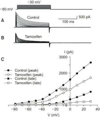
Fig. 1. The effects of tamoxifen (10μM) on the voltage-dependent K+ currents. Membrane potential is stepped from -80 mV to +30 mV with 10 mV increments. (A) Control group. (B) Tamoxifen group.
Tamoxifen inhibites voltage-dependent K+ currents in whole test voltage ranges. (C) Current-voltage relationship in early peak currents and late currents in both groups.
40 V (mV)
0 1000 2000 3000
Control (late) Tamoxifen (late)
-40 -60 -80
-30 mV
Control
Tamoxifen
500 pA 100 ms
0 20
-20 Control (peak)
Tamoxifen (peak)
B
C
I (pA) -80 mV
A
390 The Korean Journal of Gastroenterology: Vol. 46, No. 5, 2005
MnCl2를 첨가하여 사용하였다. 막 전압-의존 칼륨 전류의 비활성화를 알아보기 위하여 -80 mV에서부터 +40 mV까 지의 4초 동안의 자극을 준 다음 이어서 +30 mV로 다시 저분극 자극을 주었다. 막전압-의존 칼륨 전류의 비활성화 에서 회복되는 과정을 알아보기 위하여 -80 mV에서 0 mV 까지 4초 동안의 자극을 선행시킨 다음 시간을 달리하면서 똑같은 자극파를 200 ms 기간으로 주었다.
3. 실험용액
막 전압-의존 칼륨 전류를 관찰하기 위한 세포외 관류용 액은 NaCl 135 mM, KCl 5 mM, MnCl2 1.8 mM, glucose 5 mM, MgCl2 1.2 mM, HEPES 10 mM를 사용하였으며 tris를 사용하여 pH를 7.4로 적정하였다. 전극 내 용액은 NaCl 10 mM, KCl 102 mM, CaCl2 1 mM, ATP 5 mM, GTP 1 mM, MgCl2 1 mM, EGTA 10 mM, pH 7.2 (KOH로 적정)인 용액 을 사용하였다. 칼슘 전류만을 기록할 경우 세포외 관류용
액은 NaCl 135 mM, KCl 5 mM, CaCl2 1.8 mM, glucose 5 mM, MgCl2 1.2 mM, HEPES 10 mM을 사용하였으며 tris를 사용하여 7.4로 적정하였다. 전극 내 용액은 cesium-aspartate 110 mM, Mg2+-ATP 5 mM, ditris creatine phosphate 2.5 mM, disodium creatine phosphate 2.5 mM, MgCl2 1 mM, HEPES 0.1, TEA-Cl (tetraethylammonium chloride) 20 mM, EGTA 5 mM, pH 7.2인 용액을 사용하였다.
결 과
1. 막 전압-의존 칼륨 전류
유지전압 -80 mV로 고정한 후 10 mV씩 30 mV까지 단계 별로 저분극 자극을 주면 자극 20 ms 내에 일시적으로 최고 크기의 전류를 보였다가 이후 서서히 감소되어 지속되는 외 향성 전류가 관찰된다(Fig. 1A). 막 전압-의존 칼륨 전류에 서 일시적으로 초반부에 최고 크기를 나타내는 전류는 칼륨 통로 차단제인 4-aminopyridine에 의해서 억제되고 이후 지 속되는 종반부 전류는 TEA에 의해서 억제되는 막 전압-의
Fig. 2. The effects of tamoxifen on the voltage dependent inacti- vation in voltage-dependent K+ currents. Membrane potential is stepped to conditioning potential of +30 mV. (A) Control group.
(B) Tamoxifen group. (C) The voltage dependence of inactivation of the voltage-dependent K+ currents is shown as a plot of the conditioning potential. Datas are fitted to the function: I/IMAX =1/1
{1+exp(V-Vlhalf)/k}. Ihalf indicates the amplitude of the current, V is the value of the test potential, Vhalf is the potential at which 50%
of the current was inactivated, and k is the slope of the curves. Vhalf
and k are -43.7±3.1 mV and 7.4 in the control, -46.2±2.8 mV and 6.1 in the presence of tamoxifen, respectively (n=5).
40 mV
Control
Tamoxifen
500 pA 1000 ms
B C
-80 mV
A
+30 mV
-100 -80 -60 -40 -20 0 20 40
Normalized current
V (mV) 0.2
0.4 0.6 0.8 1.0
Control Tamoxifen
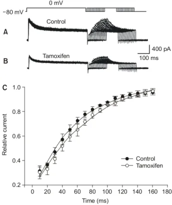
Fig. 3. The effects of tamoxifen on recovery from inactivation of voltage-dependent K+ currents. Membrane potential is stepped from -80 to 0 mM for 200 ms. (A) Control group. (B) Tamoxifen group.
(C) Recovery of the voltage-dependent K+ currents obtained by normalizing the peak current during the test step is plotted as a function of the recovery interval in the both groups. Solid lines are fitted to the data by single exponential function (n=4).
0 mV
C A
20 40 60 80 100 120 140 160 180
Relative current
Time (ms) 0.2
0.4 0.6 0.8 1.0
Control Tamoxifen Control
Tamoxifen -80 mV
B
400 pA 100 ms
0
Lee DM, et al. Effects of Tamoxifen on the Voltage-dependent Ionic Currents in Mouse Colonic Smooth Muscle Cells 391
존 칼륨 전류이다.12
2. 막 전압-의존 칼륨 전류에 대한 tamoxifen의 효과
Tamoxifen 10μM을 투여 후 농도에 비례하여 막 전압-의 존 칼륨 전류가 억제되었다. Tamoxifen은 초반부와 종반부 의 전류 크기를 모두 현저히 억제하였다(Fig. 1B). Tamoxifen 의 초반부 전류에 대한 억제효과는 10 mV에서 43%, 30 mV 에서 64%가 차단되는 양상이었다(Fig. 1C).
막 전압-의존 칼륨 전류의 동력학 특성에 대한 tamoxifen 의 효과를 알아보기 위하여 비활성화와 회복 과정에 대한 실험을 시행했다. Fig. 2A는 대조군에서 기록된 전류이며 Fig. 2B는 tamoxifen (10μM)을 투여하여 기록한 그림이다.
Tamoxifen은 막 전압-의존 칼륨 전류의 비활성화에는 효과 가 없었다. Boltzman 함수로 나타낸 비활성화-전압곡선에서 50%로 비활성화되는 전압(half-inactivation potential)은 대조 군에서 -43.7±3.1 mV를 보였으며 tamoxifen이 존재시 -46.2
±2.8 mV를 보였다. 각 군 간에 유의한 차이는 없었다(Fig.
2C).
막 전압-의존 칼륨 전류의 비활성화에서 회복되는 과정은 일차 지수함수(single exponential) 양상이었다. 비활성화 상 태에서 50%로 회복되는 시간은 대조군에서 25±3 ms였으 며 tamoxifen으로 자극할 경우 26±2 ms였다. 이 두 결과 모 두 유의한 차이가 없어 tamoxifen이 막 전압-의존 칼륨 전류 의 회복과정에 효과가 없음을 알 수 있다(Fig. 3).
3. 막 전압-의존 칼륨 전류에 대한 protein kinase C의 역할
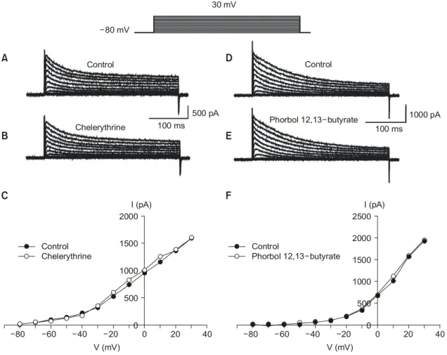
Tamoxifen 작용기전의 하나는 protein kinase C 억제이다.
따라서 막 전압-의존 칼륨 전류를 억제하는 tamoxifen의 작 용이 protein kinase C를 경유하는지 알아보기 위하여 선택적 으로 protein kinase C 차단물질인 chelerythrine과 protein kinase C 활성물질인 phorbol 12, 13-dibutyrate를 투여하였다.
Fig. 4. The effects of protein kinase C inhibitor or protein kinase C activator on the voltage-dependent K+ currents. (A) Control group. (B) In the presence of chelerythrine (1μM). (C) Current-voltage relationship for the early peak currents in the control group and in the tamoxifen group. Chelerythrine does not affect the voltage-dependent K+ currents. (D) Control group (another cell). (E) In the presence of phorbol 12, 13-dibutyrate (0.1μM). (F) Current-voltage relationship for the early peak currents in the control group and in the tamoxifen group. Phorbol 12, 13-dibutyrate does not affect the voltage-dependent K+ currents.
40 V (mV)
0 500 1000 1500 2000
-40 -60
-80 -20 0 20
Control Chelerythrine
C
I (pA)30 mV
-80 mV
Control
Chelerythrine
500 pA 100 ms
Control
Phorbol 12,13 butyrate-
100 ms
1000 pA
B A
E D
40 V (mV)
0 500 1500 2000 2500
-40 -60
-80 -20 0 20
Control
Phorbol 12,13 butyrate-
F
I (pA)1000
392 대한소화기학회지: 제46권 제5호, 2005
Chelerythrine (1μM)과 phorbol 12, 13-dibutyrate (0.1μM)는 막 전압-의존 칼륨 전류에 아무런 효과를 보이지 않아(Fig.
4) 막 전압-의존 칼륨 전류 조절과 tamoxifen 효과가 protein kinase C와는 무관함을 알 수 있었다.
G-단백질이 tamoxifen의 억제작용에 미치는 영향을 알기 위하여 G-단백질 억제제인 guanosine 5'-O-(2-thio-diphosphate) (GDPβS) 1 mM를 전극 내 투여한 후 막 전압-의존 칼륨 전 류를 기록하였다. Tamoxifen (10μM)은 GDPβS 존재하에서 도 여전히 막 전압-의존 칼륨 전류를 억제하였다(Fig. 5).
4. Tamoxifen이 막 전압-의존 칼슘 전류에 미치는 영향
유지전압 -80 mV에서 40 mV까지 단계적으로 10 mV씩 저분극 자극을 주게 되면 -40 mV에서부터 활성화되어 0 mV에서 최대 크기를 보였다가 그 이후 전압에서 다시 크기 가 감소하는 막 전압-의존 내향성 전류가 관찰된다(Fig. 6A).
이 전류는 칼슘 통로 차단제인 nifedipine (1μM)을 투여한 경우 시험전압 전 범위에서 완전히 차단되어 다른 평활근세 포에서 보고한 막 전압-의존 칼슘 전류와 동일함을 나타냈 다(Fig. 6B, C).9-11 Tamoxifen (10μM)은 nifedipine과 같이 시
험전압 전 범위에서 대조군에서 기록된 막 전압-의존 칼슘 전류를 억제하였다(Fig. 6D, E, F).
고 찰
이번 연구는 tamoxifen이 protein kinase C와는 무관하게 대장 평활근세포에서 이온 전류를 억제함을 규명한 것이다.
Tamoxifen은 항암제로 널리 쓰여지는 약물이고, 최근에는 심장 부정맥을 치료할 수 있는 약물로도 제시되나 위장관 운동 이상이 빈번하게 나타난다. 부정맥이나 위장관 운동 이상은 세포막 이온 전류의 변이와 밀접한 관계가 있다. 위 장관은 평활근으로 둘러싸여 있으며 평활근 수축으로 인해 음식물이 혼합하고 연동운동을 통해 하부 장관으로 음식물 을 보낸다. 위장관 내 평활근은 2층으로 구성되어 있으며 내측에 윤상근, 외측에 종주근이 배열되어 있다. 평활근의 수축 정도는 활동전압의 크기, 빈도, 유지되는 시간에 의해 서 결정된다. 활동전압의 발생은 평활근세포 내로 칼슘 유 입을 유발하여 결과적으로 수축이 발생한다. 칼륨 통로도 세포 내외의 높은 농도경사를 이용하여 외향 전류를 발생시 킴으로써 평활근의 전기 활동도를 형성하는 데 매우 중요한 역할을 한다. 칼륨 통로의 활성화는 세포의 안정막 전압을 유지하여 수축을 억제한다. 위장관 평활근에는 여러 종류의 칼륨 통로가 존재하는데 이는 전기 활동도가 매우 세밀하게 조정되고 있음을 시사한다.10,11
생쥐 대장 평활근에서 막 전압-의존 칼륨 통로는 약물 반 응과 생리 기능에 따라 두 종류의 통로가 존재한다. 첫 번째 는 빠르게 활성화되었다가 빠르게 비활성화되며 이 전류는 4-aminopyridine에 의해서 억제되며 A-전류라 일컬어진다.
두 번째는 서서히 활성화되었다가 비활성화 과정이 매우 서 서히 이루어지는 것으로 이 전류는 tetraethylammonium (TEA) 에 의해서 차단되며 delayed rectifier K+ 전류라 한다. 대장 조직에서 서파를 기록하면서 4-aminopyridine를 투여하면 세 포막 전압이 탈분극되어 A-전류는 안정막 전압을 유지하며 delayed rectifier K+ 전류는 전기 활동도에서 재분극을 촉진
한다.12,13 이번 연구에서 tamoxifen는 4-aminopyridine와 TEA
에 민감한 성분에 효과를 나타내어 두 종류의 이온 전류를 가역적으로 차단하는 양상을 보였다. Tamoxifen에 의해 차 단되는 정도는 막 전압의 탈분극이 커질수록 증가되어 막 전압에 비례하여 억제되었다. 약물 등에 의한 이온전류 억 제는 일반적으로 세포막에 있는 이온 통로들의 문을 단순히 차단하여 일어날 수 있으나 이온통로의 동역학(kinetics)에 영향을 미쳐 억제할 수도 있다. 이번 연구에서는 tamoxifen 이 칼륨 전류의 비활성화와 회복시간에는 전혀 영향을 나타 내지 않았다. 따라서 이온 통로의 동역학에는 변화없이 단 순히 이온통로의 문을 차단하는 것으로 보인다. 이러한 결 Fig. 5. The effects of intracellular application of GDPβS on the
tamoxifen-induced inhibition of the delayed K+ currents. Membrane potential is stepped from -80 mV to +30 mV with 10 mV incre- ments. (A) Control group. (B) Tamoxifen group. (C) Current-voltage relationship of the early peak currents and late currents in two groups. GDPβS (1 mM) does not affect the tamoxifen-induced inhibition of the voltage-dependent K+ currents.
40 V (mV)
0 500 1000 1500 2000
Control (late) Tamoxifen (late)
-40 -60
-80 -20 0 20
Control (peak) Tamoxifen (peak)
C
I (pA)
A
-80 mV
30 mV
[GDP S; 1mM]β i
Control 500 pA
100 ms
Tamoxifen
B
이동민 외 8인. 생쥐 대장 평활근세포의 막 전압-의존 이온 전류에 대한 Tamoxifen 효과 393
과는 교종 세포에서 tamoxifen이 A-currents와 delayed recti- fier K+ 전류를 억제시키고,14 심장근에서도 tamoxifen에 의 해서 delayed rectifier K+ 전류가 전압에 비례해 억제된다는 보고5와 일치하였다.
Tamoxifen은 항에스트로젠 약물이지만 주 작용기전이 세 포 내 protein kinase C 억제를 통하여 나타난다.1 Protein kinase C는 매우 다양한 생리작용을 하며 또한 여러 세포에 서 이온 통로를 조절한다.15-17 이번 연구에서 protein kinase C 억제제인 chelerythrine는 tamoxifen의 효과와는 달리 막 전 압-의존 칼륨 전류에 아무런 효과가 없었으며, protein kinase C 활성제인 phorbol 12, 13-dibutyrate 역시 효과가 없었다. 따 라서 생쥐 대장 평활근에서는 막 전압-의존 칼륨 전류 조절 과 tamoxifen의 억제 작용이 protein kinase C와는 관련이 없 다고 생각한다.
평활근이 수축하려면 세포 내에 일정 농도의 칼슘이 필수 적으로 증가되어야 한다. 세포 내 칼슘의 동원은 칼슘저장 고인 내형질 세망으로부터의 분비와 세포 밖에서 막 전압-
의존 칼슘 통로를 통한 유입을 통하여 이루어진다.10 막 전 압-의존 칼슘 통로는 동력학 특성과 약물반응에 따라 두 가 지로 분류된다.18 상대적으로 낮은 전압에서 활성화와 비활 성화가 시작되고 비활성화 정도가 빠르며 심하게 나타나는 T-type (transient)은 dihydropyridine 계통 약물인 nifedipine이 차단하지 않으며, 이와는 반대로 전류 활성화와 비활성화가 상대적으로 높은 전압에서부터 시작되는 L-type (long lasting) 은 nifedipine에 민감하게 차단된다. 혈관 평활근의 일부에서 2종류가 다 존재하지만 위장관 평활근에는 L-type만 존재한
다.10,19 이번 연구에서도 막 전압-의존 칼슘 전류는 역치전압
이 -30 mV부터 시작되고 최대전류 크기가 0 mV에서 관찰 되었으며 nifedipine에 완전히 차단되어 L-type만 존재하였 다. Tamoxifen (10μM)은 막 전압-의존 칼슘 전류를 시험전 압 전 범위에서 완전히 차단하였다. 혈관 평활근과 뇌하수 체 세포에서도 tamoxifen이 막 전압-의존 칼슘전류를 억제
한다.20,21 Tamoxifen 치료 시 혈중농도는 일반적으로 μM 농
도로 존재하고 있어 10μM은 혈중농도와 비슷한 농도로 생 Fig. 6. The effects of nifedipine or tamoxifen on the voltage-dependent inward Ca2+ currents in mouse proximal colonic smooth muscle cells.
(A) Inward control currents at various test potentials from a -80 mV of holding potential by increment of 10 mV steps. (B) In the presence of nifedipine (1μM). (C) Current-voltage relationship of the peak currents in the control group and in the presence of nifedipine. (D) Control group (another cell). (E) Tamoxifen group. (F) Current-voltage relationship for the peak currents of voltage-dependent inward Ca2+ currents in whole test voltage ranges.
40 V (mV)
-40 -60
-80 0
20 -20
Control Nifedipine
C
I (pA)
40 mV
-80 mV Control
Nifedipine
100 pA 40 ms
Control
Tamoxifen
100 pA 100 ms
B A
-300 -200 -100 0 100
40 V (mV)
-40 -60
-80 -20 0 20
Control Tamoxifen
F
I (pA) -300
-50 0 50
-250 -200 -150 -100
E
D
394 The Korean Journal of Gastroenterology: Vol. 46, No. 5, 2005
각된다. 이상을 종합해보면 tamoxifen은 대장 평활근에서 칼 륨과 칼슘 전류를 억제하며 이 작용은 protein kinase C 억제 와는 무관하게 일어나며, 이러한 이온통로의 활동도 억제가 약물 투여 시 보이는 위장관 운동 이상을 일으킬 수 있는 한 기전이 될 수 있을 것이다.
요 약
목적: 항암제인 tamoxifen의 투여는 위장관 운동에 변화 를 초래하는 이상효과를 흔히 보인다. 위장관 운동은 장관 평활근세포에 존재하는 이온통로의 활동도에 의해서 결정 되므로 대장근세포에서 막 전압-의존 이온전류를 기록하면 서 tamoxifen의 효과를 관찰하여 위장관 운동 이상에 대한 기전을 알아보고자 하였다. 대상 및 방법: 생후 20-30일 생 쥐 대장에서 평활근세포를 분리한 후 실온에서 세포막 전압 고정법을 사용하여 막 전압-의존 칼륨과 칼슘 전류를 기록 하였다. 결과: 대장 평활근에서 두 종류의 막 전압-의존 칼 륨 전류(A-type과 delayed rectifier K+ currents)가 관찰되었 다. Tamoxifen은 두 종류의 칼륨 전류에 대해 시험전압 전 범위에서 전류의 크기를 감소시켰다. 그러나 칼륨 전류의 비활성화 과정과 회복 과정에는 효과가 없었다. Protein kinase C 억제제인 chelerythrine과 활성제인 phorbol 12, 13- dibutyrate는 막 전압-의존 칼륨 전류에는 아무런 효과를 보 이지 않았다. G-단백질을 억제하는 guanosine 5'-O-(2-thio- diphosphate)는 tamoxifen의 칼륨 전류에 대한 억제작용을 차 단하지 못하였다. 막 전압-의존 칼슘 전류에 대해 tamoxifen 은 시험 전압 전 범위에서 전류의 크기를 억제하였다. 결 론: Tamoxifen이 대장 평활근에서 protein kinase C와는 무관 하게 이온 통로의 활동도를 변화시킬 수 있으며, 이러한 작 용이 tamoxifen 투여 시 나타나는 위장관 운동 이상을 설명 할 수 있는 한 기전이 되리라 생각한다.
ꠏꠏꠏꠏꠏꠏꠏꠏꠏꠏꠏꠏꠏꠏꠏꠏꠏꠏꠏꠏꠏꠏꠏꠏꠏꠏꠏꠏꠏꠏꠏꠏꠏꠏꠏꠏꠏꠏꠏꠏꠏꠏꠏꠏꠏꠏꠏꠏꠏꠏꠏꠏꠏꠏ 색인단어: 대장 평활근세포, Tamoxifen, 막 전압-의존 칼륨
전류, 막 전압-의존 칼슘 전류
참고문헌
1. O'Brian CA, Liskamp PM, Solomon DH, Weinstein IB. Inhi- bition of protein kinase C by tamoxifen. Cancer Res 1985;45:
2462-2465.
2. Valverde M, Bond TD, Hardy SP, et al. The multidrug resis- tance P-glycoprotein modulates cell regulatory volume de- creases. EMBO J 1996;15:4460-4468.
3. Zhang JJ, Jacob TJ, Valverde MA, et al. Tamoxifen blocks chloride channels. A possible mechanism for cataract forma-
tion. J Clin Invest 1994;94:1690-1697.
4. Hardy SP, deFelipe C, Valverde MA. Inhibition of voltage gated cationic channels in rat embryonic hypothalamic neu- rones and C1300 neuroblastoma cells by triphenylethylene antiestrogens. FEBS Lett 1998;434:236-240.
5. Liu XK, Alexander K, Steven NE, Woosley RL. The anties- trogen tamoxifen blocks the delayed rectifier potassium cur- rents, Ikr, in rabbit ventricular myocytes. J Pharmacol Exp Ther 1998;287:877-883.
6. Charles MH. Cancer treatment. 5th ed. Philadelphia: Saunders Company, 2001.
7. Ohba M, Sakamoto Y, Tomita T. The slow wave in the cir- cular muscle of the guinea pig stomach. J Physiol 1975;253:
505-516.
8. El-Sharkawy TY, Morgan KG, Szurszewski JH. Intracellular electrical activity of canine and human gastric smooth muscle.
J Physiol 1978;279:291-307.
9. Mitra R, Morad M. Ca2+ and Ca2+ activated K+ current in mammalian gastric smooth muscle cells. Science 1985;229:
269-272.
10. Kuriyama H, Kitamura K, Itoh T, Inoue R. Physiological features of visceral smooth muscle cells, with special refer- ence to receptors and ion channels. Physiol Rev 1998;78:
811-920.
11. Vogalis F. Potassium channels in gastrointestinal smooth mus- cle. J Auton Pharmacol 2000;20:207-219.
12. Koh SD, Ward SM, Dick GM, et al. Contribution of delayed rectifier potassium currents to the electrical activity of murine colonic smooth muscle. J Physiol 1999;515:475-487.
13. Koh SD, Perrino BA, Hatton WJ, Kenyon JL, Sanders, KM.
Novel regulation of the A-type K+ current in murine pro- ximal colon by calcium-calmodulin dependent protein kinase II. J Physiol 1999;517:75-84.
14. Smitherman KA, Sontheimer H. Inhibition of glial Na+ and K+ currents by tamoxifen. J Membr Biol 2001;181:125-135.
15. Oike M, Kitamura K, Kuriyama H. Protein kinase C activates the non-selective cation channel in the rabbit. Pflügers Arch 1993;424:159-164.
16. Takano K, Stanfield PR, Nakajima S, Nakajima Y. Protein kinase C-mediated inhibition of an inward rectifier potassium channel by substance P in nucleus basalis neurons. Neuron 1995;14:999-1008.
17. Aiello EA, Clement-Chomine O, Sontag DP, Walsh, MP, Cole WC. Protein kinase C inhibits delayed rectifier K+ current in rabbit vascular smooth muscle cells. Am J Physiol 1996;271:H109-H119.
18. Fridman MF, Kurtz GS, Kaczorowski GS, Katz GM, Reuben
Lee DM, et al. Effects of Tamoxifen on the Voltage-dependent Ionic Currents in Mouse Colonic Smooth Muscle Cells 395
JP. Two calcium currents in a smooth muscle line. Am J Physiol 1986;250:H699-H703.
19. Bean BP. Classes of calcium channels in vertebrate cells. Ann Rev Physiol 1989;51:367-384.
20. Song J, Standley PR, Zhang F, et al. Tamoxifen (estrogen antagonists) inhibits voltage-gated calcium current and con-
tractility in vascular smooth muscle from rats. J Pharmacol Exp Ther 1996;277:1444-1453.
21. Sartor P, Vacher P, Mollard P, Dufy B. Tamoxifen reduces calcium currents in a clonal cell line. Endocrinology 1988;
123:534-540.