상완골 원위 간부와 골간단의 골절
정환용・김우식・김태홍*
건양대학교 의과대학 정형외과학교실, 아주대학교 의과대학 정형외과학교실*
<국문초록>
목 적 : 원위 상완골 간부와 골간단 이행부 골절에서 외측 J형 금속판으로 치료한 환자에서 임상 결과 를 후향적으로 분석하여 보고하고자 한다.
대상 및 방법 : 1996년 1 0월부터 2 0 0 0년 5월까지 원위 상완골 간부와 골간단 이행부 골절로 관혈적 정 복술 및 외측 J 형 금속판 내 고정술을 실시한 1 2예 중 최소 1년 이상 추시가 가능한 1 1예를 대상으로 하였다. 임상적 평가를 위해 최종 추시에서 M o r r e y의 평가기준을 사용하였고 골 유합 상태, 합병증, 주 관절의 기능 평가 및 최종 운동 범위를 측정하였다.
결 과 : 전례에서 골유합을 얻었으며 수술 후 골 유합까지 기간은 평균 1 2주( 1 0 ~ 1 8주)였다. 최종 추시 에서 평균 주관절의 운동 범위는 굴곡 구축 6도( 0 ~ 2 0도), 후속 굴곡 1 2 6도( 9 0 ~ 1 5 0도) 였다. Morrey의 임상 평가에서는 우수 4예, 양호 6예, 보통 1예였다.
결 론 : J형 금속판의 내 고정술은 원위 상완골 간부 및 골간단 이행부 골절의 좋은 치료 방법으로 사 료된다.
색인 단어 : 상완골, 원위 간부와 골간단, 외측 J 형 금속판
※통신저자 : 정 환 용
대전광역시 서구 가수원동 685, 건양대학교병원 정형외과 TEL : +82.42-600-9100
FAX : +82.42-545-2373 E-mail : hy0707@unitel.co.kr
* 본논문의 요지는 2001년도 대한정형외과학회추계학술대회에서 발표되었음.
서 론
원위상완골간부및골간단이행부골절의수술적 치료는 다른 장관골 골절의 치료에서와 마찬가지로 골절부의 해부학적인 정복과 견고한 내고정이 치료 의 원칙이며, 내고정 방법으로는 전외측 혹은 후방 도달법을 이용하여, 역동적 압박 금속판, Y형 금속 판, 재형성금속판등으로고정하여왔다2 , 3 , 6 ). 그러나 골절선이 주관절직상방에서간부로이어지는긴나
선상골절인경우에는이중내고정금속판 (dual plate) 을 사용해야 하거나, 골절의 근위부 혹은 원위부로 충분한 고정을 얻지 못 하면 수술 후 조기 관절운동 의제한으로주관절운동제한이발생하는단점이있
었다2 , 4 , 7 ). 이에저자들은원위상완골간부및골간단
이행부위의 긴 나선 골절에서 외측 J 형 금속판으로 고정하여 만족스런 결과를 얻었기에 보고하는 바이 다.
연구 대상 및 방법
1 9 9 6년 1 0월부터2 0 0 0년 5월까지원위상완골간부 및골간단이행부위골절로관혈적정복 및외측 J 형 금속판 내고정술을시행한 1 2예중최소 1년이상추 시관찰이가능하였던 1 1예를대상으로하였다( T a b l e 1). 남자가 6예, 여자가 5예였으며, 평균 연령은 3 2세 ( 1 7 ~ 5 0세)였다. 손상 원인은 교통사고 6예, 추락사고
4예, 낙상사고 1예였으며, 골절의 양상은 AO 분류상 A2 3예, A3 8예였다. 수상후수술까지의기간은평균 6일( 4 ~ 1 1일)이었다.
수술 방법은 환자를 앙와위로 위치시키고 변형된 외측도달법1 0 )을이용하였다. 상완의 후외측 부위에 외과와주두골사이를양분하며주관절의원위 4 cm 까지연장된피부절개를하였다. 상완삼두근의근막 을 종절개하고 외측 근간막으로부터 삼두근을 유리 Table 1. Characteristics of the patients.
* Traffic accident / Fall down / ‡Slip down
Further Flexion
Age AO
Classification
Flexion Contracture
Functional Score
Sex Cause Complication
1 37 M TA* A3 0 110 Excellent
2 35 F TA A2 0 150 Excellent
3 17 F FD A3 0 130 Good
4 21 M FD A2 0 150 Excellent
5 33 M TA A3 10 120 Good
6 34 M FD A3 0 120 Good
7 45 F TA A3 20 120 Fair
8 50 F FD A3 Numbness on hand 15 90 Good
9 25 F TA A2 10 150 Excellent
10 31 M TA A3 0 120 Good
11 29 M SD‡ A3 10 130 Good
Serial Number
Fig. 1 : Photograph of modified lateral approach of humerus.
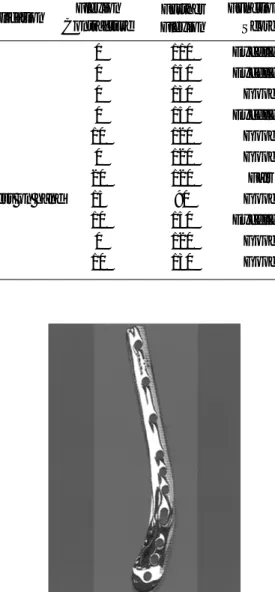
Fig. 2 : Photograph of lateral J plate.
시킨 후 주근을 종절개하였다(Fig. 1). 외측 골지주를 주관절직상부까지노출하고, 근위 상완골은 골절선 근위부로충분히노출하여, 골절부위의정복후상완 골의 외측에 J형 원위 상완골 금속판( E g i r eⓇ, Osteo Concept, Paris, France)으로내고정하였다. J 형 금속판 은외측상과의하단까지쉽게위치시켜고정할수있 도록외측 상과의형태에맞게 J 형의모양으로제작 된 해부학적인 금속판이다(Fig. 2). 분쇄 골절이 심한 A3 8예에서는동종골이식술을동시에시행하였다.
수술 후 장상지석고부목으로 평균 3주( 2 ~ 4주)간 고정하고, 이후 능동적 관절 운동을 시행하였다. 전 후면 및 측면 단순방사선 사진을 촬영하여 3부위이 상에서 골소주가 연결된 경우에 골유합으로 판정하 여 골절의유합 기간을 측정하였고, 수술 후 또는 추 시과정에서 발생한 합병증을 관찰하였다. 기능적인 평가는최소수술 1년추시후에 주관절운동범위를 측정하고, Morrey1 2 )의방법(Table 2.)을이용하여평가 하였다.
결 과
골유합 기간은 평균 1 2주( 1 0 ~ 1 8주)로 전례에서 골 유합을 얻을 수 있었다(Fig. 3). 1예에서 수술후 전완 부및수부배부에감각이상이 발생하였으나근전도 검사및신경전도검사는정상소견이었으며수술후
3개월에증상이호전되었다. 수술후 1년에주관절의 평균 운동범위는 굴곡 구축 6도( 0 ~ 2 0도), 후속 굴곡 1 2 6도( 9 0 ~ 1 5 0도)였으며, 견관절운동범위는 건측 견 관절과 비교하여 의미있는 차이를 보이지 않았다.
M o r r e y의임상평가에서는우수 4예, 양호 6예, 보통 1 예였다.
고 찰
상완골의 과간혹은 상과골절에서는양측골지주 에 대한 견고한 금속판 고정이 가장 좋은 방법으로 알려져 있고1 , 3 , 9 , 1 2 , 1 3 ), 골 지주에 대한 금속판 고정시 사용되는금속판의종류및 고정부위에대한연구는 여러 저자들에 의해보고되어왔다. 현재는 Jupiter 등
5 )이이용한재건형금속판을이용한외측및내측골 지주의후면부고정이많이사용되어지고있으며5 , 6 ), AO 그룹에서는 외측 골 지주의 후방에 재건형 금속 판을, 그리고내측골지주측면에 1/3 semitubular 금속 판 고정을 선호하고 있다7 , 8 , 1 1 , 1 3 , 1 5 , 1 6 ). 그러나 골절선 이상완골의간부혹은근위간부까지연장되어있는 경우에는 Y형 금속판이나 재형성 금속판 혹은 역동 적 압박금속판을이용한후방고정술등으로는충분 한 고정을 하기 힘들다. 또한 원위 상완골 간부와 골 간단이행부위를포함하는긴나선상골절은간부골 절만있는경우와달리역동적압박금속판을이용한 Table 2. Morrey’s functional score.
Excellent=95-100; Good=80-94; Fair=50-79; Poor=<50
Pain (30 points) None - 30 Slight - 20 Moderate - 15
Moderately severe Severe - 5 Completely disability
- 10 - 0
Motion (37 points) Extension - 0 to 8 Flexion - 0 to 17 Pronation/supination - 0 to 12
Function (12 points) 12 functions
Normal - 1 Mild compromise - 0.75
Difficulty - 0.5 With aid - 0.25 Unable - 0
Strength (15 points) Flexion, extension, pronation, supination
Normal - 5 Good - 4 Fair -3
Poor - 2 Trace - 1 None - 0
Instability (6 points) Anterior/posterior, medial/lateral
None - 3 Mild - 2
Moderate - 1 Severe - 0
전외측 고정이나 골수강내 금속정 고정술을 시행할 경우, 골절의 원위부까지 충분한 고정을 할 수 없는 단점이 있다. 즉 골절선이 주관절 직 상방에서 간부 로 이어지는 긴 나선상 골절인 경우 전외측 내고정 혹은후방내고정수술법등은금속판을골절선의근 위부와 원위부로 충분히 연장시키기가 어려워 견고 한고정을 얻기어려우므로 이중내고정금속판( d u a l p l a t e )을사용해야하는단점이있던지충분한고정이 되지 않은 경우 수술 후 관절운동의 지연으로 인한 주관절 운동제한 등이 발생되었다2 , 4 , 7 ). 이에저자들 은 긴 나선형 골절을 골절 상하에서 충분이 고정할 수있는방법으로해부학적금속판의일종인 J 형 금 속판을 이용하여 골절을 고정하였다. Schemitsch 등 1 4 )은 생역학적 연구를 통하여 골 지주내 분쇄나 골 결손이 없을 시 외측면의 J 형 금속판과 지연나사를 이용하여 고정시킨 경우 내측 골 지주와 외측 골 지 주에 금속판을 고정한 경우와 비교할 때 큰 차이가 나지 않는다고보고하였다1 4 ). 본연구의 증례에서도 골절의 근위부및 원위부에 충분한나사못의삽입으 로 견고한 내 고정을 얻을 수 있어서 조기에 주관절 운동이가능하였다.
J 형금속판을고정하기위해서는주관절부위에서 상완 근위부 1 / 3까지 상완골을 노출시켜야 하는데, 상완골 간부골절에널리사용되는전외측도달법은 상완골 원위 1 / 3의 노출을 위해서 주관절과 이를 덮 고있는연부조직의과도한박리가필요하여해부학 적으로도위험성이많다고지적되었다. 삼두근절개 를통한후방도달법도삼두근을절개하고요골신경
의근위부로충분히 박리하지않고는상완골의근위 부 노출이 어렵다는 단점이 있다2 , 1 0 ). 이를 보완하는 방법으로 변형된 외측 도달법이 소개되었는데 , M o r a n1 0 )는 근간 격막을 박리해서 도달함으로 삼두 근을 절개하여 도달하는 것 보다 출혈량이 적고, 근 육내반흔이적게남으며삼두근의내외측두를손상 시키지 않고 해부학적 위치로 금속판을 덮음으로써 치유에 도움이 된다고 하였다. 또한 요골 신경의 손 상이있는경우이를노출시켜확인하기가용이하고, 상완골골절의불유합에서도유착의박리가쉬워변 형된 외측 도달법이 유용하다고 하였다. Gerwin 등4 ) 은삼두근절개를통한후방도달법은근위상완골의 노출이 외상과를 기점으로 전체 상완골의 5 5 %까지 가능한데비해변형된외측도달법을 통해서는 전체 상완골의 9 4 %가 노출가능하다고 하였다. 따라서 원 위 상완골 간부 및 골간단 이행부위 골절의 치료에 있어서 변형된 외측 도달법이 골절선 근위부 및 원 위부로 충분한 노출을 가능하게 함으로써 J 형 금속 판고정시유용한도달법으로사료된다.
결 론
변형된 외측 도달법을 사용한 외측J 형 금속판의 내고정은견고한고정력으로수술후빠른관절운동 을 가능하게 함으로 원위 상완골 간부 및 골간단 이 행부위골절의치료에좋은치료방법의하나로사료 된다.
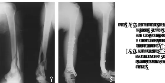
Fig. 3-A : Preoperative roentgenogram of case 5 shows distal humeral shaft and metaphyseal fracture (AO classification-A3 type).
Fig. 3-B : Postoperative 15 months roentgenogram shows lateral J plate fixation and good union.
A B
R E F E R E N C E S
1. Caja VL, Moroni A, Vendemia V, Sabato C and Zinghi G : Surgical treatment of bicondylar fracture s of the distal humerus. Injury, 25: 433-438, 1994.
2. Ebraheim NA, Andreshak TG, Yeasting RA, Saunders RC and Jackson WT : Posterior Extensile approach to the elbow joint and distal humerus.
Orthop Rev, 22: 578-582, 1993.
3. Fornasieri C, Staub C, Tourne y, Rumelhart C and Saragaglia D : Biomechanical comparative study of three types of osteosynthesis in the treatment of the supra and intercondylar fractures of the humerus in adult. Rev Chir Orthop Rep App Mot, 83: 237-242, 1 9 9 7 .
4. Gerwin M, Hotchkiss RN and Weiland AJ : Alternative operative exposures of the posterior aspect of the humeral diaphysis. J Bone Joint Surg, 78-A:
1690-1695, 1996.
5. Jupiter JB : Complex fractures of the distal part of the humerus and associated complications. In: Jackson DW ed. American Academy of Orthopedic Surgeon, Instructional Course Lectures. Vol. 44: 187-198, 1 9 9 5 .
6. Jupiter JB : Internal fixation for fractures about the elbow. Techniques Ortho, 4: 31-48, 1994.
7. Kang SY, Lee HJ and Chung JY : Modified lateral approach to the distal humerus fractures. J Korean Orthop Assoc, 35: 705-710, 2000.
8. Kim KY, Bin SI and Kim YJ : Surgical treatment of comminuted distal humerus intercondylar fracture in
adult using transolecranon approach and AO method.
J Korean Orthop Assoc, 27: 1060-1067, 1992.
9. Kundel K, Braun W and Ruter A : Distal intra- articular humerus fracture in adult. Results of surgical treatment. Unfallchirug, 95: 219-223, 1992.
10. Moran MC : Modified lateral approach to the distal humerus for internal fixation. Clin Orthop, 340: 190- 197, 1997.
11. Papaioannou N, Babis GCh, Kalavritinos J and Pantazopoulos T : Operative treatment of type C intraarticular fracture of the distal humerus: the role of stability achieved at surgery on final outcome.
Injury, 26 : 169-173, 1995.
12. Pereles TR, Koval KJ, Gallagher M and Rosen H : Open reduction and internal fixation of the distal humerus: functional outcome in the elderly. J Trauma, 43: 578-584, 1997.
13. Sanders RA, Raney EM and Pipkin S : Operative treatment of bicondylar intraarticular fractures of distal humerus. Orthopedics, 15: 159-163, 1992.
14. Schemitsch EH, Tencer AF and Henley MB : Biomechanical evaluation of methods of internal fixation of distal humerus. J Orthop Trauma, 8: 468- 475, 1994.
15. Sodergard J, Sandelin J and Bostman O : Mechanical failure of internal fixation in T and Y fractures of the distal humerus. J Trauma, 33: 687- 690, 1992.
16. Yang KH, Park SJ and Park SY : Lateral plate fixation in intercondylar fracture of the humerus. J Korean Orthop Assoc, 35: 559-563, 2000.
Fracture in Distal Humeral Shaft and Metaphyses
Whan-Yong Chung, M.D., Woo-Sik Kim, M.D., and Tae-Hong Kim, M.D.*
Department of Orthopaedic Surgery, Konyang University College of Medicine, Daejeon, Korea
*Department of Orthopaedic Surgery, Ajou University School of Medicine, Suwon, Korea
P u r p o s e : This is a retrospective study to analyze the clinical results of the usefulness of the lateral J plate fixations in distal humeral shaft and metaphyseal fractures.
Materials and Methods : From October 1996 to May 2000, eleven patients with distal humeral shaft and metaphyseal fracture were treated by open reduction and internal fixations with lateral J plate fixations. The clinical results were analyzed according to Morrey’s functional rating scale. Radiological unions, complication, and range of motion of the elbow were assessed.
Results : All fractures were united at 12 weeks (range, 10~18) in average. Finally, average range of motion of the elbow joint was flexion contracture 6 degrees in average(range, 0~20) to further flexion 126 degrees in average (range, 90~1 5 0 ) . Morrey’s functional rating scale were as follows; excellent 4, good 6, and fair 1.
C o n c l u s i o n : Lateral J plate fixations can be a good treatment method for the transitional zone of distal humeral shaft and metaphyseal fractures.
Key Words : Humerus, Distal shaft and metaphyseal fracture, Lateral J plate
Address reprint requests to Whan-Yong Chung, M.D.
Department of Orthopaedic Surgery, Konyang University Hospital,
685, Gasuwon-dong, Seo-ku, Daejeon 302-241, Korea TEL : +82.42-600-9100
FAX : +82.42-545-2373 E-mail : hy0707@unitel.co.kr
Abstract