大훌훌放射線훨學會誌 第 24 卷 第3 號 pp. 390 - 395, 1988 Journal of Korean Radiological Society, 24(3) 390 -395, 1988
신 경 섬유종 에 동반된 추골동맥의 동정맥루와 경부수막류 1 예
경북대학교 의과대학 방사선과학교실
김용주 • 서경진 • 김태헌 • 강덕식 • 박연묵 박준식” - Abstract-
Neurofibrornatosis with vertebral artery A.V fistula & cervical meningocele - A Case Report -
Yong Joo Kim, M,O., Kyung Jin Suh, M.D., Tae Heon Kim, M.O., Ouk Sik Kang, M.D.,
Yeun Mook Park, M.D .• , June Sik Park, M,D. *.
Department of Radiology, College of Medicine, Kyungpook National University
Arteriovenous fistula is a rare abnormality in patient with neurofibromatosis.
A review of English-Ianguage literature revealed only 3 reported cases.
We report a case of neurofibromatosis with vertebral artery A-V fistula and cervical meningocele
서 트응 ' -
신경섬유종은 상염색체 우성형질 (autosomal domi.
nant trait) 로 유전되는 질환으로서 1) 중배엽성파 신경 외 배 엽 성 조직 (mesodermal & neuroectodermal tissue)에 광범위한 형성장애를 초래하여 다양한 임상 증상을 냐타낸다2) Reubi3)가 1945 년 처음으로 신경 섬유증에 동반된동액혈관 명변에 판해서 기술한이후 동맥의 협착파 동액류형성에 판한 인식이 증가되었마 4 ) 저자들은 신경섬유증을 가진 43세 남자에서 우측 추골동맥에 발생한 동맥류와 동정백루( arteriovenous fistula) i。 그리고 경부 수막류 (cervical meningocele) 를 경험하였기에 운현고찰파 함께 보고하는 바이마.
• 경북대학교 의파대학 신경외파학교실
• Department of Neurosurgery, College of Medicine, Kyungpook National University
• 경북대학교 의과대학 。l비인후과학교실
’ Department of ENT, College of Medicine, Kyung.
pook National University
이 논문은 1988 년 4월 27 일에 접 수하여 1988년 5월 30 일에 채택되었음.
증례보고
1. 입상소견
43세 남자로써 1 여년천부터 운동시에 경한 경부통 증이 있었으며 1개월전부터는 오른쪽 경부에 운동성 이며 우홍인 연성종피와 견통이 있없으며 우측상지에 운동쇠약이 있었마. 연성종괴는 박동이 있었으며 청 진상 잡음이 들렸다. 체간 (trunk) 에 담갈색 색소침착 (cafe au lait spots) 파 파하섬유종이 흩어져 있었다.
2. 방사선학적 소견
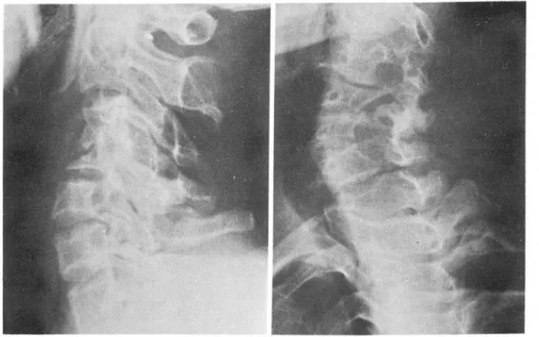
경부 단순 x- 선상 제 2
,
3 경추 추간공에서 제5, 6 경추추간공까지 추간공의 확장이 있었으며 제 4 경추 의 높아 가 낮아져 있고 우측 척 추얀곡과 척 추전굴이 있었다(Fig. 1). 경부척수조영상 제 3에서 제 6 경추 사이의 경추 천내측에 연부조직 종괴에 의해 밴행된 경 부수악류가 보이 며 이 경 부 수막류는 제 3, 제 4 경 추 체부 사이에서는 하인두 뒤부분까지 연장되어 있었다(Fig 2). 경부전산화 단층촬영상 연성종괴는 균질의
- 390 -
-김용주 외 : 신경성유종에 통반펀 추골동액의 동정액루와 경부수악츄 l예-
Fig. 1. Cervical radiograph, lateral & LAO view. There is widening of intervertebral foramen from C2-3 to C5-6 C-4 body is collapsed_ The prevertebral space widened.
Fig. 2. Cervical myelogram, AP & lateral view. There shows peculiar shaped anterior cervical meningocele, deformed by vascular mass, from C-3 to C-6 level. The meningocele extends to retropharyngeal space at hypopharyngeal level
A
B
大韓放射線훌훌學會誌 : 第 24 卷 第 3 號 1988 -
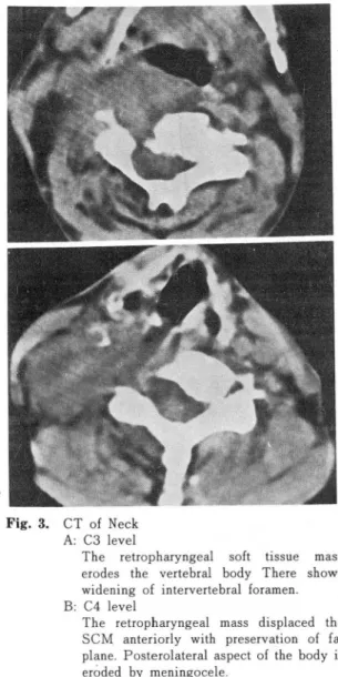
Fig. 3. CT of Neck A: C3 level
The retropharyngeal 50ft tissue mass erodes the vertebral body There shows widening of intervertebral foramen B: C4 level
The retropharyngeal mass displaced the SCM anteriorly with preservation of fat plane. Posterolateral aspect of the body is erõded by meningocele
연부조직 종괴를 보였으냐 중간중간에 석회화된 부분 이 보였마. 종괴는 우측흉쇄유돌근 (sterno.
cleidomastoid muscle) 을 전외 측으로 하인두를 천좌 측무로 밀고 있으여 경추의 앞쪽과 우측 추체와 횡챙 돌기를 첨식하고 있으맥 주위 조직과는 경계가 잘 유 지되어 있었마(Fig.3). 우측쇄골하동맥 조영상 추골 동액의 기시부에서 약 1 cm 상방에서부터 점차적으로 커지는 방추상괴-소당성 (fusiform & saccular) 동맥류 를 보이 며 중간중간에 좁은 부분을 동반하고 이 소당 성동맥류는 우측경부에 있는 커다란 땅추상 혈판과 연
결되어 있으며 주위에 있는 여러개의 정액혈관파동정 액루 형성을 하고 있었다. 오른쪽 늑경동액간 (costocervical trunk) 역 시 굵어 져 있 어 서 동정 액 루 와의 연결을 유추할 수 있었마 (Fig.4).
3. 수술소견
환자를 앙오태로 놓힌 후 어깨에 받첨대를 넣고 머 리를 좌측으로 돌려 환측 경부가 돌출되게 하였다.
피부절개는 상부는 후두부 유양돌기 후땅에서부터 시작하여 흉쇄유돌근후방을 따라하부로내려와서 쇄 골상부에서 수펑 A로 연결하여 반대펀 흉쇄유돌근 전 연부까지 하였 다. Deep cervical fascia를 통하여 천후 부로 피 부판을 만들어 흉쇄 유돌근의 전부와 흉부를 완 전히 노출시켰다. 이애 흉쇄유돌근 하부에 표연이 희 고 출혈이 쉽게 되는 큰 종괴를 확인할 수 있었마 (Fig. 5). 이 종괴는 출혈이 싱하였3며 경추에 연결 되어 유동성이 전혀 없었마. 흉쇄유돌근의 하단부를 절단하여 상부로 을어 올리 므로서 더 확실한 종괴 의 위치와 모양을 확인하고 경부하부에서 추골동액의 정 상부위 를 확인하고 No. 5 silk로 결찰하였 마. 상부 추 골동액의 정상부위를 확인하려고 하였으나 종양과의 구벨 이 불가능하였 다. 추골동액 의 상부는 unilateral suboccipital craniectomy에서와 같이 파부절개를 가 하고 연부조직을 박리하여 후두골파 제 1경추간에서 동액 을 노출시 켜 No. 5 silk로 2중 결찰하였 다. 수술 후 혈판조영숭상 제 2경추 근처에서 우측 늑경동액간 분지와 우측 외경동액의 후두분지, 그리고 우측 추골 동맥 사이의 측부순환에 의해 동정맥루가 냐타나서 늑 경동액간분지는 Ivalon(590-1000 μ) ,외경 동액의 후 두분치는 Gelfoam을 이용 각각 폐색시켰마 (Fig.6). 수술과 폐색술후 연성종괴의 크기는 줄지 않았우나 박 동과 잡음이 완천허 소실되 었으며 우측상지 운동쇠 약 의 개선이 있었다.
고 찰
신경섬유증에 동반된 혈관뱅변은 주로 대동맥파 그 분지, 성장, 경동맥과 추골동액 동에서 보이나 특히 신동맥 에 흔히 나타난다5) 그러 나 신경 성 유증에 동안 된 동맥류와 동정액루는 문헌상 3예가 보고되어 있으 며 이중 수술천에 진단한 례는 1례에 불과하다6) 본 례와 같이 동맥류와 동정맥루, 그러고 경부수악루가
-김용주 외 : 신경성유종에 동반된 추골동액의 동정액루와 경부수악류 1예-
A F.IIj톨lr. i!""!' ‘ . . γ ‘ .ß
C
Fig. 4. A: Right subclavian angiogram. Arterial phase
。f right vertebral artery shows fusiform and saccular aneurysmal dilatation with dilatation and constrictions, connected with fusiform vein in right lateral neck
B: Late arterial phase shows multiple venous channels
C: Left vertebral angiogram Upper portion .of right vertebral artery fills the dilated tortuous vessel.
大韓放射線홈學會誌 : 第 24 卷 第 3 號 1988 -
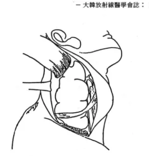
Fig. 5. UIÌder the sternocleidomastoid muscle, a large mass lesion with its white surface was found.
It gave easy bleeding during overlying fascial dissection
복합되어 연성종괴를 형성한 경우는 신경섬유증에 동 안된 신경 성유종, 수악종, 그러고 다른 척수종양으로 오인하기 쉽 다. Reubi3)가 처 음으로 보고한 신경 섬 유
증에 동반된 1 mm 이하의 작은 신동액에 생긴 조직학
적 뱅변은 혈관내막증식, 혈판내막의 섬유초자성비 후, 탄력충의 단옐 (fragmentation) • 혈관중박의 앓아짐 파 동맥류형성, 혈관중막파 외막 사이의 방추형 세포 의 결절성 종괴 이 다. Saylor 둥7)은 모든 동맥 혈판벽 의 영 변은 Schwann 세 포증식 에 기 인되 여 진천도의 차이에 의해 여러가지 명변이 나타나는 동일 명변을 갖고 있다고 보고했마. Green 둥5)은 1 mm 이 하의 소 형 혈판에 나타난 조직 학적 소견중에 Schwann 세 포가 전혀 보이지 않는 점파 혈관내막파 외막사이에 있는 결절성 종괴를 천자현미경으로 조사한 결파 펑활근세 포임을 확인하고, 이는 섬유근성 형성장애(fi bromus
cular dysplasia) 의 소견파 동일한 것으로 생 각, 소형 혈판백의 명뻔을 중배엽성 형성장애라고 보고했마.
그러냐 이보다 큰 혈판에서의 t영변은 Saylor 둥7)의
Fig. 6. Post op angiogram. The vertebral artery proximal to distal surgical ligation is filled through collateral vesssels from occipital and costocervical arteries. The branches of costocervical trunk &
。ccipital artery was embolized with Ivalon(590-1000 μ), Gelfoam(2 X2 mm Cubes) respectively
- 394-
-김용주 외 · 신경섭유종에 동안된 추골동액의 동정액루와 경부수악류 1예
의견과 같았마5) Itzchak 등4 )은 신경섭유증에 동반된 신동액의 뱅변의 혈판조영술상 소견은 섬유근성 형성 장애와 차이는 없으나 첨벙하는 위치가 신경섬유증은 근위 신동맥 , 섬 유근성 형 성 장애 는 원위 신동액 (distal 2/3)8)으로 서로 마르으로 강벨에 도웅이 되는 것우로 보고했마. 뇌강내동액에 생기는 협착성영변은 많은 보고가 있으나9.10.11) 본예에서는 나타나지 않았마.
Deans 둥6)은 여 러 가지 t영 리 학적 소견을 종함하여 동정맥루가 생기는 원인을 2 가지로 요약했마. 첫째는 이 형 성 (dysplastic) 평 활근 혹은 신경 섬 유증식 이 동액 렬판벽에 동맥류를 초래하여 누출이 얼어냐며 이 누출 이 주위에 있는 정맥혈판에 파열되어 발생하는 것파,
둘째는 선천석인 중배엽성 형성장애에 의해 냐타냐는 소견으로 생 각렀다. 신경 섬 유증에 동반된 경부수막류 는 이 등12)이 호고한 흉강내수악류와 비슷한 형태이
나 본례에서는 혈판종괴에 의해 변형되어 있었다. 경 부수막류가 딸생하는 원인으로는 흉강내수막류와 같 이 신경근판 (nerve root sleeve) 의 연장, 신경성유종 의 낭성변형, 외상, 경막형성장애, 골형성장애, 국소 형성장애, 선천성장애 둥으로 추정된마13) 신경섬유 종은 여러가지 마양한 영변을 나타내는 잘환이마. 연 성종괴가 인접장기를 압박하여 증상을 나타내며 박동 파청진상잡음이 들리연 혈판조영술을 반드시 시행하 고이와동시에 척수조영술을시행하여 마른연부조직 종괴와 강t영 진단해야 할 것으로 생각된다.
결 .%. 「」
경북대학교 의과대학 부속영원에 내원한 신경섬유 증을 가진43세 남자에서 우측추골동액에서 발생한동 액류를 동반한 동정액루와 경부 수막류를 경험하였기 에 보고하는 바이다.
REFERENCES
1. Wander JV, Das Gupta TK: Neurofibromatosis Curr. Probl Surg 14:1-81, 1977
2. Hilal SK, Solomon GE, Gold AP et al: Primary cerebral arterial occlusive disease in children Radiology 99:87-93, 1977
3. Reubi F: Neurofibromatose et lesions vasculaires Schweiz Med Wochenschr. 75:463-465, 1945 4. Itzchak Y, Katznelson D, Boichis H et al: Angiog-
raphic leatures 01 arterial lesions in neurolibroma tosis. A]R 122:643-647, 1974
5. Greene JF, Fitzwater JE, Burgess J: Arterial le- sions associated with neurofibromatosis. Am ] Clin Patho1. 62:481-487. 1974
6. Deans WR, Bloch S, Leibrock L et al: Arteriove- nous !istula in patients with neurolibromatosis Radiology 144.'103-107, 1982
7. Saylor WR, Saylor DC: The vascular lesions 01 neurQlibromatosis. Angiology 25:510-519, 1974.
8. Harrison EG, Hunt JC, Bernztz PE: Morphology 01
!ibromuscular dysplasia 01 the renal artery in re- novascular hypertension. Am ]. Med 43:97-112, 1967
9. Taboada D, Alonso A, Moreno J et al: Occlusion 01 the cerebral arteries in Recklinghausen’'s disease Neuroradiology 18:281-284, 1979
10. Leone RG, Schatzki SC, Wolpow ER: Neuro!ibro- matosis with extensive intracranial arterial occlu- sive disease. A]NR 3:572-576, 1982
11. Gebraski SS, Gabrielsen TO, Knake JE et al: Post- erior circulation intracranial occlusive disease in nero!ibromatosis. A]NR 4:1245-1246, 1983
12 이기만, 우성구, 김옥배, 서수지 : 흉강내 수막류 (Intrathoracic meningocele) 의 Metrizamide CT 소 견. 대한방사선의학회지 21 : 415-418, 1985.
13. Miles J. Pennybacker J, Sheldon P: Intrathoracic meningocele. ] Neurol Neurol Neurosurg Psychiat 32,99-11 0, 1969