대한소화기학회지 2008;52:183-187
접수: 2008년 5월 6일, 승인: 2008년 7월 30일
연락처: 전태주, 139-707, 서울시 노원구 상계7동 761-1 인제대학교 상계백병원 소화기내과
Tel: (02) 950-1990, Fax: (02) 950-1955 E-mail: [email protected]
Correspondence to: Tae Joo Jeon, M.D.
Division of Gastroenterology, Department of Internal Medicine, Sanggye Paik Hospital, Inje University College of Medicine, 761-1, Sanggye 7-dong, Nowon-gu, Seoul 139-707, Korea Tel: +82-2-950-1990, Fax: +82-2-950-1955
E-mail: [email protected]
장출혈로 진단된 혈관종에 의한 성인 소장 장중첩증 1예
인제대학교 의과대학 상계백병원 내과학교실
김선영ㆍ전태주ㆍ홍진희ㆍ김광실ㆍ오태훈ㆍ서동대ㆍ신원창ㆍ최원충
An Adult Case of Small Bowel Intussusception Caused by Hemangioma Presenting with Intestinal Bleeding
Sun Young Kim, M.D., Tae Joo Jeon, M.D., Jin Hee Hong, M.D., Gwang Sil Kim, M.D., Tae Hoon Oh, M.D., Dong Dae Seo, M.D., Won Chang Shin, M.D., and
Won Choong Choi, M.D.
Department of Internal Medicine, Sanggye Paik Hospital, Inje University College of Medicine, Seoul, Korea
Intussusception is primarily a disease of children, and is relatively rare in adults. Unlike childhood intussu- sception, adult intussusception has an identifiable leading lesion such as malignant or benign neoplasm. However, intussusception caused by hemangioma is very rare. There were few cases of small bowel intussusception caused by hemangioma in adults, but those reports were presented with abdominal pain. This report describes a 65-year-old female who suffered from small bowel intussusception caused by hemangioma presenting with in- testinal bleeding. Upper gastrointestinal endoscopy and colonoscopy were performed, but bleeding focus was not found. Abdominal computed tomography showed the target sign of small bowel with a leading point of mass.
This mass turned out to be a hemangioma after the small bowel resection. Therefore, small bowel intussusception by hemangioma should be also considered as a bleeding focus when an adult patient presented intestinal bleeding without bleeding focus in the stomach and colon. Herein we report a case of small bowel intussusception caused by hemangioma presenting with intestinal bleeding. (Korean J Gastroenterol 2008;52:183-187)
Key Words: Adult intussusception; Small bowel intussusception; Hemangioma; Bleeding
서 론
장중첩증은 장관의 근위부가 인접한 원위부의 관강으로 함입하는 것이다. 주로 소아에서 발생하며 성인에게 발생하 는 경우는 전체 장중첩증의 5-16%로 드물다.1,2 성인의 장중 첩증은 소아와는 달리 대부분 유발 원인이 있다. 소장의 경 우 양성 질환에 의한 경우가 63%, 악성 종양에 의한 경우가 14%이며, 원인을 모르는 경우가 23%이다.2 이 중 혈관종에
인해 발생한 성인 소장 장중첩증은 매우 드물며, 전체 성인 양성 소장 종양의 3-4%를 차지한다.3
장중첩증은 중첩된 장관의 위치에 따라 다양하고 비특이 적인 임상증상을 나타내어 복통과 압통이 가장 흔하며,4 소 아에서 흔히 관찰되는 혈변은 성인에서는 드물다.5 성인에 서는 유발 원인을 제거해 주어야 하므로 수술로 치료하는 경우가 대부분이다.
혈관종에 의한 소장 장중첩증은 세계적으로 드물다. 국내
184 대한소화기학회지: 제52권 제3호, 2008
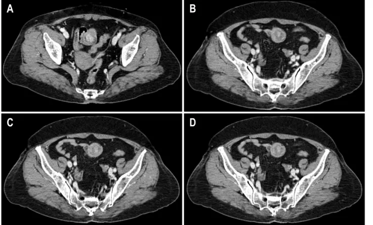
Fig. 1. Abdominal CT findings. (A) About 2 cm sized intraluminal enhancing mass is seen at a leading end of the intussuceptum. (B, C, D) The serial images of abdominal CT scan show alternating low- and high-attenuation layers surrounded by enhanced rim, which is compatible with target sign.
에도 혈관종에 의한 소장 장중첩증에 대한 일부 보고가 있 으나, 대부분 복통으로 발견되었으며, 장출혈에 의해 발견 된 경우는 없었다. 이에 저자들은 장출혈로 내원한 65세 여 자 환자에서 혈관종에 의한 소장 장중첩증 1예를 경험하였 기에 문헌 고찰과 함께 보고한다.
증 례
65세 여자 환자가 2주 간의 흑색변으로 외래에 방문하였 다. 과거력에서 고혈압 및 빈혈이 있었고, 수술 받은 과거력 은 없었다. 현재 혈압약 이외에 복용 중인 약은 없었으며 흑 색변의 과거력은 없었다. 1달 전부터 명치 부위에 간헐적인 통증이 있었고, 2주 전부터 3일 간격으로 1-2회 가량의 흑색 변이 있었지만 그 외 다른 증상은 없었다. 외래 방문 당시 활력 징후는 혈압 110/70 mmHg, 맥박 88회/분, 체온 37.3oC, 호흡수 20회/분이었으며, 탈수증 및 만성 병색을 보였으나 의식은 명료하였다. 결막은 창백하였고, 공막에 황달은 관 찰되지 않았다. 복부는 부드러웠고, 장음은 정상이었다. 명 치 부위에 경한 압통이 있었으나 반발통은 없었고, 만져지 는 종괴는 없었다. 직장수지검사에서 흑색변이 관찰되었다.
혈액검사에서 백혈구 6,430/mm3, 혈색소 6.0 g/dL, 헤마토 크리트 22.5%, 혈소판 299,000/mm3, 혈청 철 14μg/dL, 페리 틴 2.97 ng/mL, 총 철결합능 514μg/dL, 말초 혈액 도말 검사 에서 저색소 소구 빈혈 소견을 보였다. Na 138 mEq/L, K 4.4 mEq/L, Cl 106 mEq/L, 총 빌리루빈 0.2 mg/dL, AST 13 IU/L, ALT 8 IU/L, 총 단백 8.0 g/dL, 알부민 4.1 g/dL, BUN 14.2 mg/dL, Cr 0.8 mg/dL, PT INR 1.0, aPTT 29.0초, CRP 1.4 mg/dL였다. HBsAg, HBsAb, anti-HCV는 모두 음성이었다.
상부위장관 출혈 의심하에 적혈구 수혈 후 상부내시경 및 대장내시경을 시행하였다. 상부내시경 검사에서 표재 위염 소견이 보였으나, 명확한 출혈 병소는 발견되지 않았다. 대 장내시경에서 말단 회장부부터 상행 결장까지 다발성의 헤 마틴이 관찰되었으나 명확한 출혈 병소는 발견되지 않았다.
소장 출혈 의심하에 복부전산화단층촬영과 소장조영술을 시행하였다. 복부전산화단층촬영에서 골반강 안의 소장 내 부에 2 cm 크기의 조영이 잘 되는 종괴(Fig. 1A)와 이 종괴 를 유발점으로 표적 징후(target sign)가 관찰되어 소장 장중 첩증으로 진단하였다(Fig. 1B∼D). 그러나, 소장 장중첩증으 로 인한 장관 폐쇄는 관찰되지 않았다. 소장 조영술에서 원 위부 회장이 점차 좁아져 이 부위가 감입부(intussusceptum)
김선영 외 7인. 혈관종에 의한 성인 소장 장중첩증 185
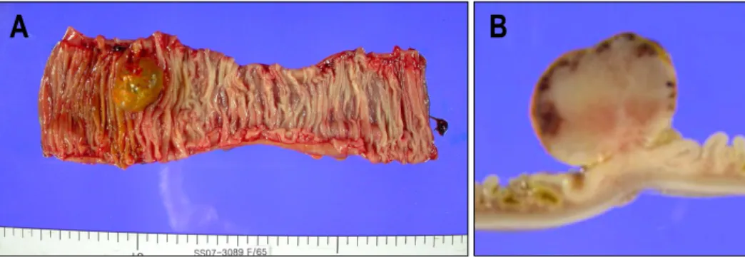
Fig. 3. Gross findings of the small bowel mass. (A) A well-defined round fungating mass measuring 2.5×2.5×2.2 cm is noted upon longitudinal opening of the ileum. The surface of the mass is eroded and covered by greenish yellow fecal material. The remaining mucosa is grossly unremarkable. (B) The cut surface of the mass is homogeneously gray white, solid and somewhat myxoid with multi- focal areas of reddish tan subepithelial hemorrhage.
Fig. 4. Microscopic findings of the small bowel mass. (A) The topmost surface of the mass shows total denudation of the mucosa with submucosal proliferation of irregularly shaped blood vessels of various sizes (H&E, ×10). (B) The proliferative vessels are mostly com- posed of small-sized thin-walled vessels lined by bland looking plump and ovoid endothelial cells (H&E, ×40).
Fig. 2. Small bowel series finding. The round filling defect is noted when spontaneous reduction is instituted. The polypoid mass is suggestive of a leading point of the intussusception.
인 장중첩증이 의심되었고, 자발적으로 정복이 되었을 때 소장 내부에 2 cm 크기의 둥근 충만 결손이 관찰되어(Fig.
2) 이 부분이 선두(leading point)가 되는 종괴로 의심되었다.
조영제의 통과 장애는 없었다.
검사 후에도 환자는 흑색변 및 혈변이 지속되어 수술 치 료를 시행하였다. 소장 종괴 및 소장 장중첩증을 의심하여 전신마취 하에 정중선으로 개복하였다. 회장에 소장이 중첩 된 부위를 확인하였으며 그 부위의 괴사는 없었다. 장중첩 된 부위의 회장을 22 cm 가량 절제하고 단단 문합술을 시행 하였다. 육안 소견은 소장 내부에 경계가 명확한 2.5×2.5×
2.2 cm 가량의 폴립 모양의 종괴가 관찰되었고(Fig. 3), 조직 병리 검사에서 소혈관의 증식이 관찰되어(Fig. 4) 혈관종의 일종인 모세혈관종으로 판명되었다. 환자는 장출혈의 원인 으로 모세혈관종에 의한 소장 장중첩증으로 진단 되었고, 수술 후 현재 외래 추적 관찰 중이며 특별한 합병증 없이
186 The Korean Journal of Gastroenterology: Vol. 52, No. 3, 2008
경과 관찰 중이다.
고 찰
혈관종은 혈관의 비정상적인 증식에 의해 발생한 병변으 로 전통적으로 다양한 혈관의 병변을 지칭한다.6 위장관 혈 관종은 위장관 어느 곳에서나 발생할 수 있는 드문 양성 혈 관 종양으로 단일 종괴 또는 다발성으로도 발생할 수 있다.
이중 45%가 소장에서 발생한다는 보고가 있으며, 소장에 발생하는 양성 종양 중에서 7-10%를 차지한다.7,8 혈관종에 의해 장중첩증이 발생한 경우는 드물며 이번 증례와 같이 복통 없이 장출혈 증상만 있는 경우는 기존 보고에 없었다.
장중첩증은 대부분 소아에서 발생하며, 성인의 장중첩증 은 드물고 증상이 다양하여 때로는 진단이 어렵다. 한국 성 인 장중첩증에 대한 한 연구에서 간헐적인 복통(94.3%)이 가장 흔했고, 복부 압통(63%), 오심 및 구토(54.7%), 복부 종 괴(51%) 순이었다.5 소아에서 흔히 관찰되는 혈변은 성인의 경우에는 25.5%로 비교적 낮았고,5 소장 장중첩증에서 혈변 이 나오는 경우는 4% 정도로 대장 장중첩증의 33%보다 드 물었다.2 소아 장중첩증은 대부분 원인을 알 수 없는 반면, 성인의 경우는 90% 이상에서 장관 내에 종양과 같은 명확 한 병변이 관찰된다. 1992년까지 보고된 한국의 장중첩증은 회장 결장형 42.5%, 소장 소장형 23.8%, 회장 맹장형 20.1%, 대장 대장형이 13.2%였다.5 소장 장중첩증은 대부분 지방종, 평활근종, 선종 등 양성 종양에 의해 발생하며, 그 외에도 선암과 같은 악성 종양이나, 장내 유착, 멕켈 게실, 림프과 다형성, 샘염, 장 중복증, 만성 소화 장애증, Henoch-Schon- lein 자반증 등에 의해 발생할 수 있다.9 최근 혈관종에 의해 생긴 회장부의 장중첩증은 국내에서 2예가 보고되었다.3,7 기존 보고된 예와는 달리 이번 증례는 복통이나 폐쇄 증상 없이 흑색변 및 혈변의 임상 양상을 나타내었다. 또한 출혈 을 동반한 성인 소장 장중첩증은 대부분 위절제술 후 발생 한 공위장형 장중첩증이거나, 전이암,10 멕켈 게실11,12에 의 한 경우였으며 이번 증례와 같이 혈관종에 의해 유발된 소 장 장중첩증에 대한 보고는 없었다.
장중첩증은 복부초음파나 복부전산화단층촬영에서 감입 부 내의 압박과 정맥 울혈로 인해 층이 구분되어 나타나는 표적 징후가 관찰되는 것이 특징적이다.13 이번 증례에서도 전산화단층촬영에서 소장의 표적 징후가 관찰되어 소장 장 중첩증을 진단한 경우로, 전산화단층촬영은 소장의 병변과 원인을 찾는데 도움이 되어 흔히 이용된다. 그 외에 지연 전 산화단층촬영이나 소장 조영술 등이 장 폐쇄 여부 및 수술 여부 결정에 도움이 된다.14
장출혈이 의심되는 환자에서 상하부 내시경 검사 후에 출 혈 병소를 찾을 수 없을 경우에는 소장 출혈을 의심해야 한
다. 소장에 대한 검사로 복부전산화단층촬영이나 소장 조영 술의 시행은 중요하며, 혈관종에 의한 소장 장중첩증도 원 인 중 하나로 고려해 볼 수 있다. 검사에서 소장 종양이 의 심되면 수술 치료로 진단 및 치료를 할 수 있으며 최근 연 구가 활발히 진행되고 있는 캡슐 내시경과 이중 풍선 소장 내시경도 향후 중요한 역할을 할 것으로 생각한다.15 특히 이중 풍선 소장내시경은 조직 검사 및 용종절제술을 할 수 있 있어 이번 증례와 같이 크기가 크지 않은 양성 종양에 대해서는 내시경 절제와 같은 치료 목적의 시술을 고려해볼 수 있다는 점에서 유용하다. 저자들은 소장 출혈 이외에는 특이 증상 없는 혈관종에 의한 소장 장중첩증 1예를 경험하 였으며, 문헌 고찰과 함께 보고한다.
참고문헌
1. Azar T, Berger DL. Adult intussusception. Ann Surg 1997;226:134-138.
2. Begos DG, Sandor A, Modlin I. The diagnosis and manage- ment of adult intussusception. Am J Surg 1997;173:88-94.
3. Jeong CY, Jung EJ. An ileocolic intussusception caused by small bowel hemangioma. J Korean Surg Soc 2004;67:490- 492.
4. Chang CC, Chen YY, Chen YF, Lin CN, Yen HH, Lou HY.
Adult intussusception in Asians: clinical presentations, diag- nosis, and treatment. J Gastroenterol Hepatol 2007;22:1767- 1771.
5. Park SH, Moon HY. Adult intussusception in Korea. J Korean Surg Soc 1998;55:713-718.
6. Drolet BA, Esterly NB, Frienden IJ. Hemangiomas in children. N Eng J Med 1999;341:173-181.
7. Chang SJ, Kang WK, Kim JY, et al. A case of intussuscept- ing hemangioma of the ileum. J Korean Surg Soc 2002;
63:437-440.
8. Levy AD, Abbott RM, Rohrmann CA, Frazier AA, Kende A.
Gastrointestinal hemangiomas: imaging findings with patho- logic correlation in pediatric and adult patients. Am J Roent- genol 2001;177:1073-1081.
9. Kim JH, Lee KM, Yun SH, et al. Ileocecal intussusception in an adult: a case report. Turk J Gastroenterol 2007;18:50-52.
10. Hofflander R, Beckes D, Kapre S, Matolo N, Liu S. A case of jejunal intussusception with gastrointestinal bleeding caused by metastatic testicular germ cell cancer. Dig Surg 1999;16:439-440.
11. Lu CL, Chen CY, Chiu ST, Chang FY, Lee SD. Adult in- tussuscepted Meckel's diverticulum presenting mainly lower gastrointestinal bleeding. J Gastroenterol Hepatol 2001;16:
478-480.
Kim SY, et al. An Adult Case of Small Bowel Intussusception Caused by Hemangioma Presenting with Intestinal Bleeding 187
12. Sogutlu G, Yilmaz M, Karadag N, Isik B, Olmez A, Cinpolat O. Lower gastrointestinal tract bleeding caused by in- flammatory pseudotumour in Meckel's diverticulum: report of a case. Inonu Universitesi Tip Fakultesi Dergisi 2006;13:
269-270.
13. Balik AA, Ozturk G, Adynli B, et al. Intussusception in adults. Acta Chir Belg 2006;106:409-412.
14. Frager D, Medwid SW, Baer JW, Mollinelli B, Friedman M.
CT of small-bowel obstruction: value in establishing the diag- nosis and determining the degree and cause. Am J Roentgen- ol 1994;162:37-41.
15. Eun CS. Current state of double balloon enteroscopy. Korean J Gastrointest Endosc 2005;31:42-46.