Abstract (J Korean Assoc Oral Maxillofac Surg 2010;36:427-33)
Ⅰ. 서 론
임플란트의 성공을 위한 가장 중요한 전제조건은 식립 할 부위에 충분한 양의 치조골이 존재하는 것이다. 그러나 외상이나 조기 치아소실, 치주질환, 종양에 의한 외과적 절제술 등에 의한 치조골 소실은 임플란트 식립을 어렵게 한다1-5.
부분 무치악 환자에서의 수평적 골소실은 적절한 직경 의 임플란트 식립을 방해한다. 식립한 임플란트의 협설측 으로 최소 1 mm의 골폭이 확보되지 않는다면, 임플란트 주변의 수직적 골흡수가 발생하며, 결과적으로 예지성있 는 결과를 기대할 수 없다. 수직적 골소실은 악골의 해부 학적 한계에 의해 임플란트 식립 시 임상가에게 어려움을 준다. 수직적으로 충분한 골고경이 확보되지 않는다면, 상 악전방부에서는 비강, 상악구치부에서는 상악동, 하악구
치부에는 하치조신경관에 의해 충분한 길이의 임플란트 를 식립하기가 어렵다. 또한 수직적 골소실이 심한 부위는 악간 거리의 증가로 인해 임플란트 식립 후 최종 보철물의 비심미적인 형태와 부적절한 치관-치근 비율을 야기할 수 있다1,5.
이러한 골소실로 인한 문제들을 해결하기 위해 많은 방 법이 제시되었다. 1990년대 초반에는 티타늄 임플란트와 연계하여 골유도재생술에 티타늄 강화 비흡수성 차단막을 이용하여 수직적 골증강술을 시행하는 방법이 제시되었 다. 그러나 이는 술자의 기술 의존성이 매우 높다는 한계를 가지고 있다6. 또한 하악구치부의 경우 신경전위술이 소개 되었으나 이 또한 술자의 기술 의존도가 매우 높으며, 신경 손상의 위험도가 매우 높다고 보고되었다7. 짧은 임플란트 를 식립하는 술식이 소개되었으나 이것은 예지성있는 결 과를 얻기에는 한계가 있으며, 최종 보철 시 이상적인 치 관-치근 비율을 얻을 수가 없다8,9. 또한 신연장치를 이용한 골신장술이 등장하였으며 현재까지 많이 사용되고 있으 나, 인접치의 치주 상태에 악영향을 미쳐 인접치의 발치 및 신연장치의 파절 등 다양한 합병증이 보고되었으며, 제한 적인 증례에 적용 가능하다는 단점이 있다10. 이외에도 자 가골 및 동종골, 이종골이식술과 치조제분할술 등 많은 술 이 백 수
130-701 서울시 동대문구 회기동1번지
경희대학교 치과대학 부속 치과병원 구강악안면외과 Baek-Soo Lee
Department of Oral and Maxillofacial Surgery, Kyunghee Dental Hospital 1 Hoegi-dong, Dongdaemun-gu, Seoul, 130-701, Korea
TEL: +82-2-958-9440 FAX: +82-2-966-4572 E-mail: [email protected]
심하게 흡수된 치조제의 증강을 위한 골막하 터널링기법을 이용한 장골이식술에 대한 임상연구
박 숭∙정준호∙김여갑∙권용대∙최병준∙오주영∙이백수 경희대학교 치의학전문대학원 구강악안면외과학교실
A clinical study of iliac bone graft using subperiosteal tunneling method for alveolar ridge augmentation
Soong Park, Jun-Ho Jung, Yeo-Gab Kim, Yong-Dae Kwon, Byung-Joon Choi, Joo-Young Ohe, Baek-Soo Lee Department of Oral and Maxillofacial Surgery, School of Dentistry, Kyunghee University, Seoul, Korea
Purpose:The objective of this study was to evaluate the efficacy of the subperiosteal tunneling technique with iliac block bone graft for bone aug- mentation in an edentulous alveolar ridge.
Patients and Methods:Total of 8 sites in 7 patients were included in this study. The bone height was evaluated by CBCT preoperatively and 4 months after operation. Total of 11 implants were inserted and evaluated clinically and radiographically.
Results:Mean value of the increased bone height was 6.29 mm and no implant failure was observed. There were no complications such as soft tissue dehiscence, exposure of the grafted bone and infection.
Conclusion:We have achieved excellent clinical outcomes by this technique, so we concluded that it is useful for augmentation of severely deficient alveolar ridge.
Key words:Iliac bone graft, Tunneling technique, Bone augmentation, Alveolar ridge, Bone resorption
[paper submitted 2010. 5. 10 / revised 2010. 9. 24 / accepted 2010. 10. 15 ]
장골부에서는 피질골뿐 아니라 다량의 해면골이 존재한 다. 임상적으로는 피질-해면골 복합체를 골괴 형태로 채취 하여 심하게 흡수된 치조제의 재건에 사용할 수 있다. 그러 나 장골이식술을 이용한 온레이 골이식술은 장기간 추적 관찰 시 최고 50%에 이르는 골흡수가 발생한다고 보고되
었으므로11,12, 임상가는 장골이식에서 필요한 골량과 흡수
량을 고려하여 과량의 골을 채취해야 한다. 과량의 골괴이 식은 구강 내 연조직의 부족을 야기하여 1차 폐쇄를 어렵 게 할 수 있으며, 술후 장력에 의한 봉합부 열개를 야기하 여 결과적으로 골소실이 발생한다.
골막하 터널링기법(또는 포켓기법)은 수산화인회석이나 무기질 우골(Bio-Oss, Geistlich), 동종골이식재 등 다양한 골이식재를 이용한 골증강술 시 최소한으로 침습적인 피 판의 도안으로 소개된 바 있다13,14. 이 술식은 작은 수직 절 개와 최소한의 조직 박리를 통하여 수술할 때 외상을 최소 화하며 치조골능 절개를 시행하지 않아“텐트 효과”를 얻 을 수 있는 장점이 있다. 또한 술후 불편감과 부종이 적으 며 골이식술에서 가장 흔한 합병증의 하나인 피판 열개가 적다고 보고된 바 있다14-16. 본과에서는 수직적으로 심하게 흡수된 치조골에서 골막하 터널링기법을 이용한 온레이 장골이식술을 시행하여 성공적인 치험례를 얻었기에, 문 헌 고찰과 함께 보고하고자 한다.
Ⅱ. 연구대상 및 방법
2007년 11월에서 2009년 3월까지 본과에서 터널링기법 을 이용한 자가골이식술을 시행한 위축된 악골을 가진 7명 의 환자와 8부위의 수혜부를 대상으로 술전 골고경과 술후 4개월경의 골고경을 후향적으로 cone-beam computed tomography (CBCT)로 비교하였으며, 골이식술 후 임플란 트 식립까지의 기간 및 식립한 임플란트의 개수와 직경 및 길이를 조사하였다. 골채취부는 모두 좌측 장골능 부위였 으며, 모든 수술은 전신마취를 하고 진행하였다.
7 명의 환자 중 2명은 당뇨병을 가지고 있었으며, 1명은 C형 간염 보균자였다. 7명 중 3명은 하악골의 병소로 인해 골결손이 발생한 환자로 각각 치성각화낭종, 법랑아세포 종, 편평상피세포암종에 대한 하악골 부분 절제술을 시행 받았다. 이중 편평상피세포암종 병력을 가진 환자의 경우 장골이식술 시행 3년 전 방사선치료를 받았었다. 골이식을 시행한 8부위 중 7부위는 하악후방부였으며, 상악전방부
의 중간점을 계측하여 그 부위에서 측정하였다.
Ⅲ. 수술방법
먼저 실혈량 감소 및 약물이 주입되는 압력을 이용한 골 막 박리를 위해 1:100,000 에피네프린을 함유한 2% 리도케 인(1:100,000 epinephrine 함유, Zeyco S.A. de C.V., Mexico) 을 하악우측 소구치부에서 후구치부까지 주입하였다. #45 의 원심 우각부 및 후구치부 치조정에서 시작하여 점막치 은경계를 넘어서 전정까지 이르는 2개의 수직 절개를 시행 하였다. 이공의 위치를 확인하면서 작은 골막기자를 이용 하여 골막하 터널을 형성하였다. 골막의 박리는 치조제 상 방을 넘어 약간 설측까지 진행하였다. 이때 이공의 위치를 확인하여 이신경손상을 각별히 주의하여야 한다. 이어서 아이리스를 이용하여 골막 이완 절개를 시행하고, 피판을 신장시켜 이식 가능한 골편의 크기를 추정하였다. 장골에 서 채취한 골괴를 round bur나 reciprocating saw를 이용하여 날카로운 부위는 제거하고 원하는 크기와 형태로 다듬어 미리 형성한 골막하 터널에 삽입하였다. 이때 술후 장골이 식편의 흡수량을 고려하여야 하며, 이를 최소화하기 위해 이식편의 망상골을 둔한 기구를 이용하여 압축하였다. 이 식편 삽입 후, 삼점 접촉을 통한 골괴의 안정을 얻어야 한 다. 이식편의 고정을 위해 �2.0 ×16 mm, �2.0 × 10 mm 스크류(Osteomed)를 각각 골괴의 근심부와 원심부를 관통 하여 식립하였다. 이식편과 수혜부 사이의 빈 공간은 장골 에서 채취한 해면골과 탈회동결건조골(Orthoblast II, IsoTis Orthobiologics Inc., Irvine, CA)을 채워 넣었다. 절개 부위에 대하여 3-0 비흡수성 실크 봉합사를 이용하여 key suture를 시행하고 나머지 부위는 4-0 비흡수성 실크 봉합사를 이용 하여 단속봉합을 시행하였다. 봉합할 때는 장력이 발생하 지 않음을 반드시 확인해야 하며, 필요하면 골괴를 추가로 다듬거나 이완 절개를 연장할 수 있다. 수술 중 출혈이 많 았거나, 술후 부종 또는 혈종 형성이 예상되어 고무 배농관 을 삽입하였으며, 다음날 제거하였다. 수술 후 항생제 및 소염 진통제를 정맥주사 하였으며, 48시간 동안 냉찜질 및 술부의 압박을 시행하였다. 술후 3일간은 총정맥 영양공급 을 시행하였으며, 이후 유동식을 섭취하도록 하였다. 발사 는 7일에서 10일 경과 후 시행하였으며, 술후 4개월간은 술 부에 어떤 보철물도 제작하지 않았다.(Figs. 1-13)
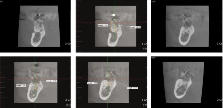
Fig. 4.Computed tomography (CT) scan showing deficient area before iliac bone graft. It shows close approximation to inferi- or alveolar nerve due to severe vertical alveolar bone resorption.
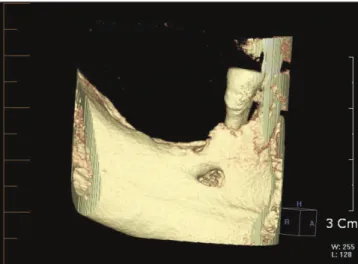
Fig. 3.Preoperative 3 dimensional view cone-beam comput- erized tomography (CBCT) scan showing the right mandibular alveolar ridge.
Fig. 2. Clinical photo of deficient right mandibular alveolar ridge.
Fig. 1. Preoperative panoramic view showing atrophic ridge on right edentulous mandibular molar area.
Fig. 10. Postoperative panoramic view showing grafted area of right posterior mandibular ridge.
Fig. 9.Immediate postoperative view of the grafted alveolar ridge.
Fig. 8. Fixation of block bone with microscrew.
Fig. 7. Iliac corticocancellous block bone graft being inserted under the elevated sub- periosteal pocket. Before insertion of the block, periosteal releasing incision was made with iris scissors. After insertion, the space between the recipient site and block was filled with bone chips mixed with FDBA (orthoblast II).
(FDBA: freeze-dried bone allograft)
Fig. 6. Dissection and formation of subperiosteal pocket using sharp periosteal elevator.
Fig. 5.Two vertical incisions are made.
Fig. 12. Two Implants were placed with more than 35 NCm torque on grafted sites with trans- gingival type fixture.
Fig. 13. The final prosthesis fabricated after 5 months of healing period of the implants in the graft.
Fig. 11.Postoperative cone-beam computerized tomography (CBCT) scan after 4 months of healing of the graft showing aug- mented alveolar ridge on right posterior mandible.
Ⅳ. 결 과
초기 고경은 평균 7.35 mm (range: 3.8-14 mm)였으며, 8부 위 모두 Cawood/Howell 분류상 IV형 또는 V형의 잔존치조 제 형태를 보였다17. 장골이식술 후 4개월경 CBCT를 이용 해 증강된 골고경을 평가한 결과 평균 13.64 mm (range:
11.5-18.6 mm)로 평균 6.29 mm의 골획득을 얻을 수 있었다.
골이식 후 임플란트 식립까지 이식부의 창상 열개 또는 감염 등의 합병증은 발생하지 않았다. 술후 일시적인 보행 장애가 관찰되었으나, 1개월경 추적 조사 결과 모든 환자 에서 보행장애는 완전히 회복된 양상을 보였다. 7명의 환 자 중 5명의 환자가 본과에서 총 11개의 임플란트를 식립 하였으며, 임플란트 식립에 골괴 고정을 위해 사용한 microscrew를 제거하였다. 이때 이식된 골괴와 수혜부간 부정 유합이나 비유합은 관찰되지 않았다. 임플란트 식립 후 단 1명에서 임플란트 주위염이 발생하였으며, 실패는 한 증례도 없었다. 식립한 임플란트의 평균 고경은 10.7 mm였다.(Table 1)
Ⅴ. 고 찰
치아 소실 후 상악에서는 구심성으로, 하악에서는 원심 성으로 점진적인 수직적 및 수평적 치조골 흡수가 진행한 다. 이런 흡수가 기저골까지 진행되면, 임플란트를 이용한 구강 재건은 더욱 어려워진다. 이러한 상황에서 가장 예지 성있는 해결책은 구외 부위에서 자가골을 채취하여 재건 술을 시행하는 것이다. 구외 공여부로는 주로 장골 부위와 두개골이 사용되어 왔는데, 특히 장골 부위는 심각하게 위 축된 상악골과 하악골 재건술에 공여부로 오랫동안 이용
되어 왔다. 장골능에서 채취한 골은 해면골이 풍부한데, 이 것은 골전도, 골유도, 골증식능을 가진 많은 생활세포(vital cells)를 함유하고 있다.
장골에서는 해면골뿐만 아니라 피질골-해면골 복합 골괴 (corticocancellous block)의 채취가 가능하며, 골괴이식 후 장기간 흡수율에 대한 많은 연구가 있다12,18-20. Verhoeven 등20 은 흡수된 하악골에 장골이식술과 임플란트 식립을 동시 에 시행 후 29개월간 추적 관찰한 연구에서 술후 첫해 이식 골 고경의 평균 36%가 흡수되었음을 보고하였으며, Maiorana 등21은 이식편 상방으로 무기질 우골(Bio-Oss, Geistlich)을 얇게 덮어주고, 흡수성 콜라겐 차단막으로 피 개하였을 때, 리모델링에 의한 생리적 흡수율이 약 50%까 지 감소하였음을 보고하였다.
이식편의 노출을 동반한 창상 열개와 이식편의 감염은 골이식 술식의 가장 주된 합병증이다22. 약간의 수직 절개 만을 동반하는 터널링기법은 연조직 피판에 혈류 공급을 보존시켜 피판괴사의 위험성을 감소시킨다. 이 술식은 Rothstein 등23에 의해 처음 소개되었으며, Khoury24에 의해 더욱 발전되었다. 이 술식은 피판이 장력 없이 봉합되는데 유리한 장점이 있으며, Ponte 등25은 술후 치유과정에서 수 평 절개를 동반하는 술식에 비해 터널링기법의 경우 연조 직의 열개의 유병률이 더 적었음을 보고하였다. 그들의 연 구에서 창상 열개의 발병률이 낮은 것은 치조능에 수평절 개를 시행하지 않았기 때문이며, 이러한 점에서 터널링기 법은 수직적 골증강 시 특히 이점이 있음을 보고하였다. 본 과에서 총 7명의 8 부위에 장골이식술을 시행한 증례에서 술후 창상 열개 및 감염 등의 합병증은 단 한 증례도 존재 하지 않았다. 이식부에 식립한 임플란트의 예후는 양호하 였으며, 단 한 증례에서 임플란트 주위염이 발생하였을 뿐,
47=4.1×12
#5 F 19 Rt. Mn. 7.8 13.9 45=3.7×8
5 months 46=4.1×10
#6 F 58 Lt. Mn. 9.6 16.1 33=3.8×14
5 months 35=4.3×14
34=4.3×12
#7 F 42 Lt. Mn. 6.7 12.4 36=4.3×10 6 months
38=4.3×8
(CBCT: cone-beam computerized tomography, T1: preoperative bone height (mm), T2: bone height when measured 4 months after bone graft (mm), T3: implant location and dimension, T4: period during bone graft and implant placement)
실패는 관찰되지 않았다. 식립한 임플란트의 평균 고경은 10.7 mm였으며, 충분한 1차 안정성을 얻을 수 있었다. 그러 나 터널형성기법은 기본적으로 맹검법이기 때문에 시야확 보가 어려우며, 해부학적으로 중요한 구조물을 침범할 수 있는 가능성도 상대적으로 크다. 따라서 숙련된 술자에 의 해 시행되지 않는다면 예지성있는 결과를 기대하기 어려 우며, 원치않는 합병증이 발생할 수도 있다.
Ⅵ. 결 론
본 논문에서는 7증례의 터널링기법을 이용한 장골이식 술을 통하여 적절한 골고경을 얻을 수 있었으며, 이식부에 대하여 임플란트 식립 시 예지성있는 결과를 얻을 수 있었 음을 보고하였다. 그러나 자료의 부족과 증례의 소수성, 추 적 관찰 기간이 길지 않다는 한계가 존재하였다. 추후 더 많은 표본을 대상으로 한 장기간의 추적 관찰과 대조군과 비교를 통한 임상통계학적 연구 및 생검을 통한 조직학적 분석 등 다각적인 연구를 통하여 터널링기법을 이용한 장 골이식술 시 합병증의 발생률과 골흡수율 등에 대한 연구 가 필요할 것으로 사료된다.
References
1. Rocchietta I, Fontana F, Simion M. Clinical outcomes of vertical bone augmentation to enable dental implant placement: a system- atic review. J Clin Periodontol 2008;35(8 Suppl):203-15.
2. Felice P, Marchetti C, Piattelli A, Pellegrino G, Checchi V, Worthington H, et al. Vertical ridge augmentation of the atrophic posterior mandible with interpositional block grafts: bone from the iliac crest versus bovine anorganic bone. Eur J Oral Implantol 2008;1:183-98.
3. Cordaro L, Amade′DS, Cordaro M. Clinical results of alveolar ridge augmentation with mandibular block bone grafts in partial- ly edentulous patients prior to implant placement. Clin Oral Implants Res 2002;13:103-11.
4. Chiapasco M, Zaniboni M, Rimondini L. Autogenous onlay bone grafts vs. alveolar distraction osteogenesis for the correction of vertically deficient edentulous ridges: a 2-4-year prospective study on humans. Clin Oral Implants Res 2007;18:432-40.
5. Mecall RA, Rosenfield AL. The influence of residual ridge re- sorption patterns on implant fixture placement and tooth position.
Part I. Int J Periodontics Restorative Dent 1991;11:8-23.
6. Simion M, Trisi P, Piattelli A. Vertical ridge augmentation using a membrane technique associated with osseointegrated implants.
Int J Periodontics Restorative Dent 1994;14:496-511.
7. Rosenquist B. Implant placement in combination with nerve transpositioning: experiences with the first 100 cases. Int J Oral Maxillofac Implants 1994;9:522-31.
8. das Neves FD, Fones D, Bernardes SR, do Prado CJ, Neto AJ.
Short implants - an analysis of longitudinal studies. Int J Oral
Maxillofac Implants 2006;21:86-93.
9. Esposito M, Grusovin MG, Kwan S, Worthington HV, Coulthard P. Interventions for replacing missing teeth: bone augmentation techniques for dental implant treatment. Cochrane Database Syst Rev 2008;(3):CD003607. Review. Update in: Cochrane Database Syst Rev 2009;(4):CD003607.
10. McCarthy JG, Schreiber J, Karp N, Thorne CH, Grayson BH.
Lengthening the human mandible by gradual distraction. Plast Reconstr Surg 1992;89:1-8; discussion 9-10.
11. Keller EE, Tolman DE, Eckert S. Surgical-prosthodontic recon- struction of advanced maxillary bone compromise with autoge- nous onlay block bone grafts and osseointegrated endosseous im- plants: a 12 year study of 32 consecutive patients. Int J Oral Maxillofac Implants 1999;14:197-209.
12. Nystro¨m E, Ahlqvist J, Legrell PE, Kahnberg KE. Bone graft re- modeling and implant success rate in the treatment of the severe- ly resorbed maxilla: A 5 year longitudinal study. Int J Oral Maxillofac Surg 2002;31:158-64.
13. Kent JN, Quinn JH, Zide MF, Guerra LR, Boyne PJ. Alveolar ridge augmentation using nonresorbable hydroxylapatite with or without autogenous cancellous bone. J Oral MaxilloFac Surg 1983;41:629-42.
14. Block MS, Degen M. Horizontal ridge augmentation using hu- man mineralized particulate bone: preliminary results. J Oral Maxillofac Surg 2004;62(9 Suppl 2):67-72.
15. Hasson O. Augmentation of deficient lateral alveolar ridge using the subperiosteal tunneling dissection approach. Oral Surg Oral Med Oral Pathol Oral Radiol Endod 2007;103:e14-9.
16. Kfir E, Kfir V, Eliav E, Kaluski E. Minimally invasive guided bone regeneration. J Oral Implantol 2007;33:205-10.
17. Cawood JI, Howell RA. A classification of the edentulous jaws.
Int J Oral Maxillofac Surg 1988;17:232-6.
18. Yerit KC, Posch M, Hainich S, Turhani D, Klug C, Wanschitz F, et al. Long-term implant survival in the grafted maxilla: results of a 12-year retrospective study. Clin Oral Implants Res 2004;15:
693-9.
19. Reinert S, Ko¨nig S, Bremerich A, Eufinger H, Krimmel M.
Stability of bone grafting and placement of implants in the se- verely atrophic maxilla. Br J Oral Maxillofac Surg 2003;41:249- 55.
20. Verhoeven JW, Cune MS, Terlou M, Zoon MA, de Putter C. The combined use of endosteal implants and iliac crest onlay grafts in the severely atrophic mandible: a longitudinal study. Int J Oral Maxillofac Surg 1997;26:351-7.
21. Maiorana C, Beretta M, Salina S, Santoro F. Reduction of auto- genous bone graft resorption by means of BioOss coverage: a prospective study. Int J Periodontics Restorative Dent 2005;
25:19-25.
22. Levin L, Herzberg R, Dolev E, Schwartz-Arad D. Smoking and complications of onlay bone grafts and sinus lift operations. Int J Oral Maxillofac Implants 2004;19:369-73.
23. Rothstein SS, Paris DA, Zacek MP. Use of hydroxylapatite for the augmentation of deficient alveolar ridges. J Oral Maxillofac Surg 1984;42:224-30.
24. Khoury F. Augmentation osseuse et chirugie implantaire. Implant 1999;5:221-37.
25. Ponte A, Khoury F. The tunnel technique in bone grafting proce- dures : a clinical study[abstract]. Int J Oral Maxillofac Implants 2004;19:766.