c cc
2017 The Korean Academy of Prosthodontics
This is an Open Access article distributed under the terms of the Creative Commons Attribution Non-Commercial License (http://creativecommons.org/licens- es/by-nc/3.0) which permits unrestricted non-commercial use, distribution, and reproduction in any medium, provided the original work is properly cited.
*Corresponding Author: Jae-Min Seo
Department of Prosthodontics, School of Dentistry and Institute of Oral Bio-Science, Chonbuk National University, 567 Baekjedaero, Dukjin-gu, Jeonju 54896, Repulic of Korea +82 (0)63 250 2696: e-mail, [email protected]
Article history: Received December 12, 2016 / Last Revision January 5, 2017 / Accepted February 2, 2017
Introduction
The function and esthetics of a prosthesis are important aspects of a successful implant prosthesis. The prosthesis should be harmonious with the neighboring teeth. To achieve successful implant prosthesis, preserving soft tissues, such as the marginal gingiva and interdental papilla, in addition to the shape and color of the prosthesis, is crucial.1,2The loss of the interdental papilla can result in the formation of a "black triangle," leading to esthetic defects, phonetic problems, and food impaction.3Soft tissues are supported by bony substruc- ture. Therefore, efforts should be made to minimize the absorption of the alveolar bone and gingival recession for implantation on the esthetic zone.
The interdental or interimplant papilla can be influenced by the height of the interdental bone, the horizontal/vertical distance of the gingival embrasure, the position of the contact point, and the con- tour of the soft tissue.4If the distance from the interproximal con- tact point to the crest of the interdental bone is ≤5 mm, the prox- imal gingival embrasure can be filled with the interimplant papil- la.3However, the gingival embrasure space between two adja- cent implants is more difficult to be filled with the interimplant papil- la than with the interdental papilla of natural teeth.3,5,6
Therefore, methods to preserve the interproximal tissue should be considered in treatment planning for adjacent implant placement.
Strategic serial extraction, immediate implantation, and provisio- nalization are effective procedures to preserve both hard and soft tis-
치간 유두 보존을 위한 전략적 연속발치술과 즉시 임플란트 식립: 증례보고
최근배
1∙이정진
2∙안승근
2∙서재민
2*
1
한빛치과의원,
2전북대학교 치의학전문대학원 치과보철학교실 및 구강생체과학연구소
Strategic serial extractions and immediate implantation for interdental papilla preservation:
A case report
Geun-Bae Choi
1, Jung-Jin Lee
2, Seung-Geun Ahn
2, Jae-Min Seo
2*
1Hanvit Dental Clinic, Wonju, Republic of Korea
2Department of Prosthodontics, School of Dentistry and Institute of Oral Bio-Science, Chonbuk National University, Jeonju, Republic of Korea
Maintaining the blood supply of the interdental alveolar bone is crucial for preserving the interdental papilla. Rebuilding the interimplant papilla between adjacent implants is more difficult than rebuilding the interdental papilla between the natural tooth and implant. Therefore, preserving the interimplant tissue is necessary when adjacent implants are closely placed. In this case report, three effective methods for maintaining the surrounding tissue, namely strategic serial extraction, immediate implantation, and provisionalization of adjacent maxillary central incisors, were performed. The marginal gingiva and interimplant papilla were well maintained for 24 months. (J Korean Acad Prosthodont 2017;55:286- 91)
Keywords: Interdental papilla; Immediate implant; Serial extraction
sues while reducing the duration of patient discomfort.7,8 This clinical report describes a case that required extraction and implant placement in both maxillary central incisors. For pre- serving the interdental bone and papilla, strategic serial extraction instead of the simultaneous extraction of adjacent teeth with imme- diate implantation and provisionalization was performed. The patient demonstrated a satisfactory result in terms of both esthetic and functional aspects.
Case report
A 58-year-old woman presented to the Chonbuk National University Hospital with both maxillary central incisors fractured (Fig. 1). Both central incisors were subjected to endodontic treat- ment and restored by metal ceramic restoration 30 years ago. A sub- gingival fracture of the crown accompanied with pain was observed in the left maxillary central incisor. As an equigingival fracture of the crown was observed, endodontic treatment was attempted for the restoration. However, because severe caries was observed without the verification of apical patency, extraction and implantation were decided.
In the right central incisor, a provisional post crown was fabricated and luted with temporary cement (Tempbond, Kerr Corp., Romulus,
MI, USA). Immediate implantation of the left central incisor was planned initially because of pain. The left central incisor was atraumatically extracted using a periotome, and the implant (4.5 × 14 mm, Superline, Dentium Co., Seoul, South Korea) was imme- diately placed at the minimum distance of 2.0 mm from the cervix of the adjacent tooth. The platform of the implant was placed approximately 3.0 mm apical to the zenith of the adjacent tooth. Bone graft material (Bio-Oss, Geistlich AG, Wolhusen, Switzerland) was filled between the implant and the alveolar bone. The absorbable collagen membrane with a hole was covered to prevent spillage of the bone graft material.9As the Implant Stability Quotient (ISQ) val- ue measured using a Resonance Frequency Analysis (RFA) device (Osstell Mentor, Integration Diagnostic AB, Göteborg, Sweden) was 73, screw-retained provisional restoration (Titanium temporary abutment, Dentium Co., Seoul, South Korea/Tokuso Curefast, Tokuyama Dental, Tokyo, Japan) was fabricated indirectly, with the distance from the interproximal contact point between the provisional restoration and the central incisor to the crest of the interdental bone being about 4.5 mm, and connected (Fig. 2). The periapical radiograph was obtained using a paralleling technique. The distance from the contact point to the bone crest was evaluated using a linear mea- suring tool of Picture Archiving and Communication System (PACS) software (Marosis M-view, Marotech Inc., Seoul, Korea).
A B
Fig. 2. (A) Immediate implant was placed and provisional restoration was connected on the left maxillary central incisor, (B) Periapical radiograph after implantation on the left maxillary central incisor.
A B
Fig. 1. (A) Intraoral photograph, (B) Periapical radiograph at the first visit.
The anatomic magnification and distortion in the radiograph were calibrated with the length and diameter of the implant as reference.
After 3 months, the right central incisor was extracted and the implant (4.5 × 12 mm, Superline, Dentium Co., Seoul, South Korea) was immediately placed at the mesiodistal distance of 3.0 mm from the adjacent implant.10Provisional restoration of the right implant (Fig. 3) was fabricated in the same manner as described previous- ly. The distance from the contact point to the bone crest was eval- uated using the periapical radiograph. After 5 months of the right implant placement, soft tissue was stably maintained (Fig. 4). The ISQ value was measured at 80 in both implants. Final impression was taken with a customized pick-up impression coping by dupli- cating the contour of the provisional restoration. Customized tita-
nium-based zirconia abutment (Myplant, Raphabio Co., Seoul, Korea) and non-splinted zirconia crown (ST ziconia Block, Upcera Co., Guangdong, China) were fabricated using Computer-Aided Design/Computer-Aided Manufacturing (CAD/CAM) (Dental system, 3Shape, Copenhagen, Denmark/Arum 5X-100, Doowon, Daejeon, Korea). Abutments were connected with 30 Ncm.
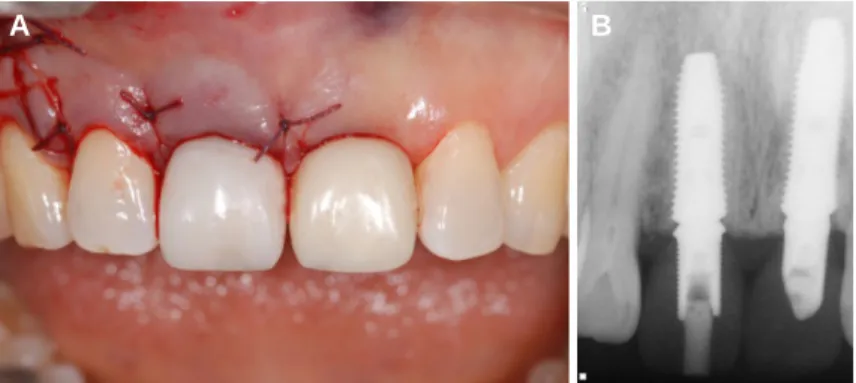
Occlusal contact of the restorations was adjusted and cemented with dual cured resin cement (Permacem 2.0, DMG, Hamburg, Germany) (Fig. 5). The distance from the contact point to the bone crest was about 4 mm in the periapical radiograph. Periodic recall check was arranged. After 24 months of implant placement, the buccal mar- ginal gingiva and interproximal papilla were stable (Fig. 6).
A B
Fig. 3. (A) Immediate implant was placed and provisional restoration was connected on the right maxillary central incisor, (B) Periapical radiograph after provisional restora- tions were connected.
Fig. 4. The buccal marginal gingiva and interimplant papil- la were stably maintained after 5 months of implantation.
Fig. 5. (A) Zirconia abutments were connected, (B) Zirconia crowns were cemented.
A B
A B
Fig. 6. (A) No marginal gingiva or interimplant papilla recession was observed after 24 months, (B) The marginal gingiva and interimplant bone around the implant were well maintained.
Discussion
Periodontal disease and surgical procedures such as flap surgery and tooth extraction are crucial factors for the loss of the interdental papilla.4Simultaneous extraction of adjacent teeth can lead to flattening of the interproximal bone, resulting in the loss of the inter- dental papilla as the infrabony support is absent.11Besides, the inter- dental papilla between adjacent implants is more difficult to rebuild than the interdental papilla between the natural tooth and the implant.12 Several studies have reported that atraumatic extrac- tion, immediate implantation, and provisionalization are favor- able procedures to preserve the interdental bone and the papilla.9,13 In the present case, strategic serial extraction (instead of simul- taneous extraction) of the adjacent teeth and immediate implanta- tion were performed and provisional restorations were fabricated.
If multiple teeth were extracted simultaneously, blood supply from the interdental bone and periodontal ligament might be inter- rupted. Consequently, the interdental papilla might be missed.
Because surgical or orthodontic method for rebuilding the lost interdental papilla is difficult and unpredictable, preservation of the interdental papilla is more important and effective in tooth extrac- tion and implant placement.14,15Serial extraction and immediate implan- tation can preserve the dentogingival complex (DGC) consisting of the gingival sulcus, epithelial attachment, and connective tissue attach- ment. When the blood supply from the DGC is maintained, the loss of the interdental bone and papilla can be minimized.16,17In addition, since the distance from the contact point to the crest of the interdental bone between the natural teeth and the implant was less than the dis- tance between adjacent implants, serial extraction and immediate implantation were favorable for the fabrication of provisional restoration.12
To preserve the interdental papilla, the right implant was mesiodis- tally placed at 3 mm away from the left implant and the provisional and final prosthesis were designed such that the distance from the contact point to the crest of the interdental bone was less than 5 mm. In addition, continuous recall check and adjustment of provisional restoration were conducted to evaluate the presence of interdental papilla.
These procedures require technical sensitivity in both implantation and laboratory procedures. However, it is considerable in pre- serving both hard and soft tissues around the implant in the esthet- ic zone. If the adjacent teeth are simultaneously extracted in the esthet- ic zone, it will be difficult to preserve the ideal interproximal bone and papilla. Strategic serial extraction, immediate implanta- tion, and provisionalization are important techniques for success- ful implant treatment.
ORCID
Jung-Jin Lee https://orcid.org/0000-0002-7381-5230 Seung-Geun Ahn https://orcid.org/0000-0002-9105-931X Jae-Min Seo https://orcid.org/0000-0001-5095-4046
References
1. Belser UC, Schmid B, Higginbottom F, Buser D. Outcome analysis of implant restorations located in the anterior maxilla:
a review of the recent literature. Int J Oral Maxillofac Implants 2004;19:30-42.
2. Phillips K, Kois JC. Aesthetic peri-implant site development. The restorative connection. Dent Clin North Am 1998;42:57-70.
3. Tarnow DP, Magner AW, Fletcher P. The effect of the distance from the contact point to the crest of bone on the presence or ab- sence of the interproximal dental papilla. J Periodontol 1992;63:995-6.
4. Chen MC, Liao YF, Chan CP, Ku YC, Pan WL, Tu YK. Factors influencing the presence of interproximal dental papillae between maxillary anterior teeth. J Periodontol 2010;81:318-24.
5. Salama H, Salama M, Kelly J. The orthodontic-periodontal connection in implant site development. Pract Periodontic Aesthet Dent 1996;8:923-32.
6. Tarnow D, Elian N, Fletcher P, Froum S, Magner A, Cho SC, Salama M, Salama H, Garber DA. Vertical distance from the crest of bone to the height of the interproximal papilla between adjacent implants. J Periodontol 2003;74:1785-8.
7. Greenstein G, Cavallaro J Jr. Serial extraction protocol: transi- tioning a hopeless dentition to a full-arch reconstruction.
Compend Contin Educ Dent 2008;29:526-34.
8. Attard NJ, Zarb GA. Immediate and early implant loading pro- tocols: a literature review of clinical studies. J Prosthet Dent 2005;94:242-58.
9. Arau′jo MG, Linder E, Lindhe J. Bio-Oss collagen in the buccal gap at immediate implants: a 6-month study in the dog. Clin Oral Implants Res 2011;22:1-8.
10. Tarnow DP, Cho SC, Wallace SS. The effect of inter-implant dis- tance on the height of inter-implant bone crest. J Periodontol 2000;71:546-9.
11. Kan JY, Rungcharassaeng K. Interimplant papilla preservation in the esthetic zone: a report of six consecutive cases. Int J Periodontic Restor Dent 2003;23:249-59.
12. Salama H, Salama MA, Garber D, Adar P. The interproximal height of bone: a guidepost to predictable aesthetic strategies and soft tissue contours in anterior tooth replacement. Pract Periodontic Aesthet Dent 1998;10:1131-41.
13. Cooper LF, Raes F, Reside GJ, Garriga JS, Tarrida LG, Wiltfang J, Kern M, de Bruyn H. Comparison of radiographic and clini- cal outcomes following immediate provisionalization of single- tooth dental implants placed in healed alveolar ridges and extraction sockets. Int J Oral Maxillofac Implants 2010;25:1222-32.
14. Beagle JR. Surgical reconstruction of the interdental papilla: case report. Int J Periodontic Restor Dent 1992;12:145-51.
15. Salama H, Salama M. The role of orthodontic extrusive re-
modeling in the enhancement of soft and hard tissue profiles pri- or to implant placement: a systematic approach to the manage- ment of extraction site defects. Int J Periodontic Restor Dent 1993;13:
312-33.
16. Gargiulo AW, Wentz FM, Orban B. Dimensions and relations of
the dentogingival junction in humans. J Periodoltol 1961;32:261- 7.
17. Kois JC. Altering gingival levels: the restorative connection part I: biologic variables. J Esthet Restor Dent 1994;6:3-7.
치간 유두 보존을 위한 전략적 연속발치술과 즉시 임플란트 식립: 증례보고
최근배
1∙이정진
2∙안승근
2∙서재민
2*
1
한빛치과의원,
2전북대학교 치의학전문대학원 치과보철학교실 및 구강생체과학연구소
치간유두의 보존을 위해서는 치간골의 혈액공급이 매우 중요하다. 인접한 임플란트 사이의 치간유두를 재생하는 것은 치아와 임플란트 사이의 치간유두보다 어렵다. 그러므로 인접한 임플란트를 식립할 경우 임플란트 사이 조직을 보존하는 것이 필요하다. 이를 위해서 전략적 발치술, 즉 시 임플란트 식립 및 임시 보철물 제작은 임플란트 주위 조직을 보존하는데 효과적인 방법으로 소개되었다. 본 증례는 손상된 양측 상악 중절치 를 전략적 연속 발치술 및 임플란트 즉시 식립을 통해 회복한 환자로 24개월 뒤 임플란트 주위 조직 및 치간 유두가 안정적으로 보존되었기에 이 를 보고하는 바이다. (대한치과보철학회지 2017;55:286-91)
주요단어: 치간유두; 즉시 임플란트 식립; 순차적 발치술
*교신저자: 서재민
54896 전북 전주시 덕진구 백제대로 567 전북대학교 치의학전문대학원 치과보철학교실 063 250 2696: e-mail, [email protected]
원고접수일: 2016년 12월 12일 / 원고최종수정일: 2017년 1월 5일 / 원고채택일: 2017년 2월 2일
2017 대한치과보철학회
이 글은 크리에이티브 커먼즈 코리아 저작자표시-비영리 3.0 대한민국 라이선스에 따라 이용하실 수 있습니다.
c cc