Address reprint requests to Jae-Lim Cho, M.D.
Department of Ortopaedic Surgery, Hanyang University College of Medicine 17 Haengdang-dong, Sungdong-gu, Seoul, 133-792, Korea
Tel: 82-2-2290-8485, Fax: 82-2-2299-3774, E-mail: [email protected]
Journal of Korean Spine Surg.
Vol. 12, No. 4, pp 331~337, 2005
협부결손형 전방전위증에서 정복의 유지를 위한 후방추체간 유합술의 효과
박예수・조우진・김석환・조재림
한양대학교 의과대학 정형외과학교실
Effect of Posterior Lumbar Interbody Fusion for Maintaining the Reduction in Isthmic Spondylolisthesis
Ye-Soo Park, M.D., Woo-Jin Cho, M.D., Suk-Hwan Kim, M.D., and Jae-Lim Cho, M.D.
Department of Orthopaedic Surgery, Hanyang University College of Medicine
– Abstract –
Study Design: This is a retrospective study on the effect of posterior lumbar interbody fusion for maintaining the reduction in isthmic spondylolisthesis patients.
Objectives: We evaluated the efficacy of performing posterior lumbar interbody fusion for maintaining the reduction in isthmic spondylolisthesis.
Summary of the Literature Review: There have been many reports regarding the surgical treatment of spondylolisthesis.
Although there are many reports that the clinical results have nothing to do with the reduction, many surgeons have tried to maintain the reduction. However, the question about what kind of fusion modality is the most effective for maintaining the reduction is still controversial.
Material and Method: Between August 2002 and January 2004, 24 patients with isthmic spondylolisthesis were operated on.
14 underwent posterolateral fusion alone (group A) and 10 underwent additional posterior interbody fusion (group B). These two groups were compared in terms of the clinical results, the radiological changes and fusion rates.
Results: the reduction rate were 11.81% and 7.32% in the PLF and PLF+PLIF groups, respectively (p>0.05). The reduction losses were 0.19% and 0.35% in the PLF and PLF+PLIF groups, respectively (p>0.05). The changes after fusion were 0.11% and 0.10%
in the PLF and PLF+PLIF groups, respectively (p>0.05). There was no case of nonunion. The satisfaction rates were 86% and 83% in the PLF and PLF+PLIF groups, respectively (p>0.05).
Conclusions: In our study, the addition of posterior interbody fusion showed no benefit in maintaining correction. If solid fusion can be obtained, then posterolateral fusion seems to be sufficient enough to maintain correction in isthmic spondylolisthesis. The authors think that further studies are mandatory because of the small number subjects in our study.
Key Words: Isthmic spondylolisthesis, Posterior interbody fusion, Maintenance of reduction
서 론
척추전방전위증의 수술적 치료시 전위된 추체의 정복 에 대해서는 확실한 감압만 이루어 진다면 정복 자체는 크게 문제되지 않는 것으로 보고되고 있고, 최근까지 무 리한 정복의 시도 보다는 수술적 치료시 자세에 의한 정 복 후 감압을 시행하는 것이 원칙으로 받아 들여지고 있 다. 특히 정복후 재전위에 대해서는 본 교실에서도 86례 의 환자를 대상으로 정복의 소실에 의한 재전위가 증상 의 발현과는 무관하다고 보고한바 있다14)
.
그러나 일부에서는 재전위의 방지를 위해 정복의 유 지에 관심을 기울이고 있으며1-9)
, 최근에는 후측방 유합
술에 전방추체간 유합술을 추가로 시행하여 정복의 유 지에 효과를 볼 수 있었다는 보고도 있는 실정이다1-3). 이
에 저자들은 협부결손형 전방전위증 환자들을 대상으 로 정복의 유지에 있어 후측방 유합술과 더불어 시행한 후방추체간 유합술의 효과를 알아 보고자 하였다.연구대상 및 방법
2002년 8월부터 2004년 1월까지 협부결손형 전방전위
증으로 수술적 치료를 시행 받은 환자 중 1년 이상 추시 가 가능하였던 24례를 대상으로 하였으며, 수술 방법에 따라 두 군으로 나누어 A군은 14례로 감압 및 후측방 유 합술 시행 군, B군은 10례로 A군의 술식에 후방추체간 유합술을 추가적으로 시행한 군으로 분류하였다. 두 군 을 나눈 기준은 무작위로 선정하였다. 유합분절은 전위 된 추체의 상하로 퇴행성 척추관 협착증이 동반되었을 경우 이를 포함하여 수술하였다.모든 환자에서 수술 전, 수술 직후, 수술 후 1개월, 2개 월, 3개월, 6개월 및 최종 추시 시에 요천추부 단순 측면 방사선촬영을 시행하였으며, 술전 전위의 정도는 Mey-
erding 방법으로
10),전위의 변화는 Taillard 방법11)을 사용 하여 측정하였다. 전위 정복율은 술전 전위율에서 술후 전위율을 뺀 값을 술전 전위율로 나눈 값의 백분율로 계 산하였고, 정복 소실율은 최종 추시 시의 전위율에서 수 술 직후 전위율을 뺀 값으로 측정하였다.골유합은 L e n k e의 기준을 이용하였는데(Table 1), 두
군에 똑같이 적용하였으며 B군의 추체간 유합의 정도는 그 판단이 힘들어 추가로 시행하지는 않았다. 임상적 평 가는 외래 추시 시의 문진 및 이학적 검사 기록을 근거 로 하였으며, 일부에서는 전화를 이용한 설문 결과도 참 조하여 Katz 등의 주관적 만족도를 분석하였다(Table 2).
A군에서는 수술 수기로 복와위에서 정중절개를 통한
후방 도달법으로 이환 부위를 노출 시킨 후 필요에 따라 추궁판 전 절제술 및 비후된 관절 돌기와 척추경의 일 부, 추궁 협부결손부의 섬유연골 조직 등을 제거하는 철Table 1. Lenke’s fusion grade
A Solid, big trabeculated fusion bilaterally (definitely solid)
B Solid, big fusion mass unilaterally with a small fusion mass on the contralateral aspect (possibly solid) C Small, thin fusion masses bilaterally with apparent crack (probably not solid)
D Graft resorption bilaterally or fusion mass with obvious bilateral pseudoarthrosis (definitely not solid) Table 2. Katz’s satisfaction scale
How Satisfied Are You With:
The overall result of back operation?
Very satisfied Somewhat satisfied Somewhat dissatisfied Very dissatisfied
Relief of pain following the operation?
Very satisfied Somewhat satisfied Somewhat dissatisfied Very dissatisfied
Your ability to walk following the operation?
Very satisfied Somewhat satisfied Somewhat dissatisfied Very dissatisfied
Your ability to do housework, yard work, or job following the operation?
Very satisfied Somewhat satisfied Somewhat dissatisfied Very dissatisfied
Your strength in the thighs, legs, and feet?
Very satisfied Somewhat satisfied Somewhat dissatisfied Very dissatisfied
Your balance, or steadiness on your feet?
Very satisfied Somewhat satisfied Somewhat dissatisfied Very dissatisfied
저한 감압술을 시행하고, 척추경 나사를 이용한 내고정 후 충분한 양의 자가 장골을 이용한 후측방유합술을 시 행하였다. B군에서는 이러한 술식에 c a g e를 이용한 추 체간유합술을 추가하였다.
술후 처치로는 수술직후부터 감압된 부위가 혈종에 의해 압박되는 것을 방지하기 위해 주로 측와위를 취하 면서 체위 변동을 허용하였고, 단계적으로 좌위, 보행을 허용하였으며 흉요천추 보조기는 골유합이 완성될 때 까지 착용할 것을 권유하였다.
결 과
1. 환자의 분포
대상 환자는 A군의 경우 남자 7 례, 여자 7례 이었으 며, 평균 연령 4 2세(19~67), 평균 추시 기간은 2 6개월
(12~30) 이었다. B군은 남자 3 례, 여자 7례 이었으며, 평
균 연령 4 4세(19~64), 평균 추시 기간은 2 3개월( 1 2 ~ 3 0 ) 이었다. 두 군간의 유의한 차이는 없었다(P>0.05).2. 이환 부위 및 전위의 정도
전위된 추체는 A군은 제 4요추 8례, 제 5요추 6례 이었 으며, B군에서는 제 4요추 7례, 제 5요추 3례 이었다. 두 군 모두 제 4요추가 가장 많았다. 평균 유합 분절은 A군
1 . 3 5분절(1~2), B군 1 . 4 0분절(1~2) 이었다. 방사선 소견
상 전위는 Meyerding 방법으로 A군은 grade I이 1 0례,
grade II가 4례로 1.29(1~2), B군은 grade I이 8례, grade II
가 2례로 1 . 2 2 ( 1 ~ 2 )등급이었다. 두 군간의 차이는 없었 다(p>0.05).3. 전위의 변화
Taillard 방법으로 측정한 전위의 변화는 A군의 경우
술전 평균 22.76%(10~48), 술후 평균 10.89%(6.5~25), 추 시 평균 10.97%(6.5~27), 최종 추시 평균 1 1 . 0 8 %( 6 . 5 ~ 2 7 . 2 )이었고, B군에서는 술전 20.28%(10~33.3), 술
후 12.96%(6.4~22), 추시 13.21 %(6.3~22), 최종 추시13.31% (6.4~22) 로(Table 3), A 군은 1 1 . 8 1 %의 정복 , 0.19%의 정복 소실, 골 유합 후 최종 추시까지 전위의 변
화는 0 . 1 1 %를 보여 주었고, B군의 경우 각 각 7 . 3 2 % ,0.35%, 0.10%를 보여 주었다(Table 4). 정복의 정도, 정복
소실률 및 추시 기간 중의 변화는 두 군간의 큰 차이는 없었다(p>0.05).4. 방사선학적 소견
Lenke의 기준을 이용한 골유합은 A군의 경우 Lenke A
등급 9례, B등급 5례 이었으며, 기간은 평균3.6개월(2~5) 이었다. B군은 A등급 6례, B등급 4례, 기간은 3 . 8개월( 3 ~ 7 )이었다 . 두 군간의 큰 차이는 없었다 ( p > 0 . 0 5 ) (Table 5).
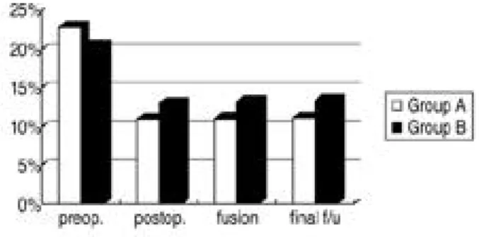
Table 3. Loss of Reduction
Preop. Postop. Fusion Final F/U Reduction Rate Reduction Loss
A 22.76% 10.89% 10.97% 11.08% 11.81% 0.19%
B 20.28% 12.96% 13.21% 13.31% 07.32% 0.35%
P-value 00.241 0.2810
Table 4. The diagraph shows the grade of spondylolisthesis in group A and group B respectively at preoperative, postoperative, com- plete bone union and last follow up. There is minimal change from postoperative to final follow up.
5. 임상적 평가
Katz 등의 주관적 만족도를 이용하여 분석한 임상적
평가로는 A군 매우 만족 7례, 만족 5례, 불만족 2례 이었 으며, B군의 경우 매우 만족 5례, 만족 3례, 불만족 2례를 보여 주었다. 임상적 평가도 두 군간의 큰 차이는 없었 다(p>0.05)(Table 6).고 찰
척추전방전위증의 수술적 치료에 있어서 감압 및 전 위의 정복에 있어서 전위된 추체의 정복은 그 동안 많은 논란이 있어왔다. Bradford12)는 도수 정복술 혹은 수술적 정복술 후 좋은 결과를 얻었음을 보고하였고, Peek 등13) 은 감압술이나 정복술 없이도 좋은 결과를 가져온다고 하였다. 하지만 확실한 감압만 이루어 진다면 정복자체 가 크게 문제되지 않는 것으로 보고되고 있다. 본 교실 에서도 8 6례의 척추전방전위증 환자를 대상으로 후측 방 유합술(Fig. 1)만을 시도한 결과 정복 후 재전위가 일 어나기는 하지만, 술후 임상 결과와는 아무런 연관이 없 음을 발표한 바 있다14)
.
이처럼 재전위는 감압만 확실히 되어진다면 크게 문 제되지 않는다는 것도 이미 잘 알려진 사실이지만, 최근 재전위를 줄이고자 하는 시도에서 후측방 유합술에 전 방추체간 유합술을 추가로 시행해야 된다는 주장이 있 다1-3)
. 이러한 시도는 정복의 유지를 통해 안정적인 시상
면상의 균형을 유지하고 이에 따른 인접분절의 변화를 최소화 하고자 하는 노력으로 보여진다15).
Laus 등은 저자들의 경우처럼 척추전방전위증의 장기
추시에서 후측방 유합술만으로도 좋은 결과를 보여주 었다고 보고하였으며16), McAfee등은 120례의 척추전방
Table 5. Radiologic Finding
Lenke A Lenke B Lenke C Lenke D
A 9 5 0 0
B 6 4 0 0
Total 150 9 0 0
Table 6. Clinical Results
Excellent Good Fair Poor
A 7 5 2 0
B 5 3 2 0
Total 12 8 4 0
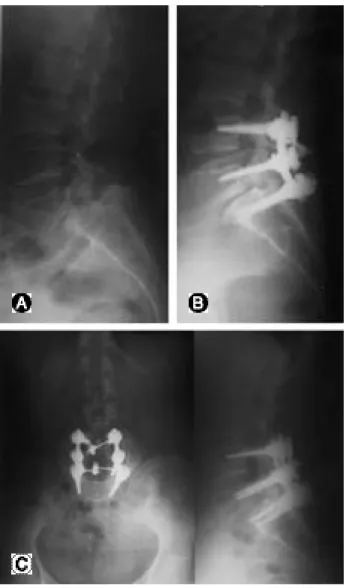
Fig. 1. (A) Preoperative lateral radiograph of 49-year-old female shows grade II isthmic spondylolisthesis at L5- S1 level. (B) Postoperative lateral radiograph shows the reduction of spondylolisthesis (48% → 20%). (C) Radi- ographs at 24 months follow up examination shows maintenance of reduction with solid bony union.
전위증을 분석하여 cage를 이용한 추체간유합술이 추간 공을 넓힐 뿐 아니라 유합율의 향상에도 기여한다고 주 장하였다17)
. Sears
18), Grob 등
19)과 Na 등20)은 후측방유합술 과 병행한 c a g e를 이용한 후방추체간 유합술로, Spruit 등21)은 전방추체간 유합술로 좋은 결과를 보고하였으며,Shin
1), Suk
2), Choy 등
3)은 titanium mesh cage를 이용한 추 가적인 후방 추체간 유합술(Fig. 2)은 기존의 후측방 유 합술만을 시행하는 것보다 방사선학적인 결과와 임상 적 결과에서 우수하다고 보고하였다. 그러나 한편으로 는 후측방 유합술에 있어 비록 골유합이 되더라도 전단 력과 전방 굴곡력이 잔존하기 때문에 재전위가 발생할 수 있다는 주장도 제기되고 있는 실정이다4 - 6 , 8 ). 그러나
많은 저자들이 특히 경도의 전방전위증에서는 추체간 유합술을 추가로 시행한 경우와 비교하여 임상적인 결 과가 큰 차이를 보이지 않는다고 주장하였다2 2 , 2 3 ). Mar- chetti 등은 Meyerding 3등급 이상인 고도의 전위에서만
전후방 유합술을 시행할 것을 제안하였고24)
, Dehoux 등
도 경도의 전방전위증에서는 후측방 유합술만을 시행 할 것을 제안하였다2 5 ). 저자들의 경우에도 후측방 유합
술만으로도 좋은 방사선학적, 임상적 결과를 확인할 수 있었다. 저자들의 경우 좋은 결과를 얻을 수 있었던 가 장 중요한 요인은 증례가 모두 경도의 전방전위 이었으 며, 이식골의 유합율이 다른 보고들에 비해 높았다는 것 을 요인으로 지적할 수 있다. 횡고정기기는 가급적 사용 하려 하였으나 보험급여기준이 까다로워진 이후부터는3분절이상에서만 시행되었기에 본 연구에서는 제외되
었다. 이전에 사용된 횡고정기기는 A군에서 총 3례로 유합율에 큰 영향을 주지 않은 것으로 사료된다26).
요약 및 결론
협부형 전방전위증 환자를 대상으로 후측방 유합술 시행 군과 후방추체간 유합술 추가 군에서 정복의 유지 를 비교한 결과 두 군 모두 재전위의 정도는 미미하였으 며 임상 결과에 있어서도 우수한 결과를 얻었다. 충분한 감압술 후 유합만 확실히 된다면 후측방유합술 만으로 도 정복을 유지하는데 충분하리라 사료된다. 그러나 본 연구의 증례가 적고 경도의 전방 전위증만을 대상으로 하여 추후 이에 대한 추가적인 연구가 필요하리라 사료 되는 바이다.
참고문헌
01) Shin BJ, Min KD, Kwon H, et al: Surgical result of isth - mic spondylolisthesis -comparison of posterolateral fusion vs. PLIF, J Kor Spine Surg 1996; 3:61-68.
02) Suk SI, Lee CK, Kim WJ, Kim HG: Adding posterior lumbar interbody fusion to pedicle screw fixatin and pos - terolateral fusion after decompression in spondylolytic spondylolisthesis, J Kor Orthop Assoc 1995; 30:1638- 1646.
03) Choy WS, Kim WJ, Kim KH, et al: The results of the posterior lumbar interboby fusion using titanium mesh cage for spondylolisthesis, J Kor Spine Surg 1999; 6:129- 134.
04) Freeman BJ, Licina P, Mehdian SH: posterior lumbar interbody fusion combined with instrumented posterolater - al fusion: 5-year results in 60 patients, Eur Spine J 2000;
9:42-46.
05) Csecsei Gl, Klekner AP, Dobai J, Lajgut A, Sikula J:
Fig. 2. (A) Preoperative lateral radiographs of 46-year-old female shows grade II isthmic spondylolisthesis at L4-5 level. (B) Postoperative lateral radiographs shows the reduction of spondylolisthesis (33% → 13%). (C) Radi- ograph and CT scan at 26 months follow up examination shows maintanace of reduction with solid bony union.
Posterior interbody fusion using laminectomy bone and transpedicular screw fixation in the treatment of lumbar spondylolisthesis, Surg Neurol 2000; 53;2-6.
06) James C.H. Goh, Hee-kit Wong, Ashvin Thambyah, Chun-Sing Yu: Influence of PLIF cage size on lumbar spine stability, Spine 2000; 25:35-39.
07) Jeon TS, Moon SH, Kim NH, Park KB, Lee HM: Com - parison between posterior lumbar interbody fusion with pedicle screw fixation and posterolateral fusion with pedi - cle screw fixation in spondylolytic spondylolisthesis in adults, J Kor Spine Surg 2003; 10:104-112.
08) Laursen M, Thomsen K, Eiskjaer SP, Hansen ES, Bunger CE: Functional outcome after partial reduction and 360 degree fusion in grade III-V spondylolisthesis in adolescent and adult patient. J Spinal Disord 1999;
12:300-306.
09) Madan S, Boeree NR: Outcome of posterior lumbar interbody fusion versus posterolateral fusion for spondy - lolytic spondylolisthesis, Spine 2002; 15;27:1536-1542.
10) Mererding HW: Spondylolisthesis: surgical treatment and results. J Bone Joint Surg 1943; 25:65-77.
11) Taillard WF: Etiology of spondylolisthesis, Clin Orthop 1976; 117:30-39.
12) Bradford DS: Closed reduction of spondylolisthesis. An experience in 22 patients, Spine 1988; 13:580-587.
13) Peek RD, Wiltse LL, Reynolds JB, Thomas JC, Guyer DW, Widell EH: In situ arthrodesis without decompres - sion for grade-III or IV isthmic spondylolisthesis in adults who have severe sciatica, J Bone Joint Surg 1989;
71A:62-68.
14) Cho JL, Yoon WK, Park YS, Choi KJ, Han JH: Redis - placement after operative reduction of spondylolisthesis - comparison between pedicle screw system and Luque ring system-, J Kor Orthop Assoc 1997; 32:1162-2333.
15) Cho JL, Park YS, Han JH, Lee CH, Roh WI : T h e changes of adjacent segments after spinal fusion - Follow- up more than three years after spinal fusion -, J Kor Spine Surg 1998; 5:239-246.
16) Laus M, Tigani D, Pignatti G, et al: P o s t e r o l a t e r a l spinal fusion: a study of 123 cases with a long-term fol - low-up, Chir Organi Mov 1994; 79(1):69-79.
17) McAfee PC, DeVine JG, Chaput CD, et al: The indica -
tions for interbody fusion cages in the treatment of spondylolisthesis: analysis of 120 cases, Spine 15;30(6 Suppl):S60-65.
18) Sears W: Posterior lumbar interbody fusion for degenera - tive spondylolisthesis: restoration of sagittal balance using insert-and-rotate interbody spacers, Spine 2005;
5(2):170-179.
19) Grob D, Scheier HJ, Dvorak J, Siegrist H, Rubeli M, Joller R: Circumferential fusion of the lumbar and lum - bosacral spine, Arch Orthop Trauma Surg 1991;
111(1):20-25.
20) Na HY, Jeong YY, Kim WS, Cho HW: Surgical treat - ment of isthmic spondylolisthesis: pedicle screw fixation, posterolateral fusion, and posterior lumbar interbody fusion with cage after wide decompression, J Kor Spine Surg 2003; 10:1199-1207.
21) Spruit M, Pavlov PW, Leitao J, De Kleuver M, Ander- son PG, Den Boer F: Posterior reduction and anterior lumbar interbody fusion in symptomatic low-grade adult isthmic spondylolisthesis: short-term radiological and functional outcome, Eur Spine J 2002; 11(5):428-433.
22) Kim NH, Lee JW: Anterior interbody fusion versus pos - terolateral fusion with transpedicular fixation for isthmic spondylolisthesis in adults. A comparison of clinical results, Spine 1999 15;24(8):812-816.
23) La Rosa G, Conti A, Cacciola F, et al: Pedicle screw fix - ation for isthmic spondylolisthesis: does posterior lumbar interbody fusion improve outcome over posterolateral fusion?, J Neurosurg 2003; Sep;99(2 Suppl):143-150.
24) Marchetti PG, Binazzi R, Briccoli A, et al: The surgical treatment of spondylolisthesis, Chir Organi Mov 1994;
79:85-91.
25) Dehoux E, Fourati E, Madi K, Reddy B, Segal P: Pos - terolateral versus interbody fusion in isthmic spondylolis - thesis: functional results in 52 cases with a minimum fol - low-up of 6 years, Acta Orthop Belg 2004; 70(6):578-582.
26) Arand M, Wilke HJ, Schultheiss M, et al: Comparative stability of the “Internal Fixator” and the “Universal Spine System” and the effect of crosslinking transfixating systems. A biomechanical in vitro study, Biomed Tech 2000; 45(11):311-316.
연구계획: 협부 결손형 전방전위증에서 정복의 유지를 위한 후방추체간 유합술의 효과에 대한 후향적 연구
연구목적: 협부결손형 척추전방전위증 환자들을 대상으로 정복의 유지에 있어 후측방 유합술과 더불어 시행한 후방 추체간 유합술의 효과를 알아 보고자 하였다.
대상 및 방법: 2002년 8월부터 2004년 1월까지 협부결손형 척추전방전위증으로 수술적 치료를 시행 받은 환자 24례 를 대상으로, A군은 14례로 감압 및 후측방유합술 시행 군, B군은 10례로 A군의 술식에 후방추체간 유합술을 추가적 으로 시행한 군으로 분류하였다. 전위의 정도는 Meyerding 방법, 전위의 변화는 Taillard 방법을, 임상적 평가는 Katz 등의 주관적 만족도, 골유합은 Lenke 등의 기준을 이용하였다.
결과: 전위된 추체는 A군은 제 4 요추 8례, 제 5 요추 6례 이었으며, B군에서는 제 4 요추 7례, 제 5요추 3례 이었다. 평 균 유합 분절은 A군 1.35분절, B군 1.40분절 이었다. 방사선 소견상 전위는 A군은 1.29(1~2), B군은 1.22(1~2)등급이 었다. 전위의 변화는 A군의 경우 술전 평균 22.76%(10~48), 술후 평균 10.89%(6.5~25), 추시 평균 10.97%(6.5~27), 최 종 추시 평균 11.08%(6.5~27.2)이었고, B군에서는 술전 20.28%(10~33.3), 술후 12.96%(6.4~22), 추시 13.21 %(6.3~22), 최종 추시 13.31% (6.4~22) 로, A군은 11.81%의 정복, 0.19% 의 정복 소실, 골 유합 후 최종 추시까지 전위의 변화는 0.11%를 보여 주었고, B군의 경우 각 각 7.32%, 0.35%, 0.10%를 보여 주었다.
골유합은 A군의 경우 Lenke A등급 9례, B등급 5례 이었으며, 기간은 평균3.6개월(2~5)이었다. B군은 A등급 6례, B등 급 4례, 기간은 3.8개월(3~7)이었다. 임상적 평가로는 A군 86%의 만족도를, B군의 경우 83%의 만족도를 보여주었다.
결론: 본 연구에서는 정복의 유지에 있어 후방추체간 유합술은 효과가 없었으며, 골유합만 확실히 된다면 후측방 유 합술만으로도 충분하리라 사료된다. 그러나 연구 대상 증례가 적어 추가적인 연구가 필수적이라 사료되는 바이다.
색인단어: 협부형 척추전방전위증, 후방추체간 유합술, 정복의 유지
※ 통신저자 : 조 재 림
서울특별시 성동구 행당동 산17번지 한양대학교병원 정형외과
Tel: 82-2-2290-8485 Fax: 82-2-2299-3774 E-mail: [email protected]
국 문 초 록