2007, Vol. 51, No. 5
Printed in the Republic of Korea
DNA 고정 탄소나노튜브 페이스트전극의 물고기 세포속 테트라싸이클린에 정량
이수영*·이장현†·정영삼‡
서울산업대학교 자연생명과학대학 정밀화학과
†평택대학교 교양학부
‡고려대학교 이과대학 화학과
(2007. 6. 12 접수)
Measuring Oxytetracycline Using a Simple Prepared DNA Immobilized on a Carbon Nanotube Paste Electrode in Fish Tissue
Suw Young Ly*, Chang Hyun Lee†, and Young Sam Jung‡
Biosensor Research Institute, Seoul National University of Technology, Seoul 139-743, Korea
†Division of General Education.Pyongtaek University, 111-Yongyi dong, Pyongtaek-Si, Gyeonggido, 450-701, Korea
‡Department of Chemistry, Korea University, Anam Dong Seoul 136-704, South Korea (Received June 12, 2007)
요 약. DNA를 고정한 탄소나노튜브 페이스트 전극으로 옥시테트라싸이클린 항생제의 분석조건을 연구하였다. 사각
파형 벗김 전압전류법과 순환 전압 전류법을 사용하여 최적조건에서 1-10 ngL−1의 정량 가능 범위에 도달하였다. 벗김법
0.1 mgL−1농도에서 15번 반복 측정하여 0.068%의 상대표준편차였으며, 벗김법 최적 분석 조건에서 0.4 ngL−1의 검출 한계를 얻었다. 개발된 결과를 양식어류의 세포에서 응용하였다.
주제어: Oxytetracycline, Square Wave Stripping, Cyclic Voltammetry, Carbon Nanotube, DNA, Paste Electrode
ABSTRACT. A simple prepared paste electrode (PE) of DNA immobilized on a carbon nanotube was utilized for monitoring the antibacterial agent oxytetracycline (OTC), using square-wave anodic stripping voltammetry (SWASV) and cyclic voltammetry (CV). Given these conditions, SWASV and CV working ranges were observed within 1-10 ngL-1 OTC.
In the SWASV and CV for OTC concentrations of 0.1 mgL-1, the relative standard deviations (n=15) were 0.068 and 0.067, respectively. At the optimized condition, the detection limit was found to be 0.4 ngL-1 OTC. This method was applied to the hatchery fish tissue.
Keywords: Oxytetracycline, Square Wave Stripping, Cyclic Voltammetry, Carbon Nanotube, DNA, Paste Electrode
INTRODUCTION
Antimicrobial OTC drugs are essential for the treatment of diseases in humans and animals. OTC is widely found in hatchery fishes, calf veals, and cow’s milk from animals with low concentration ranges in their body and fluid systems, in the medi-
cal and environmental sciences. Sensitive detection methods are in demand in their vivo or vitro sys- tems. Various analytical techniques for the drug and its analogs have been researched on. Most of the common methods employ separation and photo- metric systems, as, for example, high-performance liquid chromatography,1-6 mass spectrometry,7-10 near-
infrared spectroscopy,11 second-derivative synchro- nous spectrofluorimetry,12 and the capillary elec- trophoresis system.13 Some of these methods are composed of a temperature control system, a separate column, an injection system, and other photometric or electrochemical detection systems.
In analytical chemistry, more sensitive and simple methods are in demand. Stripping voltammetry is particularly useful and popular for trace analy- sis.14-17 since this accumulation technique is sim- ple and sensitive and is thus applicable with chromatography, electrophoresis and other analyt- ical detection systems. Voltammetric methods have rarely been researched on for OTC analysis, however, except in a few recent reports. Plastic- membrane OTC-hydrochloride-selective electrodes arrived at the detection limit of 1.0 × 10⁻⁷M (0.03 ppm),18 amperometric boron-doped diamond (BDD) thin films electrodes arrived at a 10-nM detection limit,19 and the potentiometric and cyclic voltam- metric titration methods20 and capillary electro- phoresis with the reductive fast cyclic voltammetric detection method arrived at a 1.5 × 10⁻⁶-mol/L detection limit.21 Capillary electrophoresis of some tetracycline antibiotics coupled with reductive fast cyclic voltammetric detection. In vivo or vitro sys- tems, OTC has very low concentration ranges, making more sensitive detection limits in demand.
This study researched on a DNA immobilized onto a carbon nanotube electrode for OTC analy- sis, employing CV and SWASV in a stripping mode, which is relatively less expensive, more sensitive than other convenient voltammetric results, and can be used for tissue analysis with low detection limits.
On the other hand, DNA and carbon nanotubes have a specific affinity in biosensors.22-26 Many researches have examined the interaction of DNA with a carbon nanotube and their immobilization effects.27-30 In this study, the properties of DNA and a carbon nanotube paste electrode were com- bined, and low detection ranges of OTC analysis were arrived at. The optimized results were applied to antibacterial agent analysis in the fish tissue and can be applied in any other field of OTC analysis.
EXPERIMENT Apparatus, Reagents and Procedure
Voltammetric measurements were taken using a CHI660A from Systems, Inc. A handmade PE electrode with a 0.5-mm diameter and a 10-mm length was used as the working electrode; an Ag/
AgCl electrode, as the reference electrode; and a platinum wire, as the auxiliary electrode. All elec- trolyte solutions were prepared from double- distilled water (~18-M Ohm cm-1). A 0.1-M ammo- nium phosphate solution served as the supporting electrolyte. All other reagents were of analytical grade. The PE working electrode was handmade by mixing a 40% carbon nanotube (0.4 gram, from Nanostructured & Amorphous Materials, Inc.) and a 40% double-stranded DNA (0.4 gram, prepared from calf thymus sigma) with 20% mineral oil (0.2 gram) in a mortar for 30 minutes. The paste was deposited inside the body of a capillary tube glass (2 mm in diameter and 1 cm long), and the electric contact between the electrode and the potentiostat was established with a 1-mm-diame- ter copper wire. The electrode surface was pol- ished using a cloth felt pad with a 0.05-mm alumina slurry. The three-electrode system was immersed in a stirred solution of 0.1-M phosphate buffer (pH 4.5). DNA immobilization was per- formed through a cyclic scan with an initial potential of +1.6 V, a switching potential of -1.6 V, and a scan rate of 0.5 mVs-1 with a tan cyclic repeat to stabilize the electrode surface.
Pre-concentration prior to cyclic and SW strip- ping was carried out at an open circuit. CV and SWASV were then performed, with the common parameter for CV being a scan rate of 100 mVs-1. The common parameters for the stripping were as follows: an accumulation potential of -1.7 V (vs.
that of the Ag/AgCl reference electrode); a final potential of 1.0 V; a deposition time of 500 s; a quiet time of 2 s; an SW amplitude of 35 mV; an SW frequency of 357 Hz; and an increment poten- tial of 9 mV. Background voltammograms were recorded with the phosphate buffer in the absence of OTC.
RESULTS AND DISCUSSION
Cyclic Voltammetric Behavior of OTC on the PE First, various electrolyte acid, base and buffer solutions were searched at a high OTC strength.
Phosphate solutions of 0.1 M were found to have good sensitivity. Thus, various OTC concentration effects were examined using CV. Fig. 1 shows the cyclic voltammograms of the background electro- lyte solutions for the 0.1-M ammonium phosphate with pH 4.5. The latter’s raw and 10 given concen- tration ranges were 1, 2, 3, 4, 5, 6, 7, 8, 9 and 10 ngL-1 OTC at a sweep rate of 100 mVs-1 on the PE. At the oxidation scan, an anodic peak and a shoulder at 0.18 V were obtained, which were well extracted from the background discharge and sensitively appeared. Upon scan reversal, no corresponding reduction peak emerged in any potential. Other SW stripping conditions were examined with this anodic peak current. At these conditions, the sloop ratio of
∆x/∆y = 7.8556 and the maximum peak current of 78.18×10⁻⁷ A were sensitively obtained with very low detection ranges, which can be applied in tis- sue and body fluid analysis. Other concentration ranges at the mg and ug levels were also examined,
and their results appeared well linearly. Although not shown here, in the nanogram working ranges, an analytical application was performed with the known content of 60 mgL-1 of the N company’s drug samples. The results were obtained using stan- dard addition methods by extrapolating the linear curve. A 59.0 mgL-1 OTC concentration was detected at 99 % recovery. At this condition, more sensitive SW stripping techniques were examined at an 0.18 V peak potential. Moreover, the electrode lifespan was examined at CV conditions, for which a one- month-long time was used. The electrode surface was cleaned each time simply with weighing paper.
Effects of Hydrogen Ionic Strength, SW Ampli- tude, and Accumulation Time
The electrochemical behavior of OTC was dependent on the electrolyte solutions and the hydrogen ionic strength. Thus, various concentra- tions of the phosphate electrolyte solutions, from 0.01 to 0.5 mol, were tested as possible supporting electrolytes. The 0.1-M phosphate buffer solution was found to have the best peak sensitivity. Then the same solutions were examined for their hydro- gen ionic strength, and the results are shown in Fig.
2(A). The peak current did not change from pH 3.7 to pH 4.6, reached its maximum at pH 5.5, and decreased at a higher pH range. At the other pH ranges peak width are broad and sensitivity is weak.
A pH of 5.5 was assumed at a maximum current of 6.53 × 10⁻⁵ A. This solution was used for the fixed concentration of 10 mgL-1 of OTC. Thus, all other solutions were used with this 5.5 pH strength, after which other SW amplitude parameters were exam- ined. The results are shown in Fig. 2(B). For the range of 0.01-0.25 V, the peak high responded very slowly, whereas 0.35 V was obtained at the maxi- mum peak current of 278 × 10-8 A. At the higher ranges, the peak width broadened and the peak cur- rent decreased, their amplitude effects were poorer than those of the pH peak high, and all their peak widths broadened. Then the accumulation potential was examined in the negative range from -1.0 V to -2.0 V with a positive direction. The -1.7 V accu- mulation potential turned out to be sensitive, and a Fig. 1. Various concentration effects of CV for 1-10-ngL-1
OTC, at a scan rate of 100 mVs-1 in a 0.1-M ammonium phosphate buffer (pH 5.5).
sharp peak width appeared at the maximum peak current of 8.722 × 10⁻⁵ A that was more sensitive than the SW frequency and the solutions’ pH effects.
Moreover, the positive potential was examined for a cathodic reaction, and no peak signals appeared.
Effects of Various SW Voltammetric Parameters of Frequency and Increment Potentials
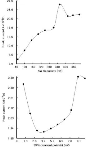
In Fig. 3(A), various SW frequencies are shown in the range of 50 to 500 Hz with 10 points at the fixed concentration of 10 mgL-1 of OTC, in which other experimental conditions in Fig. 2 were held constant. Their peak current increased as their fre- quency increased, exhibiting a maximum at about 357 Hz with a 26.05 × 10⁻⁶ A peak high. Thus, the
Fig. 2. (A): Electrolyte solutions’ pH effect for 2.7, 3.2, 3.7, 4.1, 4.6, 5.1, 5.5, 5.9, 6.4 and 7. (B): SW amplitude of 0.01, 0.04, 0.07, 0.12, 0.16, 0.2, 0.25, 0.29, 0.3 and 0.35. (C): SW accumulation potentials of -1.0, -1.2, -1.3, -1.4, -1.6, -1.7, -1.8, -1.0 and -2.0 for the 10-mgL-1 fixed OTC concentration.
Other conditions in Fig. 3 were held constant.
Fig. 3. (A): SW stripping voltammetric peak current at the frequency levels of 50, 100, 150, 200, 250, 312, 357, 416, 454 and 500 Hz. (B): Increment potentials of 1, 2, 3, 4, 5, 6, 7, 8, 9 and 10 mV for the 10-mg-1 OTC solution. Other conditions in Fig. 2 were held constant.
optimal frequency was determined to be 357 Hz, and at the other ranges, the peak width broadened and slowly disappeared. At the maximum condi- tions, other parameters of the SW increment poten- tials were examined. Fig. 3(B) illustrates the stripping peak current of OTC as a function of the SW incre- ment potential range of 1-10 mV with 10 points at a 10-mg-1 fixed OTC concentration, with an accumu- lation time of a 500 s and a deposition potential of -1.7 V. At the range of 1-3 mV, the peak currents quickly decreased. At the higher range of 9 mV, the peak width sharply appeared and the peak response increased much more significantly. At this poten- tial, a peak high of 2.398 × 10⁻⁵ A was obtained, and the results were more sensitive than the SW fre- quency effects. At these maximum conditions, vari- ous possible interference species with metals and other antimicrobial analogies were tested, with a fixed OTC concentration of 1 mgL-1. The criterion used for the presence of interference was a 5 % or greater change in the peak current of OTC. The fol- lowing levels did not show any interference: 10-mgL-1 doxycycline and 10-mgL-1 chlorotetracycline. On the other hand, 10-mgL-1 Ba(II), 10-mgL-1 Co(II), 10-mgL-1 Ca(II), 10-mgL-1 Hg(II), 10-mgL-1 Bi(II), 10-mgL-1 Fe(II), 10-mgL-1 Ge(IV), 10-mgL-1 Cr(III), and 10-mgL-1 tetracycline yielded -77.4%, 428.2%, -91.0%, 1,233.9%, -49.8%, 3779.9%, 41.85%, -10.9%
and 12.7% interference, respectively. The % means depression in the OTC peak current. At this time, mercury and iron ions quickly interfered in the peak current high. Other interference ions can be cor- rected using standard addition methods.
Effects of Concentration, Linear Range, Detection Limits, and Application
At the optimized conditions, various SWASV work- ing ranges were examined. mg and ug working rev- els were obtained although not shown here, and more sensitive nanogram ranges were arrived at.
The raw results are shown in Fig. 4(A) for 1, 2, 3, 4, 5, 6, 7, 8, 9 and 10 ngL-1 at the optimum condi- tions. After the background current was subtracted from the measured currents, a sloop ratio of ∆x/∆y
= 1.8835 sensitively responded, and a regression
yield of the y: current in amperes and the x: concen- tration at the ngL-1 levels with a correlation coeffi- cient of 0.9945 was obtained. The precisions for the 15th repeated determination of the 0.1-mgL-1 OTC solutions yielded relative standard deviations of 0.067%. The peak current turned out to be highly reproducible. At these conditions, the detection limit of 0.4-ngL-1 of OTC was estimated based on the signal-to-noise (S/N) characteristics of the data for the S/N = 3 given optimum conditions. Finally, using this condition, the developed methods can be applied for the determination of OTC concentra- tions in food samples of hatchery fish tissue. The Fig. 4. (A): Various concentration effects of SW with a 1-10-ngL-1 OTC spike. (B): Analytical application for the five curves for the electrolyte blank, then the 0.5-mL diluted tissue spike, then standard solutions of the 5-, 10- and 15-ngL-1 OTC spike. Other conditions in Fig. 3 were held constant.
results are shown in Fig. 4(B). The five curves show the electrolyte blank solution as not having obtained any noise signal. The 0.5-mL diluted tis- sue was spiked at this voltammogram, and a sensitive peak signal was obtained, after which other standard solutions of 5, 10 and 15 ngL-1 of OTC were spiked, their peaks linearly increased, and the standard addi- tion methods were applied and used to calculate the OTC values of 15.5-ng/Kg body weight and R2= 0.9969 OTC that were obtained. All the CV and SWASV applications obtained satisfactory values.
CONCLUSION
To detect the antimicrobial drug OTC using vol- tammetry, a DNA immobilized on a carbon nano- tube paste electrode was examined, as were optimum analytical conditions, and finally, very low concen- tration ranges were used that were found to have more sensitive detection limits than other common voltammetric methods.18,19,21 The optimized conditions can be applied in hatchery fish tissue. The results can be used in commercial drug quality control or any other OTC analysis. The use of a PE electrode is much simpler and more stable over a longer time.
REFERENCES
1. Papadoyannis, I. N.; Samanidou, V. F.; Kovatsi, L. A.
Journal of Pharmaceutical and Biomedical Analysis.
2000, 23, 275.
2. Hisao Oka.; Yuko Ito, & Hiroshi Matsumoto. Journal of Chromatography A. 2000, 882, 109.
3. Paul, A. Blackwell.; Hans-Christian Holten Ltzhøft.;
Hai-Ping Mab:, Bent Halling-Sørensen.; Alistair B. A.
Boxall, & Paul Kay Fast and robust. Journal of Chro- matograph A. 2004, 1045, 111.
4. Xiaojing Ding, & Shifen Mou. Journal of Chromatog- raphy A. 2000, 897, 205.
5. Rosie Coyne.; Øivind Bergh, & Ole Bent Samuelsen.
Journal of Chromatography B. 2004, 810, 325.
6. Cinquina, A. L.; Longo, F.; Anastasi, G.; Giannetti, L.; Coz- zan, R. Journal of Chromatography A. 2003, 987, 227.
7. Zhu, J.; Snow, D. D.; Cassada, D. A.; Monson, S. J.;
Spalding, R. F. Journal of Chromatography A. 2001, 928, 177.
8. Anne Kruse Lykkeberg, Bent Halling-Sørensen, Claus Cornett, Jette Tjørnelund, & Steen Honor Hansen. Journal
of Pharmaceutical and Biomedical Analysis. 2004, 34, 325.
9. Marc Cherlet, Mario Schelkens, Siska Croubels, & Patrick De Backer. Analytica Chimica Acta. 2003, 492, 199.
10. Anne Marie Jacobsen, Bent Halling-Sørensen, Flemming Ingerslev, & Steen Honor Hansen. Journal of Chroma- tography A. 2004, 1038, 157.
11. Nataa Smolaa, & Uros Urleb. Analytica Chimica Acta.
2000, 410, 203.
12. Fernandez-Gonzlez, R.; Marcia-Falcn, M. S.; Simal- Gndara, J. Analytica Chimica Acta. 2004, 455, 143.
13. Nozal, L.; Arce L.; Simonet, B. M.; Rios, A.; Valcrcel, M. Analytica Chimica Acta. 2004, 517, 89.
14. Joseph Wang.; Beatriz Serra.; Suw Young Ly.; Jianmin Lu, & Jose M. Pingarron. Talanta. 2001, 54, 147.
15. Joseph Wang.; Jianmin Lu.; Suw Young Ly.; Maika Vuki.; Baomin Tian.; William K. Adeniyi, & Ruben A.
Armendariz. Anal. Chem. 2000, 72, 2659.
16. Suw Young Ly.; Jung Ik Chae.; Young Sam Jung.; Woon Won Jung.; Hye Jeong Lee, & Seong Ho Lee. Nahr- ung/Food. 2004, 48. 201.
17. Suw Young Ly.; Duck Ho Kim, & Myung Hoon Kim.
Talanta. 2002, 58, 919.
18. Xian Xiang Sun.; Xu Zhang, & Hassan, Y.; Aboul- Enein. IL FARMACO. 2004, 59, 307.
19. Wangfuengkanagul, N.; Siangproh, W.; Chailapakul, O.
Talanta. 2004, 64, 1183.
20. Marzanna Kurzawa, & Agnieszka Kowalczyk-Marzec.
Journal of Pharmaceutical and Biomedical Analysis.
2004, 34, 95.
21. Jinkui Zhou.; Geoff C. Gerhardt.; Andrej Baranski, &
Richard Cassidy. Journal of Chromatography A. 1999, 839, 193.
22. Eugenii Katz, & Itamar Willner. Chem Phys Chem. 2004, 5, 1084.
23. Wanga, S. G.; Ruili Wang.; Sellin, P. J.; Qing Zhang.
2004, 325, 1433.
24. Randhir, P. Deo.; Joseph Wang. Electrochemistry Com- munications. 2004, 6, 284.
25. Ningning Zhu.; Zhu Chang, Pingang He.; Yuzhi Fang.
Analytica Chimica Acta. 2005, 545, 21.
26. Shifeng Hou.; Jiahai Wang.; Charles R. Martin. J. AM.
CHEM. SOC. 2005, 127, 8586.
27. Manli Guo.; Jinhua Chen.; Dengyou Liu.; Lihua Nie, &
Shouzhuo Yao. Bioelectrochemistry. 2004, 62, 29.
28. Maria L. Pedano, & Gustavo A. Rivas. Electrochemistry Communications. 2004, 6, 10.
29. Kagan Kerman.; Yasutaka Morita.; Yuzuru Takamura.;
Mehmet Ozsoz, & Eiichi Tamiya. Electroanalysis. 2004, 16, 1667.
30. Dovbeshko, G. I.; Repnytska, O. P.; Obraztsova, E. D.;
Shtogun, Y. V. Chemical Physics Letters. 2003, 372, 432.