서론
경동맥 스텐트 설치술(CAS)은 중증의 경동맥 협착을 가진 환자에서 경동맥내막 절제술(CEA)을 대신하여 뇌졸중을 줄일 수 있는 치료법이다(1-3). 경동맥 스텐트 설치술의 결과는 시 술자의 경험, 판단 및 기구 등에 의해 영향을 받으며 환자의 위 험인자, 플라크의 특성 또한 영향을 미친다(4-6). 대부분의 경 동맥 스텐트 설치술은 총경동맥과 내경동맥 사이에서 이루어 지고 있으며, non-tapered stent를 사용한 경동맥 스텐트 설치 술은 총경동맥과 내경동맥의 직경의 차이에 의해 내경동맥에 더 큰 직경의 스텐트 삽입(stent oversizing)이 발생할 수 있다.
일부 연구에서 신생내막의 증식을 유발하는 요인으로 더 큰 직 경의 스텐트 삽입보다는 스텐트를 삽입하는 동안의 혈관 손상 을 주 요인으로 보고하였으나(7, 8), Wallstent를 사용하여 경 동맥 스텐트 설치술을 시행한 한 보고에서는 혈관 직경의 차이
가 delayed stent shortening을 유발할 수 있으며, 이는 재협착 의 한 요인이 될 수 있다고 보고하기도 하였다(9). Tapered stent는 해부학적으로 적합한 형태를 갖추고 있어 이 혈관의 직 경의 차이에 따른 더 큰 직경의 스텐트 삽입에 의한 효과를 줄 일 수 있으며, 장기적인 동맥벽면에 전달되는 반지름 방향의 힘 (chronic outward radial force), 목 움직임시에 발생하는 기계 적 스트레스(mechanical stress)와 이로 인한 strut migration을 줄일 수 있다는 판단하에 최근 사용이 증가하고 있으나 이에 대 한 정확한 연구는 아직 부족한 상황이다(10). 이에 본 연구의 목적은 내경동맥 팽대부에 중증의 협착을 가진 환자에서 ta- pered stent를 이용한 치료 결과를 분석하고 시술 후 발생한 합병증과 추적관찰을 통해 재협착 및 폐쇄, 임상적으로 무증 상을 보인 기간에 대해 분석해 보고자 하였으며, 기존에 보고 된 연구를 바탕으로 non-tapered stent의 치료성적과 비교해 보고자 하였다.
J Korean Soc Radiol 2011;65(4):365-372
Received May 17, 2011; Accepted August 15, 2011 Corresponding author: Dong Hyun Kim, MD Department of Radiology, Chosun University College of Medicine, 588 Seoseok-dong, Dong-gu, Gwangju 501- 717, Korea.
Tel. 82-62-220-3163 Fax. 82-62-228-9061 E-mail: [email protected]
Copyrights © 2011 The Korean Society of Radiology
Purpose: To analyze the results of carotid artery stenting using a tapered stent and
to evaluate the effectiveness of the tapered stent compared to previously reported studies using non-tapered stents.Materials and Methods: From October 2008 to August 2010, elective carotid ar-
tery stenting using a tapered stent was attempted in 39 lesions from 36 consecu- tive patients. Post-procedural complications were evaluated by neurologic symp- toms and magnetic resonance imaging. Restenosis or occlusion was evaluated by carotid Doppler ultrasound and computerized tomography with angiography. Newly developed neurologic symptoms were evaluated clinically.Results: The self-expandable tapered stent was placed across the carotid artery ste-
nosis. A total stroke was noted in 3 patients, while a major stroke was noted in 1 patient. On diffusion weighted imaging, new lesions were observed in 15 patients, but 13 patients were clinically silent. Follow-up imaging studies were performed in the 13 clinically silent lesions, and no evidence of restenosis or occlusion was found any of the 13 lesions. During clinical follow-up in 34 lesions from 31 patients, there were newly developed neurological symptoms in only 1 patient.Conclusion: Carotid artery stenting using a tapered stent may be safe and useful
for the treatment of carotid artery stenosis.Index terms
Carotid Arteries Carotid Artery Stenosis StentsTransluminal Angioplasty
Preliminary Report of Carotid Artery Stenting Using a Tapered Stent
1Tapered Stent를 사용한 경동맥 스텐트 설치술에 대한 예비 보고
1Chang Woo Jeong, MD
1, Dong Hyun Kim, MD
1, Seong Hwan Ahn, MD
2, Dong Uk Kim, MD
2, Seung Jeong Hong, MD
1, Young Suk Kim, MD
1, Joo Nam Byun, MD
1, Jae Hee Oh, MD
1Departments of 1Radiology, 2Neurology, Chosun University College of Medicine, Gwangju, Korea
bral angiography)을 시행하였으며, NASCET criteria에 따라 협착의 정도를 평가하였다.
전처치 및 스텐트 설치술
환자는 최소 시술 7일 전부터 aspirin 100 mg과 clopidogrel 75 mg을 복용하였다. 모든 시술은 국소마취하에 시행하였으 며, 시술 중 환자의 동맥산소 포화도와 혈압을 적절히 감시하였 다. 환자의 활성 혈액응고 시간(activated clotting time)을 250~300초 정도로 유지하기 위해 hepatin을 일시 주사로 3,000 IU를 투여한 후 시간당 1,000 IU를 추가로 정맥주사 하 였다. 8F 유도관(Guider SoftipTM XF, Boston Scientific Corp., Maple Grove, MN, USA)을 총경동맥에 위치시킨 후 Filterwire (FilterWire EZ™, Boston Scientific Corp., Maple Grove, MN, USA)와 delivery sheath를 협착부위를 넘어서까지 진행하고 sheath를 제거하고 Filterwire를 설치하였으며, Filterwire를 따 라서 스텐트를 진행하여 외경동맥의 기시부를 가로질러 총경 동맥 분지부에 설치하였다. 스텐트는 자가팽창성(self-ex- pandable)의 tapered stent를 사용하였다. 스텐트 설치 후 풍선 카테터(Ultra-softTM SV MONORAILTM, Boston Scientific Corp., Maple Grove, MN, USA)를 이용하여 규정압력에 따라 풍선확장술을 시행하였으며, 경동맥 확장에 의한 서맥, 일시적 인 뇌혈류 차단에 의한 경련 등에 유의하였다. 스텐트 설치가 끝나면 Filterwire는 retrieval sheath를 이용해 제거하였다. 20 분 후에 뇌혈관 조영술을 시행하여 잔여협착(residual stenosis) 여부와 색전증에 의한 뇌혈관 폐쇄 여부를 확인하였다.
시술 후 평가
스텐트 설치술 직후 모든 환자는 신경학적 검사를 시행하였 다. 중증 뇌졸중(major stroke)은 24시간 이상 지속하는 신경 학적 결손이 있으며 National Institutes of Health Stroke Scale (NIHSS)(12)이 3점 이상 증가한 경우로 정의하였으며, 신경학 적 결손이 24시간 이상 지속하나 NIHSS가 3점 이상 증가하지 않은 경우는 경증 뇌졸중(minor stroke)으로, 신경학적 결손이 24시간 이내에 완전히 회복된 경우는 일과성 허혈증(transient ischemic attack)으로 정의하였다. 모든 환자는 시술 3일 이내 에 T2 강조영상(T2 weighted image), 액체감약반전회복영상 (fluid-attenuated inversion recovery: FLAIR) 그리고 확산강 조영상(diffusion weighted image)을 포함하는 뇌 자기공명영 상을 시행하여 시술 전 영상과 비교하여 새롭게 발생한 병변의 유무를 확인하였으며, 이는 색전병변으로 정의하였다. 총 12명 환자에서 13개 병변에 대해 영상검사를 통한 추적관찰을 시행 하였으며, 11예에서 경동맥 도플러 초음파(carotid doppler ul-
대상과 방법
환자군
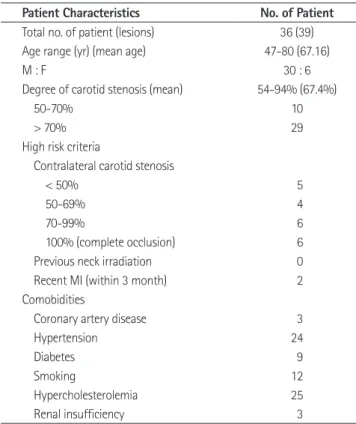
2008년 10월부터 2010년 8월까지 일률적으로 tapered stent (PROSTE′GE′TM RX Tapered, ev3 Inc., Plymouth, MN, USA) 를 이용하여 경동맥 팽대부에 스텐트 설치술을 시행한 36명의 환자에서 39개 병변을 후향적 연구방법으로 분석하였다. 환자 는 남자가 30명, 여자가 6명이었으며, 평균 나이는 67.4세 (47~80세)였다. North American Symptomatic Carotid End- arterectomy Trial (이하 NASCET) criteria (11)에 따른 평가에 서 29예가 70% 이상의 협착이 있었으며, 10예는 증상을 동반 한 50% 이상의 내경동맥 팽대부의 협착이 있었다. 6명의 환자 는 반대편 경동맥이 이미 폐쇄되었던 환자였다. 1명의 환자는 스 텐트 설치술 시행 후 반대편 경동맥에 경동맥내막 절제술을 시 행하였다(Table 1).
시술 전 평가
모든 환자는 시술 전에 신경과 의사에게 신경학적 검사를 받 았으며, 혈관촬영술을 포함하는 전산화단층촬영(CT angiog- raphy) 및 자기공명영상(MR angiography)을 시행하였다. 이 후 정확한 협착범위와 두개강외 동맥과 두개강내 동맥에 대한 평가를 위해 대퇴동맥 경유 뇌혈관 조영술(transfemoral cere-
Table 1. Patient Demographic Characteristics
Patient Characteristics No. of Patient
Total no. of patient (lesions) 36 (39)
Age range (yr) (mean age) 47-80 (67.16)
M : F 30 : 6
Degree of carotid stenosis (mean) 54-94% (67.4%)
50-70% 10
> 70% 29
High risk criteria
Contralateral carotid stenosis
< 50% 5
50-69% 4
70-99% 6
100% (complete occlusion) 6
Previous neck irradiation 0
Recent MI (within 3 month) 2
Comobidities
Coronary artery disease 3
Hypertension 24
Diabetes 9
Smoking 12
Hypercholesterolemia 25
Renal insufficiency 3
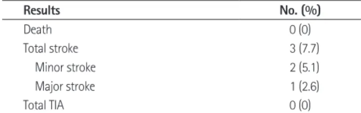
용하여 총경동맥 분지부에 설치하였다. 총 3예에서 뇌졸중이 발생하였으며, 1예에서 중증 뇌졸중이 발생하였다. 이 환자는 반대쪽 경동맥의 완전폐쇄 및 반대측 중뇌동맥영역의 경색이 있었던 환자였다. 스텐트 삽입술 후 NIHSS가 3점 이상 증가하 였으며, 1일 후 시행한 확산강조영상에서 반대측 중뇌동맥영역 에 새롭게 발생한 뇌경색 병변이 관찰되었다. 5일 후 퇴원시 증 상호전을 보였으며 19일 후 복부 대동맥류에 대한 수술을 시행 하였다. 수술 4일 후 양측 전하소뇌동맥영역 및 반대측 중뇌동 맥에 경색이 발생하였다(Fig. 1). 경증 뇌졸중이 발생한 2예 중 trasound; DUS)를 통해, 2예에서는 혈관촬영술을 포함하는
전산화단층촬영을 통해 재협착 및 폐쇄에 대한 평가를 시행하 였다. 50% 이상의 협착을 재협착으로 정의하였다. 31명의 환 자에서 34개의 병변에 대한 임상적인 추적관찰을 시행하여 새 롭게 발생한 신경학적 증상유무를 평가하였다.
결과
모든 39예에서 일률적으로 자가팽창성의 tapered stent를 이
D A
E F
B C
Fig. 1. In a 67-year-old woman patient, MRI shows focal stenosis at the right carotid bulb and complete occlusion of a left internal carotid ar- tery (arrow) (A). 3D digital subtraction angiography before the procedure shows focal stenosis with ulcerated plaque at the right carotid bulb (arrow) (B). Post-procedural angiography shows well conformable stent structure (C). Post-procedural diffusion weighted-MR image shows newly developed high signal intensity in left middle cerebral artery territory (D). Surgical repair of abdominal aortic aneurysm was performed at 1 month after carotid artery stenting. Post-operative diffusion weighted-MR images shows high signal intensity in anterior inferior cerebellar ar- tery territory, both and left middle cerebral artery territory (E, F).
총 12명에서 13개의 병변에 대해 영상검사를 통한 추적관찰 을 시행하였으며 11예에서 경동맥 도플러 초음파, 2예에서는 혈관촬영술을 포함하는 전산화단층촬영을 이용해 추적검사를 시행하였다. 평균 추적관찰 기간은 315일(2~552일)이었으며, 13예 모두에서 재협착 및 폐쇄는 없었다(Fig. 3). 또한 31명 환 자에서 34개의 병변에 대해 임상적으로 추적관찰을 시행하였 다. 평균 추적관찰 기간은 310일(12~777일)이었으며, 1예에서 신경학적 증상이 발생하였다. 이 환자는 퇴원시 NIHSS 11점에 서 16일 후 12점으로 증가하였으며 이후 추적관찰에서 삼킴곤 란, 명칭실어증(anomia)의 증상이 발생하였다. 그 외 30명의 환자에서는 추적관찰 기간 중 새롭게 발생한 신경학적 증상이 관찰되지 않았다(Table 4).
고찰
경동맥내막 절제술은 NASCET가 그 효용성을 입증한 이후 로 두개강외 경동맥 폐쇄성 질환에 대한 표준치료법으로 알려 졌다. 하지만 CAVATAS (3)는 경동맥내막 절제술과 경동맥 스 텐트 설치술이 시술과 연관된 뇌졸중이나 사망 위험도에서 유 의한 차이가 없음을 보고하였으며, SAPPHIRE (1)는 시술과 연 관된 뇌졸중이나 사망 위험도를 경동맥내막 절제술과 경동맥 스텐트 설치술에서 각각 7.3%와 4.4%로 보고하는 등 최근 여러 연구에서 경동맥 스텐트 설치술은 경동맥내막 절제술을 대체할 수 있는 적합한 치료로 부각되고 있다.
경동맥 스텐트 설치술시 고려할 사항으로 시술과 연관된 합 1예는 동측에 점 모양의 작은 고신호 강도가 보였으며, 이 환
자는 고혈압, 당뇨 및 흡연 등의 위험인자를 가지고 있던 환자 로 좌측 반맹과 촉각적 일측무시(tactile neglect)의 신경학적 증상이 발생하였다. 다른 1예는 확산강조 자기공명영상에서 색 전병변이 관찰되지 않았다. 2예 모두 5일 이내에 신경학적 증 상이 완전소실 되었으며 퇴원시 새롭게 발생한 신경학적 증상 은 보이지 않았다(Table 2).
시술 후 시행한 확산강조 자기공명영상에서 15예에서 술전 영상에서는 보이지 않던 새로운 색전병변이 관찰되었으며, 이 중 13예에서는 점 모양의 작은 고신호 강도가 보였으나 임상증 상은 없었다. 증상이 있었던 2예 중 한 명은 중증 뇌졸중이 발 생한 환자였으며, 다른 한 명은 경증 뇌졸중이 발생하였다 (Fig. 2)(Table 3).
Fig. 2. Diffusion weighted-MR image of a 69-year-old man with severe stenosis of right carotid artery.
A, B. Compared with pre-procedural image (A), post-procedural diffusion weighted-MR image (B) shows high signal intensity in right parietal lobe (arrow), the patient had neurologic symptoms such as left hemianopsia and left tactile neglect, but the neurological symptoms completely disappeared within 5 days.
A B
Table 2. The Results of Carotid Artery Stenting
Results No. (%)
Death 0 (0)
Total stroke 3 (7.7)
Minor stroke 2 (5.1)
Major stroke 1 (2.6)
Total TIA 0 (0)
Note.-TIA = transient ischemic attack Table 3. Embolic Lesions
New lesions on DWI (%) 15 (38.5)
Symptomatic (%) 2 (5.1)
Asymptomatic (%) 13 (33.3)
Note.-DWI = diffusion weighted image
창 및 과도한 혈관성형술을 시행하게 된다. 하지만 경동맥은 큰 직경의 혈관이며 현재는 자가팽창성의 스텐트를 사용하기 때문에 과도한 혈관성형술을 시행하지 않는다. 따라서 혈관의 과팽창에 따른 내막손상에 의한 부정적인 효과보다는 스텐트 삽입 중에 발생하는 혈관벽의 손상이 더 중요한 인자로 생각되 고 있다(7, 8). Non-tapered stent는 삽입 중에 더 큰 직경의 스텐트에 의한 내탄성막(internal elastic lamina)의 분절에 의 병증뿐만 아니라 스텐트 설치술 후 발생하는 재협착에 대한 문
제가 있다. 스텐트 삽입 후 재협착의 발생기전은 탄성반동 (elastic recoiling), 동맥재형성(arterial remodeling) 및 신생내 막의 증식(neointimal hyperplasia)으로 알려져 있으며, 대부분 혈관 평활근세포(smooth muscle cell; SMC)에 의한 신생내막 의 증식에 의한다(13-15). 신생내막의 증식을 유발하는 요인으 로는 혈관성형술에 의한 기계적 신전, stent struts 혹은 스텐트 를 삽입하는 동안의 혈관손상이 알려져 있다(16-18).
자가팽창성 non-taped stent를 이용하여 내경동맥 팽대부에 스텐트를 삽입하는 경우 총경동맥의 직경에 맞추어 삽입할 스 텐트의 직경을 결정하게 되며 스텐트 삽입 후 내경동맥에서 스 텐트가 “self-taper” 되게 된다. 하지만 이 과정에서 총경동맥 과 내경동맥의 직경차에 의해 내경동맥에는 더 큰 직경의 스텐 트가 삽입되게 되며 이때 혈관벽에 지속적으로 힘(wall stress force)이 작용하게 된다. 이에 대하여 관상동맥성혈술과 관련 하여 많은 연구가 보고되었는데 이러한 작은 직경의 혈관에서 는 스텐트 내 혈전 및 급성폐쇄를 예방하기 위해 스텐트 과팽
Fig. 3. Post-procedural follow-up studies of a 59-year-old man with severe stenosis of left carotid artery (A, B) and a 69-year-old man with se- vere stenosis of carotid artery (C, D, E).
Computerized tomography with angiography (A, B) and carotid Doppler ultrasound (C, D, E) show no restenosis.
D
A B
E C
Table 4. Follow-Up Examination Imaging F/U
Total no. of patients (lesions) 12 (13)
CT angiography 2 (2)
Carotid doppler ultrasound 10 (11)
Period (mean) (days) 2-552 (315)
Restenosis or occlusion (%) 0 (0%) Clinical F/U
Total no. of patients (lesions) 31 (34)
Period (mean) (days) 12-777 (310)
No. of patients with newly developed symptoms (%) 1 (2.9)
11점으로 증상이 호전된 후 16일 뒤에 시행한 신경학적 검사에 서 NIHSS 12점으로 증가하였으며, 이후 추적관찰에서 삼킴곤 란, 명칭실어증(anomia)의 증상이 발생하였다. 이는 시술 후 새롭게 발생한 뇌경색에 의한 증상보다는 이전에 있던 뇌경색 의 후유증으로 생각된다. 그 외 30명의 환자에서는 새롭게 발 생한 신경학적 증상은 보이지 않았으며, 임상적으로 새롭게 발 생한 뇌경색이 의심되는 소견은 없었다.
이번 연구는 36명(39개 병변)의 비교적 적은 환자군을 대상 으로 하였으며, 추적관찰 기간이 일정하지 않으며, 영상검사를 시행한 대상이 13예로 대상이 적다는 제한점이 있었다. 또한 적 절한 대조군이 없으며, 대부분 고위험군으로 이루어진 단일 센 터 환자군으로 구성되어 이전의 연구와 직접적인 비교에 제한 점이 있었다.
결론적으로 이전에 보고된 non-tapered stent와 비교해 ta- pered stnet의 사용은 시술에 따른 신경학적 이상(neurologic event), 즉 조기결과에서 이전에 보고된 연구와 큰 차이를 보이 지 않았다. 하지만 장기결과에서 재협착을 줄일 수 있을 것으 로 생각되나 아직 장기결과에 대한 연구는 부족한 상황이며 이 에 대해서 다수의 환자에서 장기간의 추적관찰을 통한 연구가 필요할 것으로 사료된다.
참고문헌
1. Yadav J. Stenting and Angioplasty with Protection in Pa- tients at High Risk for Endarterectomy (SAPPHIRE). Chica- go: American Heart Association, 2002
2. Roubin GS, New G, Iyer SS, Vitek JJ, Al-Mubarak N, Liu MW, et al. Immediate and late clinical outcomes of carotid 해 내경동맥의 내막에 손상을 줄 가능성이 있으나 이 기전에
대한 정확한 연구는 아직 발표되지 않았다.
경동맥 스텐트 설치술에서 재협착에 대한 연구는 아직 정확 히 보고되지 않았으며, 보고된 연구에 따라 1~24.5%까지 다 양하게 보고되고 있다(19-22). 이는 연구에 따라서 재협착을 평가하기 위한 도플러 초음파의 혈류속도에 대한 기준이 다르 며, 또한 재협착의 기준을 50% 이상, 혹은 80% 이상으로 정 의하는 등 재협착의 기준을 정의하는 데 어려움이 있기 때문으 로 생각된다(6, 23, 24). Non-tapered stent와 tapered stent 를 비교한 한 연구에서는 추적관찰시 재협착률을 각각 2.6%
와 0%로 보고하기도 하는 등(10) 장기 결과에서 좋은 결과를 보일 수 있음을 시사하였으나 tapered stent에 대한 연구는 아 직 많이 보고되지 않은 상태이다.
본 연구에서는 총 39개 병변에 대해 시술을 시행한 결과 중 증 뇌졸중과 경증 뇌졸중이 각각 2.6%와 5.1%로 이전에 보고 된 연구와 큰 차이가 없었다. 중증 뇌경색이 발생한 환자는 이 미 반대측 경동맥의 완전폐쇄 및 중뇌동맥영역에 경색이 있었 으며 스텐트 삽입술 시행 후 전신마취하에 복부 대동맥류에 대 한 수술을 시행 받았다. 이 환자에서 발생한 중증 뇌졸중은 스 텐트 삽입술 후의 과관류 증후군(25)과 전신마취에 따른 저혈 압, 장기간의 수술시간, 저체온, 수술 중 수혈에 의한 헤마토 크리트 상승(26) 등에 의한 복합적인 원인이 작용했을 것으로 생각된다. 13예에서 시행한 영상검사를 통한 추적관찰에서 모 두 재협착이 없었으며 이는 tapered stent를 이용한 연구와 비 교해 동일한 결과를 보였다(10)(Table 5). 임상적인 추적관찰 기간 중 새롭게 발생한 신경학적 증상이 1명의 환자에서 발생 하였으나 이 환자는 내원시 NIHSS 12점으로 좌측 중뇌동맥영 역에 광범위한 뇌경색이 있었던 환자로 시술 후 퇴원시 NIHSS Table 5. Previously Published Studies of Carotid Artery Stenting
References
Patient Data Results
Restenosis (%) No. of
Patients No. of CAS Stenosis (%) Mortality Rate (%)
Major Stroke (%)
Minor Stroke (%)
Roubin et al. (2) 528 604 74 1.6 1 4.8 3
Yadav (1) 159 - > 50 0.6 3.1 3.1 0.7
CAVATAS (3) 251 - > 30 3.0 4.0 4.0 14
SAPPHIRE (1) 156 - > 50 1.2 0.6 3.2 -
Brown et al. (10) Non- tapered
- 152 ≥ 80, asymptomatic or
≥ 50, symptomatic
0 1.3 (Total stroke) 5.3
Tapered - 156 ≥ 80, asymptomatic or
≥ 50, symptomatic
0.6 3.2 (Total stroke) 0
Our study Tapered 36 39 ≥ 70, asymptomatic or
≥ 50, symptomatic
0 2.6 5.1 0
Note.-CAS = carotid artery stenting
clinical examination scale. Stroke 1989;20:864-870 13. Rajagopal V, Rockson SG. Coronary restenosis: a review of
mechanisms and management. Am J Med 2003;115:547- 553
14. Schwartz RS, Henry TD. Pathophysiology of coronary ar- tery restenosis. Rev Cardiovasc Med 2002;3 Suppl 5:S4-S9 15. Hoffmann R, Mintz GS. Coronary in-stent restenosis - pre-
dictors, treatment and prevention. Eur Heart J 2000;21:
1739-1749
16. Schwartz RS, Huber KC, Murphy JG, Edwards WD, Camrud AR, Vlietstra RE, et al. Restenosis and the proportional neointimal response to coronary artery injury: results in a porcine model. J Am Coll Cardiol 1992;19:267-274 17. Hoffmann R, Mintz GS, Mehran R, Kent KM, Pichard AD,
Satler LF, et al. Tissue proliferation within and surrounding Palmaz-Schatz stents is dependent on the aggressiveness of stent implantation technique. Am J Cardiol 1999;83:1170- 1174
18. Sullivan TM, Ainsworth SD, Langan EM, Taylor S, Snyder B, Cull D, et al. Effect of endovascular stent strut geometry on vascular injury, myointimal hyperplasia, and restenosis.
J Vasc Surg 2002;36:143-149
19. Wholey MH, Al-Mubarek N, Wholey MH. Updated review of the global carotid artery stent registry. Catheter Car- diovasc Interv 2003;60:259-266
20. Christiaans MH, Ernst JM, Suttorp MJ, van den Berg JC, Overtoom TT, Kelder JC, et al. Restenosis after carotid an- gioplasty and stenting: a follow-up study with duplex ul- trasonography. Eur J Vasc Endovasc Surg 2003;26:141-144 21. Bosiers M, Peeters P, Deloose K, Verbist J, Sievert H, Sugita
J, et al. Does carotid artery stenting work on the long run:
5-year results in high-volume centers (ELOCAS Registry). J Cardiovasc Surg (Torino) 2005;46:241-247
22. de Donato G, Setacci C, Deloose K, Peeters P, Cremonesi A, Bosiers M. Long-term results of carotid artery stenting. J Vasc Surg 2008;48:1431-1440; discussion 1440-1441 23. Eskandari MK, Brown KE, Kibbe MR, Morasch MD, Mat-
sumura JS, Pearce WH. Restenosis after carotid stent placement in patients with previous neck irradiation or endarterectomy. J Vasc Interv Radiol 2007;18:1368-1374 24. Gröschel K, Riecker A, Schulz JB, Ernemann U, Kastrup A.
Systematic review of early recurrent stenosis after carotid artery stenting in patients with symptomatic and asymp-
tomatic carotid artery stenosis: a 5-year prospective anal- ysis. Circulation 2001;103:532-537
3. Endovascular versus surgical treatment in patients with carotid stenosis in the Carotid and Vertebral Artery Trans- luminal Angioplasty Study (CAVATAS): a randomised trial.
Lancet 2001;357:1729-1737
4. Bosiers M, de Donato G, Deloose K, Verbist J, Peeters P, Castriota F, et al. Does free cell area influence the out- come in carotid artery stenting? Eur J Vasc Endovasc Surg 2007;33:135-141; discussion 142-143
5. Hart JP, Peeters P, Verbist J, Deloose K, Bosiers M. Do device characteristics impact outcome in carotid artery stenting? J Vasc Surg 2006;44:725-730; discussion 730-731
6. Skelly CL, Gallagher K, Fairman RM, Carpenter JP, Velazquez OC, Parmer SS, et al. Risk factors for restenosis after carotid artery angioplasty and stenting. J Vasc Surg 2006;44:1010- 1015
7. Kirsch EC, Khangure MS, Morling P, York TJ, McAuliffe W.
Oversizing of self-expanding stents: influence on the de- velopment of neointimal hyperplasia of the carotid artery in a canine model. AJNR Am J Neuroradiol 2002;23:121- 127
8. Piamsomboon C, Roubin GS, Liu MW, Iyer SS, Mathur A, Dean LS, et al. Relationship between oversizing of self-ex- panding stents and late loss index in carotid stenting.
Cathet Cardiovasc Diagn 1998;45:139-143
9. Yoon SM, Jo KW, Baik MW, Kim YW. Delayed carotid wall- stent shortening resulting in restenosis following success- ful carotid artery angioplasty and stenting. J Korean Neu- rosurg Soc 2009;46:495-497
10. Brown KE, Usman A, Kibbe MR, Morasch MD, Matsumura JS, Pearce WH, et al. Carotid stenting using tapered and nontapered stents: associated neurological complications and restenosis rates. Ann Vasc Surg 2009;23:439-445 11. Barnett HJ, Taylor DW, Eliasziw M, Fox AJ, Ferguson GG,
Haynes RB, et al. Benefit of carotid endarterectomy in pa- tients with symptomatic moderate or severe stenosis.
North American Symptomatic Carotid Endarterectomy Trial Collaborators. N Engl J Med 1998;339:1415-1425 12. Brott T, Adams HP Jr, Olinger CP, Marler JR, Barsan WG,
Biller J, et al. Measurements of acute cerebral infarction: a
cervical arteries. Neurosurgery 2000;47:335-343; discus- sion 343-345
26. Hart R, Hindman B. Mechanisms of perioperative cerebral infarction. Stroke 1982;13:766-773
angioplasty and stenting. Stroke 2005;36:367-373 25. Meyers PM, Higashida RT, Phatouros CC, Malek AM, Lem-
pert TE, Dowd CF, et al. Cerebral hyperperfusion syndrome after percutaneous transluminal stenting of the cranio-
Tapered Stent를 사용한 경동맥 스텐트 설치술에 대한 예비 보고
1정창우
1· 김동현
1· 안성환
2· 김동욱
2· 홍승정
1· 김영숙
1· 변주남
1· 오재희
1목적: 본 연구는 내경동맥 팽대부의 협착을 가진 환자에서 tapered stent를 이용한 치료 결과를 분석하고 기존에 보고된 연구를 통해 non-tapered stent의 치료성적과 비교해 보고자 하였다.
대상과 방법: 2008년 10월부터 2010년 8월까지 tapered stent를 이용한 36명의 환자에서 39개 병변에 대해 분석하였 다. 시술 후 환자의 임상증상, 자기공명영상을 통해 합병증을 평가하였다. 경동맥 도플러 초음파 및 혈관촬영술을 포함하 는 전산화단층촬영을 통해 재협착에 대한 평가를 하였으며, 임상적인 무증상 기간을 평가하였다.
결과: 모든 39예에서 자가팽창성의 tapered stent를 총경동맥 분지부에 설치하였다. 총 3예에서 뇌졸중이 발생하였으며, 이 중 1예에서는 중증 뇌졸중이 발생하였다. 시술 후 확산강조 자기공명영상에서 15예에서 새로운 병변이 관찰되었으나 13예에서 임상증상은 없었다. 13예에서 시행한 추적검사에서 모두 재협착 소견을 보이지 않았다. 임상적인 추적관찰 기 간 동안 총 31명 중 1명에서 신경학적 증상이 새롭게 발생하였다.
결론: Tapered stent를 이용한 경동맥 협착의 치료는 안전하고 효과적인 치료법이며, 향후 더 많은 환자를 대상으로 한 추적검사가 필요할 것으로 생각된다.
조선대학교 의과대학 1영상의학과학교실, 2신경과학교실