유방 엽상종양은 모든 유방종양의 1% 미만을 차지하는 드 문 유방의 섬유상피성 병변이다(1). 임상적, 방사선학적, 심지 어 조직학적으로도 이는 섬유선종, 특히 거대 섬유선종과 매우 유사한 특징을 보이지만 호발 연령이 35-55세로 섬유선종보 다 10년 정도 늦다(2). 유방 엽상종양의 조직병리학적 진단은 10배의 고배율 시야당 세포분열 수, 기질세포질의 충실도와 기 질 과증식의 유무, 종양 가장자리의 침윤성 또는 국한성에 따 라 양성, 경계성 및 악성 엽상종양으로 나뉜다(2). 치료는 광 범위 절제술이 원칙이며, 국소 재발은 0%에서 60%까지 다양 하게 보고되고 있다(2). 수술 후 국소 재발의 대부분은 불완 전 절제 때문에 발생한다고 알려져 있으나, 그 외 알려진 다른 임상적 예후 인자는 없다(1, 3, 4). 따라서 수술 전 정확한 진 단은 임상의사에게 있어 치료 계획을 수립하는데 중요한 역할 을 차지하며, 차후 재발을 줄이는 가장 효과적인 방법이다. 임 상적으로 유방 엽상종양의 80-90%는 만져지는 종괴를 주소 로 내원하므로(1-3), 초음파 영상검사와 더불어 조직검사가 시행되는 것이 보통이지만, 조직검사의 정확도는 높지 않아 세 포검사의 경우 약 23-63%, 14게이지 핵생검의 경우 약 61- 65% 정도에 불과하며, 가양성 및 가음성의 대표적인 예로 엽 상종양과 같은 섬유상피성종양의 가장 많은 부분을 차지하는 섬유선종이 있다(4, 5). 과거에도 유방 엽상종양의 영상 소견
및 진단에 관한 보고들은 많았으나 그 증례가 적음으로 인해 한계가 있었다. 이에 저자들은 유방 엽상종양의 과거에 알려진 임상적, 영상학적 소견들과 더불어 다양한 초음파 영상 소견을 제시함으로 수술 전 엽상종양의 정확한 진단 및 치료에 도움 이 되고자 한다.
양성 엽상종양
양성 유방엽상종양은 조직학적으로 경도에서 중등도 이상의 세포이형성이 없는 증가된 세포충실성 종양으로 주변 조직의 침입이 없으며, 10배의 고배율 시야당 4개 미만의 세포분열 활 성도를 보이는 것이다(2). 현재 양성과 경계성 및 악성 엽상 종양을 감별하는 정확한 유방촬영술 혹은 초음파 영상소견은 보고된 바 없으나, 크기가 3 cm 이상인 경우에 악성일 가능성 이 높고, 8 cm 이상일 경우에는 거의 대부분 악성이라 하지만 (1, 6) 반드시 그런 것은 아니다. 유방 엽상종양은 유방촬영술 에서 대부분 석회화를 포함하지 않은, 구형 혹은 엽상형의, 비 침상형 경계의 종괴로 관찰된다(Fig. 1).
초음파 영상검사에서는(Fig. 2) 대부분 후방음향증강을 동 반하거나 후방음영의 변화가 없는 경계가 좋은 난원형의 저에 코성 종괴로 보인다(1, 2). 이러한 경우 섬유선종 등과 같은 다른 양성 종양과는 영상학적으로 구분이 되지 않는다(7).
Hassouna 등에 따르면 양성 엽상종양의 경우 조직병리학적 소 견에서 종괴 내 괴사나 출혈의 빈도(6.45%, 4/62)가 악성
유방 엽상종양의 초음파 영상의 특징: 임상 화보1
최선형1,2・김은경2・곽진영2・김민정2・오기근2
유방의 엽상종양은 여성 유방종양의 0.3-0.5%를 차지하는 드문 종양이며, 조직학적 특성에 따라 양성, 경계성 및 악성으로 나뉜다. 엽상종양은 재발이 흔하며 완치를 위한 유일한 치료법 은 광범위 국소 절제로 알려져 있다. 따라서 수술 전 정확한 진단이 치료 후 재발을 줄일 수 있는 방법이다. 초음파 영상에서 양성 엽상종양은 다른 양성 종양과 같이 경계가 뚜렷한 저에 코 혹은 등에코의 달걀형 종괴로 보이는 경우가 흔하며, 때로 내부에 구열이나 낭성 부분이 보 일 수 있다. 경계성이나 악성 엽상종양의 경우 양성 엽상종양에 비해 크기가 크며 좀 더 높은 범주에 속하는 경향이 있다. 대부분의 엽상종양은 만져지는 병변으로 내원하여 초음파 영상검 사와 더불어 14게이지 핵생검 조직검사를 받게 되는데, 세포학적 특징으로 인하여 정확한 진 단에 제한이 있어 진단의 정확도는 60 퍼센트 정도로 알려져 있다. 그러므로 조직검사 후 추 적검사가 필요하며 추적검사시 크기가 커지는 경우 엽상종양의 가능성도 생각해보아야 한다.
1국립암센터 영상의학과
2연세대학교 의과대학 영상의학교실
이 논문은 2007년 10월 4일 접수하여 2007년 12월 14일에 채택되었음.
(64.29%, 18/28) 및 경계성(25%, 4/16)에 비하여 매우 낮았 으며(2), 이러한 소견은 초음파 영상에서 낭성공간이 양성 (14%)보다 악성(44%)에서 더 자주 보인다는 이전 Liberman 등의 보고와 일치된다(1). 종괴 내 조직학적 낭성공간은 초음 파 영상에서 수평선형구열(horizontal linear cleft, slit-like
A B
Fig. 3. 19-year-old woman with benign phyllodes tumor
US (A) shows a 3.5 cm sized, lobular, heterogeneously echoic mass with posterior acoustic enhancement. There are variable intra- tumoral cystic spaces (arrows). Photomicrograph shows leaf-like pattern and well-defined intersurface with surrounding normal tissue. (Haematoxylin and eosin, ×10)
Fig. 1. Benign looking mass on mammogram
Spot compression view shows a nonspiculated soft-tissue mass without calcification, suggesting a probable benign lesion. It was confirmed as a borderline phyllodes tumor in 33-year-old woman.
Fig. 2. 19-year-old woman with benign phyllodes tumor Breast US reveals a circumscribed, oval, hypoechoic mass with posterior acoustic enhancement, suggesting a benign le- sion. It was proven to be a benign phyllodes tumor in a 19- year-old woman.
cleft) 이나 무에코의 다양한 크기의 낭성공간 (Fig. 3A, B)으 로 관찰되며, 이러한 소견들은 엽상종을 섬유선종 등과 같은 다른 유방 양성종양과 감별하는데 도움이 된다(6). 또한 종괴 는 얇은 고에코의 바깥 피막을 보이기도 하는데(Fig. 3A, Fig.
6), 피막의 유무가 양성과 악성을 감별할 수는 없다(6). 종괴 내 괴사는 낭성 변화뿐 아니라 섬유성 변화를 일으켜 전반적 으로 매우 비균질한 에코의 종괴로 보이게 하며, 이러한 전반 적인 종괴의 비균질 에코는 악성도와 관련이 있다고 알려져 있 으나 양성 엽상종양에서도 보일 수 있는 소견이다(Fig. 4A, B).
초음파 유도하 조직검사로 엽상종양을 진단할 수 있는 방법
으로 과거에는 미세침흡입검사가 이용되었으나 최근에는 14게 이지 핵생검이 이용되고, 각각의 정확도는 약 23-63%와 약 61-65%로 알려져 있다(4, 5). 핵생검시 최소 3회 이상 코어 를 얻을 것이 권유되나(7) 유방엽상종과 섬유선종과의 조직학 적 감별은 특히나 어려운 것으로 알려져 있다(4-7). 핵생검 의 양성예측도는 71-83%, 음성예측도는 96%로 보고되고 있 으나, 조직검사의 결과가 엽상종양이면 수술적 완전절제가 필 요하고, 조직검사 결과가 섬유선종 등 그 외의 양성병변일 지 라도 각각의 영상검사 소견이 다시 검토되어 영상 소견이 엽 상종양이 의심스러운 경우에는 수술적 절제가, 그렇지 않은 경
A B
Fig. 4. 39-year-old woman with benign phyllodes tumor
US (A) shows about 55mm sized, circumscribed, heterogeneously hypoechoic mass. The 14-gauge core biopsy suggested a fi- broadenomatous hyperplasia, but the final diagnosis was a benign phyllodes tumor at excision. Photomicrograph shows cellular stroma, leaf-like pattern and well-defined intersurface with surrounding normal tissue. (Haematoxylin and eosin, ×10)
A B
Fig. 5. False negative biopsy & interval volume increase
Initially there was a hypoechoic nodule of about 10 mm, which was diagnosed as a fibrocystic change (A) with 14G core biopsy.
The mass has grown up to 22 mm in diameter (B) 9 months later. Final diagnosis was a borderline phyllodes tumor in a 43-year-old woman.
우에는 추적검사가 권고된다(7). 또한 추적검사 시 크기가 커 지면(Fig. 5A, B) 엽상종의 가능성도 고려되어야 한다(5, 6).
이러한 이유는 아마도 대부분의 유방 엽상종양이 새로 (de novo) 발생을 하지만 어떤 일부는 기존의 섬유선종에서 발생 하기도 하며, 엽상종양의 조직학적 소견이 세관내성 섬유선종 과 유사하다는 점이 진단에 어려움을 주어, 흔히 알려진 바 대 로 엽상종양 자체의 조직학적 다양성 때문에(6) 총조직검사시 진단적이지 않은 부분이 핵생검조직으로 적출될 가능성이 있 기 때문이다.
경계성 및 악성 엽상종양
경계성 및 악성 엽상종양은 조직학적으로 10배의 고배율 시 야당 각각 5-9개, 10개 이상의 세포분열 활성도의 증가와 저 명한 기질세포이형성을 보이며, 기질 과증식이나 주위 조직으 로의 침윤을 보이는 경우로 일부에서는 낮은 등급의 악성과 높 은 등급의 악성으로 나누기도 한다(2). 발생빈도는 전체 유방 엽상종양에서 각각 약 9.5-20.2%와 6.0-26.4%로 다양하다 (2, 5). 경계성 및 악성 엽상종양은 양성에 비하여 크기가 크 고, 구열이나(Fig. 6) 낭성변화를 동반하는 경우가(Fig. 7) 더 흔하나 통계학적 유의는 없는 것으로 알려져 있다(1, 5, 6). 유
A B
Fig. 6. 24-year-old woman with borderline phyllodes tumor
US (A) shows a 10 cm sized circumscribed homogeneously isoechoic mass with clefts (arrows) and a thin echogenic external cap- sule (arrow heads). Photomicrograph (B) shows moderately increased cellular stroma with infiltrative margin. (Haematoxylin and eosin, ×100)
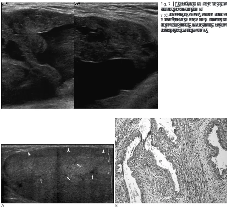
Fig. 7. 41-year-old woman with a bor- derline phyllodes tumor
US reveals a huge, circumscribed mixed-echoic mass with multiple cys- tic spaces (star). Bloody fluid was aspi- rated at the cystic portion.
방촬영술에서 악성 엽상종은 경계가 불분명한 것보다 분명한 것이 더 많으며, 초음파 영상에서도 경계성 및 악성 엽상종이 일반적으로 양성 엽상종양과 마찬가지로 난원형의 경계가 분 명하고 후방음향증가를 보이는 소엽성 고형종괴로 보인다(1, 6). 그러나 일부에서는 불규칙한 모양의 병변으로 다른 악성 유방암과의 감별이 어려운 경우도 있으며, 통계학적으로 유의 하지는 않지만 경계성 및 악성 엽상종이 양성 엽상종에 비하 여 영상학적 검사 결과에서 더 높은 범주에 속하는 경향이 있 다(1, 2). 색도플러와 엽상종양의 관계에 대하여 아직 정확하 게 보고된 바는 없으나 급작스런 크기 증가로 인해 종괴 주변
에 두드러진 정맥들이 나타나기도 하며(6), 색도플러 영상에 서 현저한 혈류 증가로 보이기도 한다(Fig. 8A, B). 종괴 내부 에 석회화가 동반될 수도 있으나(Fig. 9A, B) 이의 모양과 발 생 빈도는 적으며 악성도와 무관하다(1, 6). 최근 자기공명영 상에서 낭성 부분의 불규칙적인 벽과 T2 강조영상에서 정상 조직과 같거나 낮은 신호 강도, 그리고 낮은 현성 확산계수 (ADC: apparent diffusion coefficient) 값이 조직학적 등급과 관련이 있다는 보고가 있으나 향후 이에 대한 연구는 더 필요 할 것이다(8). 현재까지의 문헌적으로 보고된 유방 엽상종양 의 증례들에서 통계학적으로 유의한 악성과 양성을 구별하는
A B
Fig. 8. 33-year-old woman with borderline phyllodes tumor
Gray scale image reveals a circumscribed, isoechoic mass with posterior enhancement, (A) but color Doppler image shows promi- nent intratumoral vascularity (B).
A B
Fig. 9. 44-year-old woman with malignant phyllodes tumor
Cone magnification view shows a hyperdense partially obscured and spiculated mass with coarse calcifications (A). Sonography re- veals a mixed echoic mass with echogenic boundary (B). Echogenic calcifications within the mass are seen.
A B Fig. 10. 21-year-old woman with recurred benign phyllodes tumor
A 21-year-old patient visited for palpable mass in her left breast. She had taken the directional vacuum assisted biopsy (DVAB) for palpable mass and the result was benign phyllodes tumor. There was no residual mass immediate after DVAB. However, the tu- mor was palpable at the same site after 5 months later and US revealed a round shaped, indistinct margined (arrow) mass with pos- terior enhancement. Pathologic diagnoses were same as a benign phyllodes tumor at surgery.
A
B
Fig. 11. 53-year-old woman with lung metastasis
She had got a simple mastectomy for the malignant phyllodes tu- mor (A) in her left breast. After 1 year, chest radiograph (B) and CT (C) showed a large hypodense mass in Rt. lung. Right pnemonec- tomy had been done and pulmonary metastasis by breast phyl- lodes tumor was confirmed.
C
특별한 유방촬영술이나 초음파영상에서의 특징은 없다 (6).
재발 엽상종양
재발하는 경우는 불완전한 절제에 의한 국소재발이 가장 흔 하며, 국소재발율은 10%에서 60%까지 다양하나 평균적으로 15% 내외로 보고된다(2, 3, 5). 재발엽상종양의 영상소견에서 대해서는 아직 구체적으로 언급된 바가 없으나, Parker 등은 국소재발 종괴 대부분의 병리학적 소견이 본래의 종괴와 유사 하다고 보고하였다(3). 이에 따라 재발 엽상종양의 경우 원발 종양과 유사한 영상 소견을 보일 것으로 추측할 수 있겠으나 (Fig. 10A, B), 이에 대해서는 앞으로 더 연구가 되어야 할 것 이다. 최근 directional vacuum-assisted biopsy (DVAB)를 이용해 수술 대신 양성 병변을 제거하는 시술이 많아지고 있 지만, 핵생검 후 엽상종이 나온 경우에는 이 시술의 적용 대상 이 되지 아니하며 수술적 치료가 적합하다. 그러나 핵생검 후 양성병변으로 확인되어 추가적으로 병변의 제거를 목적으로 시 행한 경우에 있어서 엽상종으로 진단명이 바뀐다면, 엽상종양 은 절제 부분에서의 재발율이 높으므로 시술 후 재수술이 권 고된다(9). 재발엽상종양의 경우 원격전이나 사망률 감소와 무 관하며, 대부분 부분절제술로 치료된다.
원격전이
엽상종양의 원격전이율은 6.6%에서 70%까지 다양하나 보 통 10%로 알려져 있으며, 악성 엽상종의 경우 20%까지 원격 전이가 일어난다고 보고된다 가장 흔히 전이되는 장기로는 폐 (84.5%)와 뼈(39%)가 있으나. 이들 원격전이는 국소재발과 는 무관하며 림프절전이 없이 혈행성으로 전이된다. 드물게 양 성 엽상종의 절제 후에도 원격전이가 발생하기도 한다(2, 3).
조영증강 흉부 CT 스캔에서(Fig. 11) 비교적 균질한 저음영 의 종괴로 보인다(10).
결 론
80%이상의 유방 엽상종양들은 만져지는 유방병변으로 내원 하는 경우가 많으며, 초음파 검사와 더불어 총조직검사를 시행 하게 되는 경우가 많다. 초음파 영상에서 섬유선종 등과 같은 양성종양과 유사한 소견부터 악성 유방암을 모방하는 다양한
영상 소견들을 보일 수 있다. 병변의 크기가 크거나 임상적으 로 빨리 자라는 경우, 종괴 내부에 낭성변화가 있는 경우는 엽 상종양의 가능성을 고려해 볼 수 있다. 초음파유도하 핵생검으 로 엽상종양으로 정확히 진단될 가능성은 61-65%로 알려져 있으나 음성예측도는 96%로 낮으므로, 조직 검사 후 엽상종 으로 진단이 된다면 수술적 절제가 필요하고, 엽상종이 아닌 다른 양성 병변으로 진단되더라도 영상 소견이 의심스럽거나 크기가 큰 경우에는 수술적 절제가 권고된다. 만약 영상 소견 이 섬유선종과 같은 양성 병변과 유사하더라도 절반이상의 엽 상종이 양성병변처럼 보이므로 (7) 단기간 추적검사는 꼭 필 요하다. 만약 추적검사 시 갑자기 크기가 커진다면 엽상종양의 가능성도 고려되어야 하며 재조직검사가 요망된다.
참 고 문 헌
1. Liberman L, Bonaccio E, Hamele-Bena D, Abramson AF, Cohen MA, Dershaw DD. Benign and malignant phyllodes tumors: mam- mographic and sonographic findings. Radiology 1996;198:121-124 2. Ben Hassouna J, Damak T, Gamoudi A, Chargui R, Khomsi F,
Mahjoub S, et al. Phyllodes tumors of the breast: a case series of 106 patients. Am J Surg 2006;192:141-147
3. Parker SJ, Harries SA. Phyllodes tumours. Postgrad Med J 2001;77:428-435
4. Jacklin RK, Ridgway PF, Ziprin P, Healy V, Hadjiminas D, Darzi A. Optimising preoperative diagnosis in phyllodes tumour of the breast. J Clin Pathol 2006;59:454-459
5. Foxcroft LM, Evans EB, Porter AJ. Difficulties in the pre-operative diagnosis of phyllodes tumours of the breast: a study of 84 cases.
The Breast 2007;16:27-37
6. Stravros AT. Breast Utrasound. Philadelphia: Lippincott, Williams
& Wilkins, 2001
7. Bode MK, Rissanen T, Apaja-Sarkkinen M. Ultrasonography and core needle biopsy in the differential diagnosis of fibroadenoma and tumor phyllodes. Acta Radiol 2007;48:708-713
8. Yabuuchi H, Soeda H, Matsuo Y, Okafuji T, Eguchi T, Sakai S, et al. Phyllodes tumor of the breast: correlation between MR findings and histologic grade. Radiology 2006;241:702-709
9. Vargas HI, Vargas MP, Gonzalez K, Burla M, Khalkhali I.
Percutaneous excisional biopsy of palpable breast masses under ul- trasound visualization. Breast J 2006;12:22
10. Takahashi M, Murata K, Mori M, Kawaguchi N, Furukawa A, Kushima R, et al. Giant metastatic cystosarcoma phyllodes to the lung: CT and MR findings. Radiat Med 1992;10:210-213
J Korean Radiol Soc 2008;58:199-206
Address reprint requests to : Eun-Kyung Kim, M.D., Department of Radiology, Research Institute of Radiological Science, Yonsei University College of Medicine, 134 Sinchon-dong, Seodaemun-gu, Seoul 120-752, Korea.
Tel. 82-2-2228-7400 Fax. 82-2-393-3035 E-mail: [email protected]
Sonographic Features of Phyllodes Tumors of the Breast:
A Pictorial Review
1Seon Hyeong Choi, M.D.1,2, Eun-Kyung Kim, M.D.2, Jin Young Kwak, M.D.2, Min Jung Kim, M.D.2, Ki Kun Oh, M.D.2
1Depertment of Radiology, National Cancer Center
2Department of Radiology, Research Institute of Radiological Science, Yonsei University College of Medicine
Phyllodes tumors of the breast are rare, accounting for 0.3-0.5% of breast neoplasms. The tumors are divid- ed into benign, borderline and malignant lesions according to the histological features. Phyllodes tumors are commonly recurrent, so wide local excision is considered as the only curable treatment. Therefore, an accu- rate pre-operative diagnosis can reduce recurrence after treatment. On ultrasonography, benign phyllodes tu- mors are generally seen as well circumscribed, oval shaped, hypoechoic or isoechoic masses, and occasionally, internal clefts or cystic portions are visible. Borderline or malignant phyllodes tumors tend to be larger and more highly categorized than benign tumors. Most phyllodes tumors present as a palpable mass, which usual- ly require sonographic evaluation with a core biopsy; however, the reported diagnostic accuracy is approxi- mately 60 percent, due to limitations of the histological features. Thus, a follow-up ultrasonographic evalua- tion is essential after a core biopsy and a phyllodes tumor should be considered in cases of growth occurring after a benign biopsy.
Index words :Phyllodes Phyllodes tumor Breast
Ultrasound (US)