RAW 264.7 세포에서 투석시킨 커피 추출액의 선천면역활성화와 항염증의 동시발생
윤철수, 이석근*
강릉원주대학교 치과대학 병리학교실 및 구강과학 연구소
<Abstract>
Concurrent Innate Immunity Activation and Anti-inflammation effects of Dialyzed Coffee Extract in RAW 264.7 Cells, Murine Macrophage Lineage
Cheol Soo Yoon, Suk Keun Lee
*Department of Oral Pathology, College of Dentistry, Gangneung-Wonju National University, and Institute of Oral Science, Gangneung, Korea
Coffee ( Coffea spp.) is one of the most important agricultural commodities, being widely consumed in the world. Various beneficial health effects of coffee have been extensively investigated, but data on habitual coffee consumption and its bio-physiological effect have not been clearly explained as well as it is not proved the cause and effect between drinking coffee and its bio-physiological reactions. We made the dialyzed coffee extract (DCE), which is absorbable through gastrointestinal tract, in order to elucidate the cellular effect of whole small coffee molecules. RAW 264.7 cells, a murine macrophage lineage, were directly treated with DCE, i.e., DCE-2.5 (equivalent to 2.5 cups of coffee a day), DCE-5, and DCE-10, for 12 hours, and their protein extracts were examined by immunoprecipitation high performance liquid chromatography (IP-HPLC). RAW 264.7 cells differently expressed the inflammation-related proteins depending on the doses of DCE. RAW 264.7 cells treated with DCE showed marked increase of cathepsin C, cathepsin G, CD20, CD28, CD31, CD68, indicating the activation of innate immunity. Particularly, the macrophage biomarkers, cathepsin G, cathepsin C, CD31, and CD68 were markedly increased after DCE-5 and DCE-10 treatments, and the lymphocyte biomarkers, CD20 and CD28 were consistently increased and became marked after DCE-10 treatment. On the other hand, RAW 264.7 cells treated with DCE showed consistent increase of IL-10, an anti-inflammatory factor, but gradual decreases of different pro-inflammatory proteins including TNFα, COX-2, lysozyme, MMP-2, and MMP-3. In particular, the cellular signaling of inflammation was gradually mitigated by the reduction of TNFα, COX-2, IL-12, and M-CSF, and also the matrix inflammatory reaction was reduced by marked deceases of MMP-2, MMP-3, and lysozyme. These anti-inflammatory expressions were consistently found until DCE-10 treatment. Therefore, it is presumed that DCE may have dynamic effects of innate immunity activation and pro-inflammation suppression on RAW264.7 cells simultaneously. These effects were consistently found in the highest dose of coffee, DCE-10 (equivalent to 10 cups of coffee a day in man), that might imply the small coffee molecules were accumulated in RAW 264.7 cells after DCE-10 treatment and produce synergistic cytokine effects for innate immunity activation and anti-inflammatory reaction concurrently.
Key words : Dialyzed coffee extract, Cellular innate immunity activation, Cellular and matrix anti-inflammatory reaction
Korean Journal of Oral and Maxillofacial Pathology 2017;41(3):121-129 ISSN:1225-1577(Print); 2384-0900(Online) Available online at http://journal.kaomp.org https://doi.org/10.17779/KAOMP.2017.41.3.003
* Correspondence: Suk Keun Lee, Department of Oral Pathology, College of Dentistry, Gangneung-Wonju National University, 123 Chibyun-dong, Gangneung, 210-702, Korea
Tel: +82-33-640-2228, Fax: +82-33-642-6410 E-mail: [email protected]
Ⅰ. INTRODUCTION
If you have a daily coffee habit, here’s somethings for
you to know why the coffee consumption is associated with a lower risk of cardiovascular disease, diabetes, neurologic disease such as Parkinson’s1-5). As the coffee bean itself is loaded with many different nutrients and phytochemicals, many researchers explained these effects that some compounds in coffee such as lignans, quinides and magnesium may help to reduce insulin resistance and inflammation6,7). However, there has not been clearly proved the cause and effect between drinking coffee and its potential benefits. There are still a lot of unknowns as to what may explain the benefits. For the biological investigation to know the anti-inflammatory mechanism of coffee effects, we performed this study by using the RAW 264.7 cells treated with dialyzed coffee extract (DCE) similar to the previous study8,9). And the protein expression changes of RAW 264.7 cells were assessed depending on the dose of DCE through precision protein expression method, immunoprecipitation high performance chromatography (IP-HPLC).
As the accuracy of IP-HPLC has been improved to detect the protein expression level within 5% error range10), the molecular signaling mechanism of DCE effects in RAW 264.7 cells could be assessed and explained in this study.
As we’ve previously reported, the DCE induced remarkable changes in the expression of proliferation-related proteins in dose dependent manner9). This dose dependent modality of DCE in protein expression may be caused by the biochemical properties of DCE elements, including caffeine, chlorogenic acid, etc., which are small organic compounds interacting with various cellular substances.
This study was carried out to make plain the expression changes of inflammation-related proteins by different doses of DCE in RAW 264.7 cells and also to evaluate their molecular signaling for the regulation of inflammatory reaction. The immortalized murine macrophages, RAW 264.7 cells which were not stimulated by bacterial antigen (lipopolysaccharides) were utilized to know the real DCE
effect in cells. The resulted data of protein expression changes were discussed with the review of the literatures.
Ⅱ. MATERIALS and METHODS
Dialyzed coffee extract (DCE) production and treatment
The coffee beans (Coffea arabica L.) were roasted and treated with hot water to get the ordinary coffee drink, followed by the dialysis using cellulose bag (131492, Spectra/Por, Spectrum, CA, USA) which filtrates small molecules less than 1000 Da as done in the previous study8). Different doses of dialyzed coffee extract (DCE) equivalent to 2.5, 5, and 10 cups of coffee (DCE-2.5, DCE-5, and DCE-10, respectively) were separately treated in RAW 264.7 cell culture.
RAW cell culture treatment with dialyzed coffee extract (DCE)
RAW 264.7 cells (ATCC, USA), murine macrophage cell line, were cultured in Dulbecco’s modified Eagle’s medium (WelGene Inc. Korea) supplemented with 10% (vol/vol) heat-inactivated fetal bovine serum (WelGene Inc. Korea), 100 unit/mL penicillin, 100μg/mL streptomycin, 250ng/mL amphotericin B (WelGene Inc. Korea), at 5% CO2, 37.5°C.
The cells were tested for mycoplasma on a regular basis to ensure that only mycoplasma-free cell lines were studied in the assays.
Immunoprecipitation HPLC analysis for the protein extract obtained from RAW 264.7 cell culture
After 12 hours culture of RAW 264.7 cells treated with dialyzed coffee extract (DCE) equivalent to 2.5, 5, and 10 cups of coffee, DCE-2.5, DCE-5, and DCE-10, respectively, the RAW 264.7 cells were harvested with protein lysis buffer
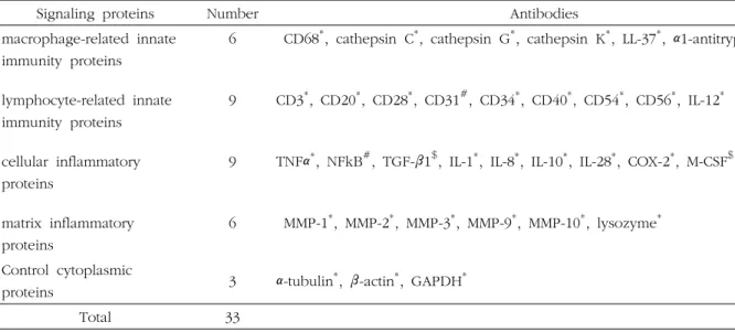
Table 1. Antibodies used in this study.
Signaling proteins Number Antibodies
macrophage-related innate immunity proteins
lymphocyte-related innate immunity proteins cellular inflammatory proteins
matrix inflammatory proteins
6
9
9
6
CD68
*, cathepsin C
*, cathepsin G
*, cathepsin K
*, LL-37
*, α1-antitrypsin
#CD3
*, CD20
*, CD28
*, CD31
#, CD34
*, CD40
*, CD54
*, CD56
*, IL-12
*TNFα
*, NFkB
#, TGF-β1
$, IL-1
*, IL-8
*, IL-10
*, IL-28
*, COX-2
*, M-CSF
$MMP-1
*, MMP-2
*, MMP-3
*, MMP-9
*, MMP-10
*, lysozyme
*Control cytoplasmic
proteins 3 α-tubulin
*, β-actin
*, GAPDH
*Total 33
* Santa Cruz Biotechnology, USA; # DAKO, Denmark; $ Neomarkers, CA, USA; @ ZYMED, CA, USA
Abbreviation: α1-AT; α1-antitrypsin, COX-2; cyclooxygenase-2, GAPDH; glyceraldehyde 3-phosphate dehydrogenase, IL-1; interleukin-1, M-CSF; macrophage colony stimulating factor, MMP-1; matrix metalloprotease-1, NFkB; nuclear factor kappa light chain enhancer of activated B cells, TGF-β1; transforming growth factor-β1, TNF-α; tumor necrosis factor-α.
(0.3% SDS, 50 mM Tris-HCl pH 8.0, 0.3% β-mercaptoethanol, 1 mM PMSF, 1 mM EDTA) containing protein inhibitor cocktail (Sigma, USA). Then, the protein extracts were preserved in -70°C deep freezer to prevent further protein degradation11).
100 μg of protein extract was applied to each immunoprecipitation procedure using protein A/G agarose column (Amicogen, Korea). The protein A/G agarose columns were separately pre-incubated with 1μg of each 33 different antisera (Table 1). Briefly, the protein samples were mixed with 5 mL binding buffer (150 mM NaCl, 10 mM Tris pH 7.4, 1 mM EDTA, 1 mM EGTA, 0.2 mM sodium vanadate, 0.2 mM PMSF and 0.5% NP-40), and incubated in the protein A/G agarose columns at 10°C for 1 hour. The columns were placed on the rotating stirrer during the incubation time.
After washing each column with sufficient amount of PBS solution (pH 7.3, 137 mM NaCl, 2.7 mM KCl, 43 mM Na2HPO4-7H2O and 1.4 mM KH2PO4), the target protein was eluted with 150 μL IgG elution buffer (Pierce, USA).
The immunoprecipitated proteins were analyzed by HPLC
(1100 series, Agilent, USA) equipped with a reverse phase column and micro-analytical detector system (SG Highteco, Korea). Elution was performed using 0.15M NaCl, 20%
acetonitrile solution at 0.4 mL/min for 30 min, and detection by UV spectroscopy at 280 nm. Control and experimental samples were run sequentially to allow comparisons. For IP-HPLC, whole protein peak areas (mAU*s) were calculated by subtracting antibody peak areas of the negative control, and square root value was calculated to normalize in the GMS unit. The proportional values (%) compared between the experimental and control groups were plotted, and the analysis was repeated two to six times until mean errors were
≤±5%. Results were analyzed using the Chi-squared test12). The expressions of cytoplasmic control proteins, i.e., β -actin, α-tubulin, and GAPDH were relatively unchanged (≤
5%) by DCE-2.5, 5, or 10 treatment. Protein expression changes were defined as non-significant for a change of evaluated as ≤±5%, slight for ±5-10 %, marked for
±10-20%, and great for ≥±20%.
Fig. 1. IP-HPCL analysis for cellular innate immunity-related proteins in RAW 264.7 cells treated with DCE. A1 and A2: A bar and line graphs, respectively, plotted with same data. The protein expressions for macrophage-related innate immunity were dramatically increased by DCE-2.5 and DCE-5 treatments but became lesser by DCE-10 treatment. B1 and B2: A bar and line graphs, respectively, plotted with same data. The protein expressions for lymphocyte-related innate immunity were dramatically increased by DCE-2.5 and DCE-5 treatments and also consistently increased by DCE-10 treatment.
Ⅲ. RESULTS
RAW 264.7 cells treated with DCE showed remarkable expression changes of inflammation-related proteins, implicative for the beneficial effect of coffee such as innate immunity activation and anti-inflammatory reaction. These protein expression changes were characteristic depending on the doses of DCE-2.5, DCE-5, and DCE-10, which were
equivalent 2.5, 5, 10 cups of coffee a day, respectively.
Innate immunity activation relevant to macrophages and lymphocytes
RAW 264.7 cells treated with DCE expressed significant increases of cathepsin C, cathepsin G, CD20, CD28, CD31, and CD68 compared to the control. The macrophage biomarkers, i.e., cathepsin G, cathepsin C, CD31, CD68, α
Fig. 2. IP-HPCL analysis for inflammatory proteins in RAW 264.7 cells treated with DCE. A1 and A2: A bar and line graphs, respectively, plotted with same data. The cellular inflammatory protein expressions were gradually decreased by DCE treatments, while an anti-inflammatory factor, IL-10 was consistently increased. B1 and B2: A bar and line graphs, respectively, plotted with same data. The matrix inflammatory protein expressions were consistently decreased by DCE treatments, while the expression of MMP-9 was markedly increased by DCE-2.5 treatment but reduced by DCE-5 and DCE-10 treatments.
1-antitrypsin, and LL-37, were markedly expressed in DCE-5 treatment, but their expressions became lesser in DCE-10 treatment. Particularly, the cathepsin G, a macrophage maturation factor, was overexpressed in the cells treated with DCE-5 and DCE-10, and the expression of macrophage protecting factor, α1-antitrypsin, was slightly increased by the DCE treatments (Fig. 1A).
The expressions of lymphocyte biomarkers, i.e., CD3,
CD20, CD28, CD34, CD40, CD54, CD56, and IL-12, were consistently increased by the DCE treatments. Among them, a T cell stimulating factor, IL-12 was overexpressed by DCE-2.5 treatment, and B and T cell activating factors, CD20 and CD28, respectively, were markedly expressed by DCE-5 and DCE-10 treatments (Fig. 1B).
The innate immunity-related proteins working at macrophages (cathepsin C, cathepsin G, and CD68) and
signaling to lymphocytes (CD3, CD20, CD28, CD34, CD40, CD54, CD56, and IL-12) gradually increased depending on the doses of DCE-2.5, DCE-5, and DCE-10, and became markedly expressed by DCE-10 treatment (Fig. 1).
Decrease of pro-inflammatory protein expressions both in the cells and matrix
RAW 264.7 cells treated with DCE-2.5, DCE-5, and DCE-10 consistently showed the increase of IL-10, an anti-inflammatory factor, but gradually showed the decreases of TNFα, TGF-β1, IL-1, IL-6, IL-8, IL-10, IL-28, M-CSF, and COX-2, which were essential for the cellular inflammatory signaling. Among them, the expressions of TNFα, IL-6, M-CSF, and COX-2 were markedly decreased by DCE-5 and DCE-10 treatments (Fig. 2A).
The matrix inflammatory proteins, MMP-1, MMP-2, MMP-3, MMP-9, MMP-10, and lysozyme were gradually decreased by DCE-2.5, DCE-5, and DCE-10 treatments, and became markedly decreased by DCE-10 treatment compared to the control. Although the expressions of MMP-9 and MMP-10 were slightly and markedly increased by DCE-2.5 treatment, the potent extracellular proteinases, MMP-2, MMP-3, and lysozyme were markedly decreased by the DCE-5 and DCE-10 treatment (Fig. 2B).
The pro-inflammatory proteins working in macrophages (TNFα, TGF-β1, IL-1, IL-6, IL-8, IL-10, IL-28, M-CSF, and COX-2) and in extracellular matrix (MMP-1, MMP-2, MMP-3, MMP-9, MMP-10, and lysozyme) were gradually down-regulated depending on the doses of DCE-2.5, DCE-5, and DCE-10, and markedly decreased by DCE-10 treatment (Fig. 2).
Ⅳ. DISCUSSION
In the present study, DCE may have different small
molecules including polyphenols, i.e., caffeine, chlorogenic acids, diterpenes (kahweol and cafestol), melanoidins, and trigonelline, low molecular mass arabinogalactan-protein (AGP), and 3-methyl-1,2-cyclopentanedione (3-MCP), which can produce anti-inflammatory effects as identified by many authors13-16). The present study also found the anti- inflmmatory effect of DCE in RAW 264.7 cells through IP-HPLC analysis for different inflammatory protein expressions. The pro-inflammatory signaling of cellular inflammation was markedly reduced by the decreases of TNFα, NFkB, IL-6, IL-28, COX-2, and M-CSF. Particularly, the inflammation inhibitory factor, IL-10 was markedly increased until DCE-10 treatment. The anti-inflammatory effect of DCE was gradually increased depending on the doses of DCE, and became strongest in DCE-10. Therefore, it was supposed that the pro-inflammatory signaling between antigen presenting cells and leukocytes would be more reduced by DCE-10 treatment than DCE-2.5 and DCE-5 treatments. These results are a little different from the previous notion that heavy coffee drink may hamper the beneficial coffee effects found in 3-5 cups coffee drink a day, however, in this study it was found that RAW 264.7 continuously showed the decreased expressions of pro-inflammatory proteins after DCE-10 treatment, which is equivalant to 10 cups coffee drink common in heavy coffee consumer.
The reduced cellular inflammatory signaling in RAW 264.7 cells concurred with the decreases of matrix inflammatory proteins, MMP-2, MMP-3, and lysozyme after DCE treatments. Generally, most of matrix-assoicated inflammatory proteins were down-regulated after DCE treatments, these expressions were continuous until DCE-10 treatment. As the anti-inflammatory role of coffee is one of the most important beneficial effects of coffee6,17), the present study further explored the DCE-based anti-inflammatory effect in dose-dependent manner. And
the result showed that the dose of heavy coffee consumer, DCE-10 (about 10 cups of coffee a day) is continuously able to induce the anti-inflammatory effect both in the cells and the matrices. These findings were indirectly supported by the fact that the long-time heavy coffee consumers were more resistent to the chronic inflammatory diseases, i.e., hepatitis, etc.18,19). It was also known that caffeine and chlorogenic acid are key elements of coffee and induce the anti-inflammatory activity and ventricular remodeling after myocardial ischemia20,21). In the in vitro experiment using lipopolysaccharide (LPS)-stimulated RAW 264.7 cells, caffeine suppressed LPS-induced inflammatory responses by regulating NFkB activation and MAPK phosphorylation22-24). However, the more precise investigation may be required to elucidate the pharmacological effects of caffeine and chlorogenic acid, which are most abundant in coffee and can be accumulated in the body of heavy coffee consumer.
RAW 264.7 cells treated with DCE expressed significant increases of cathepsin C, cathepsin G, CD20, CD28, CD31, CD68, CD3, CD20, CD28, CD34, CD40, CD54, CD56, and IL-12, compared to the control, indicating the activation of both macrophages and lymphocytes. The innate immunity- related proteins working at macrophages (cathepsin C, cathepsin G, and CD68) and signaling to lymphocytes (CD3, CD20, CD28, CD34, CD40, CD54, CD56, and IL-12) gradually increased depending on the doses of DCE-2.5, DCE-5, and DCE-10, and became markedly expressed by DCE-10 treatment. Therefore, it presumed that DCE was effective to induce the innate immunity affecting both macrophages and lymphocytes. This fact is identical to the previous reports demonstrating many clinical experiences and in vivo and in vitro experiments13,15,25,26)
. However, it is thought that the cellular effect of DCE, innate immunity activation is important for the treatment of debilitating chronic inflammatory diseases, cancers, immuno-deficiency diseases, etc15,19,21,25,27-29).
In the present study, RAW 264.7 cells derived from murine macrophages were readily responsible to DCE and induced different signaling for cellular innate immunity and anti-inflammatory reaction concurrently. As the increased innate immunity usually counteracts the anti-inflammatory reaction by accelerating the inflammation, the concurrent innate immunity activation and anti-inflammatory effects may be a characteristic pharmacological property of small coffee molecules in DCE. Additionally, these inflammatory reactions of coffee were consistently found after DCE-10 treatment which was equivalent to 10 cups coffee a day common in heavy coffee consumer, therefore, it was presumed that the small coffee molecules were accumulated in RAW 264.7 cells after DCE-10 treatment and produced synergistic cytokine effects for innate immunity activation and anti-inflammatory reaction concurrently.
ACKNOWLEDGMENTS
This study was supported by a Grant df Korea Health Technology R&D Project, Ministry of Health and welfare, and Republic of Korea (HI15C0689).