ISSN 0378-6471 (Print)⋅ISSN 2092-9374 (Online)
https://doi.org/10.3341/jkos.2019.60.7.685
Original Article
간헐외사시에서 비디오안구운동검사를 통한 주시안과 비주시안의 안구편위에 대한 분석
The Analysis of Ocular Deviations between Dominant and Non-dominant Eye Using Video-oculography in Intermittent Exotropia
반지훈1,2⋅문성혁1,2
Ji Hoon Ban, MD1,2, Sung Hyuk Moon, MD, PhD1,2
인제대학교 의과대학 부산백병원 안과학교실1, 인제대학교 의과대학 부산백병원 안과질환 T2B 기반구축센터2 Department of Ophthalmology, Busan Paik Hospital, Inje University College of Medicine1, Busan, Korea T2B Infrastructure Center for Ocular Disease, Busan Paik Hospital, Inje University College of Medicine2, Busan, Korea
Purpose: To measure and analyze ocular deviations between dominant and non-dominant eyes using video-oculography (VOG) in intermittent exotropia.
Methods: Fourteen subjects who were diagnosed with intermittent exotropia from July 2017 to July 2018 with age of 5 or more, visual acuity of 20/30 or better and corrected visual acuity of 20/25 or more and difference in vision of both eyes of 1 line or less on Snellen optotype were included. The subjects were asked to fixate on a black-on-white optotype at 1 m, which subtended a visual angle of 50 minutes of arc. The video files and data about ocular deviations were obtained using VOG with alternate cover test. We analyzed angles of ocular deviations in dominant and non-dominant eyes.
Results: Among the 14 subjects in this study, the mean age were 7.6 ± 1.7 (range 5-9 years). Seven of 14 subjects had the right eye dominance. Six of the 14 subjects were men. There was no significant difference of ocular deviations between the dominant and non-dominant eyes in VOG (p = 0.167). Additionally, there was no significant difference of the values of VOG when one eye was exodeviated or re-fixated (p = 0.244), when both eyes were deviated, and when both eyes were re-fixated (p = 0.195, 0.637).
Conclusions: In this study, there was no significant difference of ocular deviations between the dominant and non-dominant eyes, between when an eye was exodeviated or fixated using VOG. Therefore, it may not be a problem even if alternate prism cover test is performed in any eye in intermittent exotropia of more than 50 prism diopter without amblyopia or refraction abnor- mality that could affect the uncorrected visual acuity.
J Korean Ophthalmol Soc 2019;60(7):685-691
Keywords: Intermittent exotropia, Ocular deviations, Strabismus, Video-oculography
■Received: 2019. 2. 13. ■ Revised: 2019. 3. 12.
■Accepted: 2019. 6. 19.
■Address reprint requests to Sung Hyuk Moon, MD, PhD Department of Ophthalmology, Inje University Busan Paik Hospital, #75 Bokji-ro, Busanjin-gu, Busan 47392, Korea Tel: 82-51-890-6016, Fax: 82-51-890-6329
E-mail: [email protected]
*Conflicts of Interest: The authors have no conflicts to disclose.
ⓒ2019 The Korean Ophthalmological Society
This is an Open Access article distributed under the terms of the Creative Commons Attribution Non-Commercial License (http://creativecommons.org/licenses/by-nc/3.0/) which permits unrestricted non-commercial use, distribution, and reproduction in any medium, provided the original work is properly cited.
외편위는 내편위보다 잠복성이나 간헐성으로 나타나는 경우가 많아서 같은 환자라도 검사할 때마다 불변외사시, 간헐외사시, 외사위 등으로 다양하게 나타나기도 한다. 동 양인에서 외사시가 과반수 이상을 차지하고 있으며, 이 중 간헐외사시는 국내에서 가장 흔하게 관찰되는 사시의 형태 이다.1,2 사시환자에서 주시안은 정확한 목표물을 주시하지 만, 비주시안은 목표물 없이 주변부를 주시하기 때문에 불 안정하다.3 많은 연구에서 사시환자의 사시각이 다양한 것 에 대해 보고하였으며, 이는 검사자의 기술, 안구편위를 측
A B
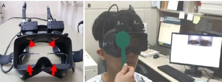
Figure 1. A video-oculography (VOG) (accuracy: 0.1 degree). (A) Two cameras installed at the top can make measurements of both
eye movements (red arrowheads). A tilted semi-transparent glass allows subjects to fixate the target (red arrows). (B) The subjects wearing the VOG goggles were asked to fixate a target point between the two eyes and were instructed to keep the head straight so that the eyes were in primary position during the examination.정하는 방법, 인구학적인 특성 및 실제 사시각의 다양성과 관련이 있다.4-9
사시환자에서 안구편위를 측정하는 방법으로 허쉬버그 검사, 크림스키검사 및 프리즘교대가림검사가 있으며, 안구 운동을 기록하고 분석하는 방법으로 안구운동사진, 공막탐 지코일법 및 비디오안구운동검사가 있다.10-12 비디오안구운 동검사(video-oculography, VOG)를 이용하여 교대가림검 사를 통해 얻어진 측정값과 프리즘교대가림검사를 이용하 여 구한 사시각이 유의하게 일치한다고 확인되었다.13
Economides et al14은 비디오안구운동검사를 이용한 연구 에서 사시가 있는 눈이 정위인 눈보다 안구 위치가 더 가변 적이며, 사시환자의 비주시안이 주시안보다 안구 위치가 가변적이라고 보고하였다. 하지만 간헐외사시환자에서 주 시안과 비주시안의 안구편위의 차이가 있는지, 안구 운동 에 따라 사시각의 변동이 있는지에 대한 보고는 없다. 따라 서 이번 연구에서 간헐외사시환자에서 비디오안구운동검 사를 이용하여 교대가림 동안 주시안과 비주시안의 안구편 위의 변화를 측정하고 그 차이를 알아보고자 한다.
대상과 방법
2017년 7월부터 2018년 7월까지 본원 안과에서 간헐외 사시로 진단받은 환자를 대상으로 전향적으로 조사하였다.
만 5세 이상이며, 스넬렌시력표를 기준으로 나안시력 20/30 이상, 교정시력 20/25 이상인 환자만 포함하였으며, 사시를 제외한 안과적 질환이 있는 경우, 신경학적 이상을 포함한 전신 이상이 있는 경우, 이전에 사시수술을 시행한 경우,
-2.00디옵터(diopter, D) 이상의 근시 혹은 +2.00D 이상의 원시 및 난시 도수가 ±2.5D 이상인 경우, 50프리즘디옵터 (prism diopter, PD) 이상의 사시각을 가진 경우, 양안의 시 력차가 2줄 이상이거나 spherical equivalent 차이가 2 이상 인 경우 및 교대주시하는 경우는 제외하였다. 또한 비디오 안구운동검사에 협조도가 떨어지거나, 검사 후 측정값이 부 정확한 대상은 제외하였다. 본 연구는 인제대학교 부산백 병원 임상연구관리규정과 헬싱키선언을 준수하였고, 모든 환자에서 본 연구 및 검사에 대해 충분히 설명을 한 뒤 서 면 동의서를 받았다(인제대학교 부산백병원 임상시험윤리 위원회 승인번호: 2016-08-0028).
비디오안구운동 장치(SLVNG, SLMED, Seoul, Korea)는 시축의 수직 방향으로 윗부분에 2개의 카메라가 각각의 눈 을 측정하도록 설치되어 있으며, 이를 통해 두 눈의 움직임 을 측정할 수 있다(Fig. 1A). 내장되어 있는 실시간 시선 추 적 체계는 지속적으로 동공을 탐색하고, 동공의 중심을 기 준점으로 인식한다. 안구의 움직임은 기준점의 변화에 맞 춰 비디오 파일로 기록되어 연동되어 있는 컴퓨터에 저장 된다. 비디오안구운동 측정은 본원의 비디오안구운동검사 실에서 진행되었으며, 스넬렌시력표로 20/200, 50분 시각에 해당하는 1 m 거리의 전자 시표를 주시하게 한 상태에서 진행되었다(Fig. 1B). 먼저, 비디오안구운동 장비를 착용한 상태로 대상자를 5초간 전자 시표를 주시하게 하여 안구정 렬이 정위가 되도록 하여 칼리브레이션(calibration)을 시행 하였다. 이후 우안을 5초간 가림 검사를 시행한 후 5초간 안가림 상태를 유지하였고, 좌안도 우안과 동일한 방법으 로 시행하였다. 5초간 휴식기를 둔 뒤 3초 간격으로 교대가
A1 A2 B1 B2 C1 C2
Figure 3. The section of alternative cover test of a patient who has the right-dominant eye. (A) Differences of ocular deviations be-
tween the dominant and non-dominant eye at the same section. Six values (A-C) of the non-dominant eye with values of the dominant eye in the same section. We compared LA1 with RA1, LA2 with RA2, LB1 with RB1, LB2 with RB2, LC1 with RC1, and LC2 with RC2. (B) Differences in values when an eye is deviated or re-fixated. In the right eye, RA1 and RA2, RB1 and RB2, RC1 and RC2.In the left eye, LA1 and LA2, LB1 and LB2, LC1 and LC2. (C) Differences in values when the non-dominant eye is deviated and when the dominant eye is deviated. Applied when the non-dominant eye and the dominant eye are re-fixated. Re-fixation: RA2 and LA1, RB2 and LB1, RC2 and LC1, deviation: RA1 and LA2, RB1 and LB2, RC1 and LC2. ‘RA1’ is defined as the values of ‘A1’
section in the right eye.
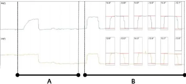
Figure 2. The graphs of video-oculography in the right-dominant eye. (A) is section of cover-uncover test in both eyes, (B) is section
of alternative cover test. Initial binocular alignment was verified with both eyes open during the first 5 seconds. Subsequently, each eye was allowed 5 seconds of covered time and 5 seconds of uncovered time, and then the alternate cover test was repeated 3 times, with each eye being covered for 3 seconds.림검사를 5회 반복하여 시행하였다(Fig. 2). 대상자가 눈을 깜박이거나 다른 곳을 주시하는 등의 오차가 발생할 경우 5회 이상의 안정된 결과가 나올 때까지 교대가림검사를 반 복하여 시행하였다.
비디오안구운동검사를 통해 얻은 자료를 이용하여 3가지
결과값을 도출하였다(Fig. 3). 안구편위는 안구 변위 전 1초 간의 값들 중 중간값과 안구 변위 후 1초간의 값들 중 중간 값의 차이로 측정하였다. 먼저, 비주시안과 주시안의 안구 편위를 구하기 위해 교대가림검사 동안 외편위-재주시 혹 은 재주시-외편위되는 6개의 구간을 선별해서 외편위와 재
Baseline characteristic Value (n = 14)
Sex (male/female) 6/8
Age (years) 7.6 ± 1.7
Non-dominant eye (OD/OS) 7/7
Spherical equivalent (D) 0.09 ± 0.76 Angle of deviation at 1 m (PD) 27.64 ± 6.10
Stereopsis (sec) 64.29 ± 29.01
Values are presented as mean ± standard deviation unless other- wise indicated.
OD = oculus dexter; OS = oculus sinister; D = diopter; PD = prism diopter; Sec = seconds of arc.
Table 1. Baseline characteristics of subjects
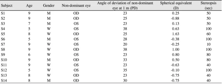
Subject Age Gender Non-dominant eye Angle of deviation of non-dominant eye at 1 m (PD)
Spherical equivalent (D)
Stereopsis (sec)
S1 9 M OD 23 0.25 50
S2 9 M OD 25 -0.88 50
S3 7 M OS 23 0.13 50
S4 5 W OS 38 0.63 100
S5 8 W OD 25 1.63 60
S6 5 M OS 28 -0.38 100
S7 9 W OS 20 -0.25 10
S8 9 W OD 38 1.00 100
S9 6 W OS 23 0.80 80
S10 9 M OD 33 0.50 80
S11 9 W OS 23 -0.63 40
S12 5 W OS 35 -0.10 100
S13 8 W OD 23 -0.75 40
S14 8 M OD 30 -0.75 40
PD = prism diopter; D = diopter; sec = seconds of arc; M = man; OD = oculus dexter; OS = oculus sinister; W = woman.
Table 2. Demographics and clinical characteristics of each participant
주시 측정값을 각각 3개씩 도출하였다. 교대가림검사를 1회 시행하였을 때 단안에서 외편위-재주시-외편위되는 과정을 한 개의 사이클로 정하였다. 또한 우안이 외편위-재주시된 다면 좌안은 재주시-외편위를 하게 되며, 이 과정을 한 개 의 구간이라고 설정하였다. 같은 구간에서 주시안과 비주 시안의 안구 변화를 비교 분석하였다(Fig. 3A). 단안의 안 구편위를 분석하기 위해서 한 사이클에서 단안의 외편위와 재주시를 비교하였다(Fig. 3B). 또한 한 사이클 내에서 주 시안과 비주시안의 외편위(RA1-LA2, RB1-LB2, RC1-LC2) 와 재주시(RA2-LA1, RB2-LB1, RC2-LC1)를 추가적으로 비교 분석하였다(Fig. 3C). 측정값은 도(°) 단위로 나타내었다.사시각 측정은 비디오안구운동검사 후 그 자리에서 같 은 검사자가 시행하였으며, 플라스틱 프리즘 세트(Luneau Technology France, Pont-de-l'Arche, France)를 이용하여 측정하였다. 프리즘은 재주시의 방향이 바뀔 때까지 점차 적으로 증량하고 재주시 움직임이 보이지 않을 때 감량하 였다. 사시각은 재주시의 방향이 바뀌는 프리즘의 중간값
혹은 편위가 중화되는 값으로 간주하였다.
사시각 및 안구편위에 영향을 줄 수 있는 인자로 성별, 나이, 구면렌즈 대응치, 입체시 등을 조사하여 분석하였다.
통계분석은 SPSS statistics 24 (IBM Corp., Armonk, NY, USA)을 이용하여 paired t-test를 시행하였으며, p값이 0.05 미만인 경우를 통계적으로 유의하다고 정의하였다.
결 과
14명의 연구 참여자의 평균 나이는 7.6 ± 1.7세(5-9)였으 며, 7명은 우안이 주시안이었고, 6명이 남자였다. 비주시안 의 평균 1 m 거리의 사시각은 27.64 ± 6.10PD (20-38)였으 며, 입체시는 64.29 ± 29.01 seconds of arc (10-100)이었다 (Table 1, 2). 비디오안구운동검사를 이용하여 모든 구간에 서 주시안과 비주시안의 안구편위의 변화를 보았을 때, 평 균 14.42 ± 3.52°, 14.19 ± 3.67°로 통계학적으로 유의한 차 이를 보이지 않았다(p=0.167). 외편위될 때 주시안과 비주 시안의 평균은 각각 14.34 ± 3.38°, 14.01 ± 3.66°, 재주시될 때 주시안과 비주시안의 평균은 각각 14.50 ± 3.70°, 14.38
± 3.72°로 통계학적으로 유의한 차이가 없었다(p=0.195, 0.637) (Table 3). 단안이 외편위될 때 측정값과 재주시될 때 측정 값은 주시안에서 14.34 ± 3.38, 14.50 ± 3.70, 비주시안에서 14.01 ± 3.66, 14.38 ± 3.72로 유의한 차이가 없었다(p=0.088, 0.244) (Table 4).
비디오안구운동검사를 통해 분석한 84개 구간 중 13개 구간(15%)에서 양안 측정값의 차이가 2° 이상이었고, 14명 중 6명이 최소 한 구간에서 2° 이상 차이를 보였다. 2° 이상 차이를 보인 6명 중 2명(14%)은 6개 구간 중 4개 구간에서
Dominant eye Non-dominant eye p-value*
Ocular deviations in the same section (°) 14.42 ± 3.52 14.19 ± 3.67 0.167
Exodeviation in the same cycle (°) 14.34 ± 3.38 14.01 ± 3.66 0.195
Re-fixation in the same cycle (°) 14.50 ± 3.70 14.38 ± 3.72 0.637
Values presented as mean ± standard deviation.
*Paired t-test.
Table 3. Comparison of ocular deviations between the dominant and non-dominant eyes
Exodeviation Re-fixation p-value* Dominant eye (°) 14.34 ± 3.38 14.50 ± 3.70 0.454 Non-dominant eye (°) 14.01 ± 3.66 14.38 ± 3.72 0.088 Total (°) 14.22 ± 3.61 14.39 ± 3.58 0.244 Values presented as mean ± standard deviation.
*Paired t-test.
Table 4. Comparison of ocular deviations of an eye when the
eye is deviated or re-fixated양안 측정값이 2° 이상 차이를 보였고, 6개 구간의 평균은 각각 2.0, 2.2였다. 나머지 4명은 1개의 구간(3명) 혹은 2개 의 구간(1명)에서 2° 이상의 차이를 보였다.
고 찰
사시환자에서 안구편위를 측정하는 방법 중 현재 임상적 으로 사시각을 측정하기 위해 프리즘교대가림검사를 주로 사용하고 있으며, 측정하는 검사자의 숙련도 및 환자의 협 조 정도에 따라 오차가 발생할 수 있다.4,5,15 한편, 비디오안 구운동검사는 검사자의 숙련도에 영향을 적게 받고, 동시 에 양안의 안구편위의 변화를 측정할 수 있으며, 비디오 파 일로 기록을 남길 수 있는 유용한 검사이다.13
간헐외사시는 안구와 주변 조직 및 외안근과 관련된 해 부학적, 기계적인 요인 및 신경분포적인 요인 및 여러 가지 요인이 복합적으로 작용하여 영향을 미치는 것으로 알려져
있으며,16-18 눈벌림 중추에서 신경분포의 불균형, 외안근 풀
리의 위치로 인해 양안의 배열 이상, 두뇌 피질의 활동이 영향을 준다는 연구가 있다.19-21 Li et al21은 두뇌자기공명 영상을 통해 두뇌의 여러 부분에서 복잡한 두뇌신경망을 형성하여 양안의 융합에 관여하며, 특히 상두 및 하두정소 엽의 피질 활동이 간헐외사시 양안의 융합 이상에 관여한 다고 보고하였다. 본 연구에서 주시안과 비주시안에서 안 구편위의 변화량이 차이가 없는 것으로 보아 눈벌림 중추 및 두뇌 피질의 활동이 간헐외사시에 관여한다는 가설에 설득력이 있는 것으로 생각된다.
Economides et al14와 Ghasia et al22는 비디오안구운동검 사를 이용한 연구에서 사시환자의 편위안이 주시가 불안정 하다고 보고하였으며, Economides et al23는 간헐외사시환
자의 눈이 편위되었을 때, 더 강하게 주시하려는 경향을 보 이며, 이는 우안을 가리거나 좌안을 가릴 때 차이는 없다고 보고하였다. 본 연구에서 편위안이 주시가 불안정하기 때 문에 사시수술량의 결정에 필요한 사시각이 두 눈에서 다 를 수 있다는 가정을 하였으나, 비디오안구운동검사를 이 용하여 주시안과 비주시안의 안구편위 변화를 비교했을 때 유의한 차이가 없었다. 또한 안구가 편위되거나 재주시될 때 유의한 차이가 없었다. 따라서 사시각을 측정할 때 편위 되거나 재주시되는 방향과 관계없이 어느 눈에 측정하여도 문제가 되지 않을 것으로 보인다.
본 연구에서 비디오안구운동검사를 통해 분석한 84개 구 간 중 13개 구간에서 양안 측정값의 차이가 2° 이상이었고, 14명 중 6명이 최소 한 구간에서 2° 이상의 차이를 보였다.
비디오안구운동검사는 잠재적인 위치 오차를 ±1°로 가지고 있으므로, 양안 측정값이 2° 이상 차이가 나면 유의한 차이 가 있다고 생각할 수 있다.23 하지만 6명 중 2명(14%)만 4개 구간에서 양안 측정값이 2° 이상 차이를 보였고, 6개 구간 의 평균은 각각 2.0, 2.2였다. 나머지 4명은 2개의 구간 이 하에서만 차이를 보였고 양안 측정값의 차이의 평균은 2° 미 만이었다. 따라서 대부분에서 양안 측정값의 차이가 2° 미 만이며, 전체 구간에서 통계학적으로 양안 측정값이 유의 한 차이를 보이지 않았으므로 비디오안구운동검사를 이용 하여 분석하였을 때 양안 측정값의 차이가 보이지 않았다 고 볼 수 있다.
본 연구에서 몇 가지 제한점이 있었다. 첫째, 연구에 사 용된 비디오안구운동검사는 나안 상태에서만 시행 가능하 므로 안경을 끼는 대상자가 안경을 끼지 않고 검사를 시행 하였다. 스넬렌시력표를 기준으로 나안시력 20/30 이상, 교 정시력 20/25 이상만 포함하고, 2.00D 이상의 근시 혹은 원 시, 2.50D 이상의 난시인 군을 배제하여 이러한 한계를 극 복하려고 하였다. 둘째, 환자의 협조도가 떨어질 때 결과값 에서 발생하는 오차를 완전히 극복하기 어려웠다. 만 5세 미만의 환아 및 비디오안구운동검사에 협조도가 떨어지거 나, 검사 후 측정값이 부정확한 경우는 배제하였으나, 검사 중 눈 깜빡임, 신속운동, 주시의 불안정성으로 인한 오차는 고려하지 못했다. 마지막으로 연구에 참여한 개체 수가 적 었다. 14명의 간헐외사시환자를 대상으로 전향적으로 연구
하면서 분석해 보았을 때, 개체 수가 증가함에 따라 통계 결과가 변하지 않았다. 따라서 이번 연구는 개체 수가 적지 만 충분히 의미 있는 결과라고 볼 수 있겠다. 향후 이러한 한계점들을 극복하여, 개체 수가 많은 연구가 추후 필요할 것으로 보인다.
결론적으로 나안시력에 큰 영향을 줄 수 있는 굴절 이상 및 약시가 없는, 50PD 미만의 사시각을 가진 간헐외사시에 서 비디오안구운동검사를 이용하여 양안을 동시간대에 분 석하였으며, 양안의 편위 차이는 보이지 않았다. 따라서 사 시각을 측정할 때 양안 중 어느 눈에 측정하여도 결과값에 문제가 되지 않을 것으로 보이며, 비디오안구운동검사는 안구운동 분석을 위한 사시 검사의 보조적인 방법으로 활 용이 가능할 것이라 생각된다.
REFERENCES
1) Romano R. Worldwide surveys of current management of inter- mittent exotropia by MD strabologists. Binocul Vis Strabismus Q 1993;8:167-76.
2) Ing MR, Pang SW. The racial distribution of strabismus. A stat- istical study. Hawaii Med J 1974;33:22-3.
3) Vail D. Worth and Chavasse's Squint. Am J Ophthalmol 1960;50:189.
4) Johns HA, Manny RE, Fern K, Hu YS. The intraexaminer and in- terexaminer repeatability of the alternate cover test using different prism neutralization endpoints. Optom Vis Sci 2004;81:939-46.
5) Holmes JM, Leske DA, Hohberger GG. Defining real change in prism-cover test measurements. Am J Ophthalmol 2008;145:381-5.
6) Hatt SR, Mohney BG, Leske DA, Holmes JM. Variability of con- trol in intermittent exotropia. Ophthalmology 2008;115:371-6.e2.
7) Yang HK, Hwang JM. The effect of target size and accommodation on the distant angle of deviation in intermittent exotropia. Am J Ophthalmol 2011;151:907-13.e1.
8) Pediatric Eye Disease Investigator Group. Inter-observer reliability of the prism and alternate cover test in children with esotropia.
Arch Ophthalmol 2009;127:59-65.
9) Pediatric Eye Disease Investigator Group, Christiansen SP, Chandler
DL, et al. Instability of ocular alignment in childhood esotropia.
Ophthalmology 2008;115:2266-74.e4.
10) Choi RY, Kushner BJ. The accuracy of experienced strabismolo- gists using the Hirschberg and Krimsky tests. Ophthalmology 1998;105:1301-6.
11) Yang HK, Han SB, Hwang JM, et al. Assessment of binocular alignment using the three-dimensional Strabismus Photo Analyzer.
Br J Ophthalmol 2012;96:78-82.
12) van der Geest JN, Frens MA. Recording eye movements with vid- eo-oculography and scleral search coils: a direct comparison of two methods. J Neurosci Methods 2002;114:185-95.
13) Park N, Park B, Oh M, et al. A quantitative analysis method for comitant exotropia using video-oculography with alternate cover.
BMC Ophthalmol 2018;18:80.
14) Economides JR, Adams DL, Horton JC. Variability of ocular devi- ation in strabismus. JAMA Ophthalmol 2016;134:63-9.
15) Hrynchak PK, Herriot C, Irving EL. Comparison of alternate cover test reliability at near in non‐strabismus between experienced and novice examiners. Ophthalmic Physiol Opt 2010;30:304-9.
16) Von Noorden GK, Campos EC. Binocular vision and ocular motility.
Theory and management of strabismus, 1st ed. vol. 6. St. Louis:
Mosby, 1990;356-76.
17) Burian H. Symposium on horizontal ocular deviations, 1st ed. St.
Louis: Mosby, 1971;235.
18) Costenbader FD. The physiology and management of divergent strabismus, 1st ed. St. Louis: CV Mosby, 1950;349-76.
19) Lee SA, Sunwoo IN, Kim KW. Divergence paralysis due to a small hematoma in the tegmentum of the brainstem. Yonsei Med J 1987;28:326-8.
20) Clark R, Demer J, Miller J, Rosenbaum A. Heterotopic rectus ex- traocular muscle pulleys simulate oblique muscle dysfunction. J AAPOS 1997;39.
21) Li Q, Bai J, Zhang J, et al. Assessment of cortical dysfunction in patients with intermittent exotropia: an fMRI study. PLoS One 2016;11:e0160806.
22) Ghasia FF, Otero-Millan J, Shaikh AG. Abnormal fixational eye movements in strabismus. Br J Ophthalmol 2018;102:253-9.
23) Economides JR, Adams DL, Horton JC. Capturing the moment of fusion loss in intermittent exotropia. Ophthalmology 2017;124:
496-504.
= 국문초록 =
간헐외사시에서 비디오안구운동검사를 통한 주시안과 비주시안의 안구편위에 대한 분석
목적: 간헐외사시환자에서 비디오안구운동검사를 이용하여 주시안과 비주시안의 안구편위의 변화를 측정하고, 그 결과값을 분석하고 자 한다.
대상과 방법: 2017년 7월부터 2018년 7월까지 간헐외사시로 진단된 환자 중 만 5세 이상, 스넬렌시력표로 나안시력 20/30, 교정시력 20/25 이상이며 양안 시력 차이가 1줄 이하인 14명을 대상으로 하였다. 대상자를 50분 시각에 해당하는 1 m 거리의 전자 시표를 주시하게 한 뒤, 비디오안구운동검사를 이용하여 교대가림 동안 안구편위에 대한 비디오 파일 및 측정값을 얻었다. 이를 통해 주시안 과 비주시안의 안구편위의 변화를 분석하였다.
결과: 14명의 연구 참여자의 평균 나이는 7.6 ± 1.7세(5-9)였으며, 7명은 우안이 우세안이었고, 6명이 남자였다. 비디오안구운동검사 를 이용하여 주시안과 비주시안의 안구편위의 변화를 보았을 때 유의한 차이를 보이지 않았다(p=0.167). 또한 단안에서 외편위와 재 주시 측정값을 비교해 보았을 때 유의한 차이가 없었으며(p=0.244), 주시안과 비주시안이 외편위될 때 각각의 측정값과 재주시될 때 각각의 측정값도 유의한 차이가 없었다(p=0.195, p=0.637).
결론: 본 연구에서 주시안과 비주시안의 안구편위의 변화를 비교했을 때 유의한 차이가 없었으며, 안구가 외편위되거나 재주시될 때 또한 유의한 차이가 없었다. 따라서 나안시력에 영향을 줄 수 있는 굴절 이상 및 약시가 없는, 50프리즘디옵터 미만의 사시각을 가진 간헐외사시환자에서 사시각을 측정할 때 주시안과 비주시안 어느 눈 앞에 프리즘을 대어도 문제가 되지 않을 것으로 보인다.
<대한안과학회지 2019;60(7):685-691>
반지훈 / Ji Hoon Ban
인제대학교 의과대학 부산백병원 안과학교실 Department of Ophthalmology,
Busan Paik Hospital, Inje University College of Medicine