212 http://www.ecevr.org/
CLINICAL
EXPERIMENTAL VACCINE
RESEARCH
Introduction
Global warming can increase the activity of mosquito carrying out infectious virus to animals and human. Japanese encephalitis (JE) considered as re-emerging disease as- sociated with climate change is an acute central nervous inflammatory disease bitten by mosquitoes containing Japanese encephalitis virus (JEV). JEV has been recognized recently as reemerging pathogen and the geographical distribution of JEV is expend- ing to southwest India and Torres Strait of Northern Australia [1]. Approximately 50,000 human cases of JE per year have been reported in southern Asia and resulted in about 10,000 death and neuropsychiatric defects among survivors [2]. JEV has caused a swine
© Korean Vaccine Society.
This is an Open Access article distributed under the terms of the Creative Commons Attribution Non-Com- mercial License (http://creativecommons.org/licenses/
by-nc/3.0) which permits unrestricted non-commercial use, distribution, and reproduction in any medium, pro- vided the original work is properly cited.
K O R E A N V A C C I N E S O C I E T Y
K O R E A N A C C I N E O C I E T Y V
S
Clin Exp Vaccine Res 2014;3:212-219 http://dx.doi.org/10.7774/cevr.2014.3.2.212 pISSN 2287-3651 • eISSN 2287-366X
Dong-Kun Yang, Jin-Ju Nah, Ha-Hyun Kim, Jae-Young Song
Viral Disease Division, Animal and Plant Quarantine Agency, Ministry of Agriculture, Food and Rural Affairs (MAFRA), Anyang, Korea Received: April 4, 2014
Revised: April 14, 2014 Accepted: April 20, 2014
Corresponding author: Dong-Kun Yang, PhD, DVM Viral Disease Division, Animal and Plant Quarantine Agency, MAFRA, 175 Anyang-ro, Anyang 430-757, Korea
Tel: +82-31-467-1783, Fax: +82-31-467-1797 E-mail:yangdk@korea.kr
No potential conflict of interest relevant to this article was reported.
This article was supported by a grant from the Animal and Plant Quarantine Agency, Korea (B- AD14-2012-13-01).
Purpose: Japanese encephalitis is a reproductive disorder caused by Japanese encephalitis virus (JEV) in swine. Recent genotype (G) shift phenomenon (G3 to G1) in the Asia-wide has posed a challenge for proper prevention by the current vaccine strain. Thus, new kinds of JEV G1 vaccines with enhanced immunogenicity have been required for pigs.
Materials and Methods: Recombinant porcine granulocyte monocyte-colony stimulating factor (reporGM-CSF) protein was expressed in Spodoptera frugiperda (Sf-9) cells using baculo- virus expression system. Two kinds of trials with inactivated JEV vaccines containing IMS1313 adjuvant (Seppic, France) were prepared with or without reporGM-CSF protein. Safety and immunogenicity of the pigs inoculated with the JEV vaccines via intramuscular route was evaluated for 28 days after inoculation.
Results: Mice, guinea pigs, and fattening pigs inoculated with the inactivated vaccine showed no signs for 14 and 21 days. Both hemagglutination inhibition and plaque reduction neutral- izing antibody titers were significantly higher in pigs immunized with the vaccine containing reporGM-CSF protein after boosting. However, on the side of vaccine efficacy, most mice (87%) immunized with the inactivated JEV vaccine survived after virulent JEV challenge. Whereas the group with the vaccine containing reporGM-CSF protein showed lower protective effects than the vaccine alone for the biological activity of the GM-CSF depending on species specific.
Conclusion: Our data indicate that animals inoculated with the JEV vaccines was safe and pigs inoculated with inactivated JEV vaccine containing reporGM-CSF protein showed higher humoral immune responses than that of inactivated JEV vaccine without reporGM-CSF protein.
Keywords: Japanese encephalitis virus, Inactivated vaccine, GM-CSF, Swine
Inactivated genotype 1 Japanese
encephalitis vaccine for swine
disease showing abortion and weak piglets and caused occa- sional fatal outcome in horses [3].
JEV belongs to a member of family Flaviviridae and genus Flavivirus containing an encapsulated 11 kb of positive single strand RNA. The envelope (E) possess three domains desig- nated I, II, and III which involved important biological func- tions such as virulence and induction of neutralizing anti- body [4]. Based on the nucleotide sequence analysis of the E gene in JEV, JEV can be clustered into five genotypes (G1-5) [5]. Since the replacement of JEV G3 with G1 was first identi- fied in 1994 in Japan, G1 has become the dominant circulat- ing JEV in many Asian countries including China, Thailand, Vietnam, and Korea [6,7]. The potential impact of JEV geno- type change on vaccine potency has been estimated using a mouse model and different JEV genotypes [8]. It was indicat- ed that the vaccine consisting of JEV G3 showed similar pro- tections against both G1 and G3, but low level of strain spe- cific cross neutralization was observed in mice and pigs. For the prevention of JEV infection in sow, live attenuated JEV vaccine containing G3 was developed and has applied to pig farms since the late 1980’s in Korea. However, the live JEV strain, Anyang 300, should be propagated in chicken or duck embryonic cell that cultivated in media adjusted to pH 8.0.
The previous study revealed that the vaccine induced low level of antibody titer in pigs [9].
Several genetic engineered vaccines have currently been reported, including a yellow fever virus-based novel JE vac- cine, recombinant adenoviruses expressing immune-domi- nant epitopes against JEV, and the plasmid based DNA vac- cine [10-12]. In order to increase the immunogenicity of the vaccine, an alternative approach is to co-deliver adjuvants with antigens to up-regulate the immune response of vac- cine, and to include interleukin-2, flagellin and granulocyte monocyte-colony stimulating factor (GM-CSF) [6,13,14]. GM- CSF is a pleiotropic cytokine and has been used as adjuvant to enhance immune response of many vaccine antigens [13].
GM-CSF is one of the discrete families of cytokines that pro- vides a link between innate and acquired immunity and plays a role as one of the first lines of the body’s defensive barriers [15].
In this study, to develop more effective JEV G1 vaccine for pigs, the humoral immune responses and efficacy of inacti- vated JEV G1 (KV1899 strain) vaccine containing recombi- nant porcine GM-CSF (reporGM-CSF) protein was evaluated in the mice, guinea pigs, and fattening pigs.
Materials and Methods
Viruses and cells
The KV1899 strain of JEV G1, which had undergone 10 serial passages in Vero cell culture, was used for the preparation of vaccine. The JEV was propagated in Vero cells and checked by indirect fluorescent assay test using monoclonal antibody (MEDIAN diagnostic, Chuncheon, Korea) against JEV (Fig. 1) [9]. Vero cells were regularly maintained in α-minimum es- sential medium (MEM) supplemented with 5% fetal bovine serum (FBS), penicilline (100 μg/mL), streptomycine (100 unit/mL) and amphotericin B (0.25 μg/mL). To propagate the JEV, Vero cells grown in α-MEM were washed three times with phosphate buffered saline (PBS; pH 7.2) and the virus was inoculated. After adsorption, α-MEM was added and in- cubated until cytopathic effect (CPE) showed 80-90%. In or- der to harvest the virus, the bulks were thawed and frozen three times and centrifuged at 5,000 ×g for 30 minutes to get rid of cell debris.
Inactivation of JEV
JEV was inactivated with binary ethyleneimine (BEI) by meth- od of Barteling and Cassim [16]. In brief, BEI was prepared from 2% 2-bromo-ethylamine hydrobromide in solution of 0.2 N NaOH and treated the solution in incubator at 37°C 1 hour, and then prepared 0.1 M BEI. The final concentration of BEI was adjusted to 0.001 M of bulk and pH of bulks also
Fig. 1. Identification of Japanese encephalitis virus (JEV) strain (KV1899) for the inactivated JEV G1 vaccine by indirect fluorescent assay (×200).
Specific cytoplasmic fluorescent was shown in the Vero cells infected with JEV.
was adjusted to 8.0 with 1 N NaOH. Inactivation was done at 37°C for 10 hours and was stopped with 2 mM sodium thio- sulfate. For verifying virus inactivation, supernatant from the final bulk was dialyzed in PBS for 24 hours and inoculated in- to Vero cells, and CPE of the cells inoculated with the super- natant were observed for 7 days. After confirming the inacti- vation of viruses, bulks were used for preparation of vaccine.
Construction and expression of reporGM-CSF baculovirus Porcine GM-CSF gene was synthesized based on the Gen- Bank accession number U67175 and cloned into pGEM-T easy vector. For the construction of reporGM-CSF baculovi- rus, Bac-N-Blue DNA (Invitrogen, Carlsbad, CA, USA) and 10 μg/μL of purified pBlueporGM-CSF plasmid DNA were mixed with Cellfectin, commercial lipid-based transfection reagent (Invitrogen), in Grace’s insect medium without supplement.
After incubation for 15 minutes at room temperature, trans- fection mixture was added into the 60 mm dish in which Spo
doptera frugiperda (Sf-9) cells had been cultivated at 27°C.
After 3 days, supernatant was harvested and the cells were incubated continuously by adding fresh medium containing FBS. Plaque assay to purify recombinant baculovirus was performed in 1% agarose medium containing 150 μg/mL of X-gal. Polymerase chain reaction (PCR) assay against repor- GM-CSF baculovirus was carried out to confirm the isolation of a pure plaque using specific baculovirus primers (Table 1).
Passage of reporGM-CSF baculovirus was conducted three times using Sf-9 cell infected with 0.1 multiplicity of infection.
The third passage number was used as viral stock for expres- sion. As for the vaccine adjuvant, the bulk from infected Sf-9 cells were frozen and thawed three times and centrifuged at 5,000×g for 30 minutes to get rid of cell debris. One dose of the reporGM-CSF baculovirus was composed of virus titer of 107.0 TCID50/mL and added to the test vaccine formula as 10%
volume (v/v).
Formula of experimental vaccines
One dose of the vaccine was composed of virus titer of 107.0 TCID50/mL. Two kinds of inactivated JEV vaccines were pre- pared with or without reporGM-CSF protein and IMS1313 adjuvant (Seppic, Paris, France) was used as adjuvant for the preparation of the inactivated vaccine against JEV. The first experimental vaccine consisting of JEV antigen and IMS1313 adjuvant were blended with 7:3 ratios under agitation. The second vaccine comprising JEV antigen, IMS1313 and repor- GM-CSF protein was 7:2:1 ratio.
Hemagglutination inhibition test
Before the hemagglutination inhibition (HI) test, the sera were inactivated at 56°C for 30 minutes. The KV1899 (G1) strain of JEV was used as the antigen for the HI test, which was isolated from Korean pig blood in 1999. An HI test was performed in 96-well microplates, using slightly modified standard methods to estimate the JEV antibody in the pig se- ra. Using a sucrose–acetone extraction method, viral antigens were prepared from the brains of suckling mice infected with the Korean isolate of JEV strain KV1899. Briefly, to remove non-specific inhibitors, 10 μL of serum and 50 μL of 4% bo- vine albumin were mixed with 40 μL of 25% kaolin (Sigma, St.
Louis, MO, USA) and it was incubated for 30 minutes. After pipetting, the kaolin was removed by centrifugation at 3,000
×g for 15 minutes. The resultant clear supernatant was mixed with 5 μL of packed goose erythrocytes to remove any natural agglutinins. After incubation at 37°C for 1 hour, the treated sera were separated from the goose erythrocytes by centrifu- gation. The treated sera (25 μL) were diluted two-fold from 1:10 to 1:10,240 in round-bottom 96-well microplates and re- acted with 8 HA units of JEV. After incubation at 37°C for 1 hour, 50 μL of 0.33% goose erythrocytes was added to the mi- croplates and they were incubated for 30 minutes at 37°C. To confirm test reliability, positive and negative JEV infection pig control sera were used in all HI tests. HI titer was expressed as the reciprocal of the highest dilution of serum showing complete HI.
Plaque reduction neutralization test by 90 percent
Plaque reduction neutralization test (PRNT) was performed by using monolayers of Vero cells in 24-well plates seeded with 3×10⁴ Vero cells per well in α-MEM (Gibco BRL, Grand Island, NY, USA) with 5% heat-inactivated FBS, and 100 units of penicillin and streptomycin (Gibco BRL). Cells were incu- bated for 2 days at 37°C. Test sera were heat inactivated at Table 1. Oligonucleotide primers to clone and identify the porcine
GM-CSF gene
Primer Oligo nucleotide sequences (5´-3´) Genomic region GMCSFF CCGGATCCT ATG TGG CTG CAG AAC CTG C Porcine GM-CSF GMCSFR CC GAATTC CTT TTT GAC TGG CCC CCA GCA
Bac F TTT ACT GTT TTC GTA ACA ACA GTT TTG Baculovirus Bac R CAA CAA CGC ACA GAA TCT AGC Multi cloning site Underlined sequences show restriction enzyme sites (BamH1 and EcoR1) and start codon.
GM-CSF, granulocyte monocyte-colony stimulating factor.
56°C for 30 minutes. The same volume (200 μL) of the test se- ra with 2-fold dilutions (from 1:10 to 1:320) and KV1899 virus diluent (200 pfu) were mixed and then it was incubated for 90 minutes at 37°C. Virus-sera mixture was inoculated (0.1 mL per well) and absorbed for 1 hour at 37°C at which point the inoculums were removed. Media for the first overlay consist- ed of 0.5 mL of 1.0% low-melting point agarose and MEM containing 2.5% heat-inactivated FBS. The second overlay contained 1.0% low-melting point agarose in α-MEM with neutral red. Plaques were counted at 2 days of the second overlay. The antibody titer in serum was determined as that reduced the number of plaques by 90% of the control without serum.
Safety and immunogenicity
The inactivated JEV vaccines were inoculated into mice, guin- ea pig and 90-day-old pigs to check safety including the change of behavior and feeding activity. Ten mice were inoculated with 0.5 mL of vaccine intraperitoneally (IP), four guinea pigs with 2 mL and 1 mL of vaccine, IP and intramuscularly (IM), respectively. Two doses (6 mL) of vaccine were inoculated in- to fattening pigs at lateral ear site. Mice, guinea pigs and pigs inoculated with vaccines were observed for 14 and 21 post inoculation days. In order to evaluate immunogenicity in pigs, one dose of two kinds of vaccine was inoculated into eight pigs twice with 2 weeks interval and then bloods were col- lected at 2 weeks after second immunization. Control pigs re- mained with any treatment except for taking blood.
Efficacy of vaccine in mice
The potency of vaccines was performed according to World Organization for Animal Health (OIE) manual [17]. In short, groups (n=10) of 3-week-old mice were immunized IM with 0.1 dose of vaccine. Booster doses were given subsequently three days later with the same dose of vaccine. A control group of mice remained without any treatment. All mice were chal- lenged eight days post-immunization with a highly lethal dose (100 LD50) of virulent JEV (KV1899 strain), which was admin- istered intracerebrally, since adult mice aren’t vulnerable to peripheral JEV infection and observed for 15 days.
Statistical analysis
All data were expressed as mean±standard deviation (SD).
Statistical significance was assessed with two-tailed Student’s t-test between two groups. Statistical significance was defined as p<0.05.
Results
Expression of reporGM-CSF protein in insect Sf-9 cells Porcine GM-CSF gene was cloned into pGEM-T and pBlu- Bac4.5/V5- His vector, which contains six-histidine tag in the C-terminal region. After transfection into Sf-9 cells, the plaque purified reporGM-CSF baculovirus was identified by CPE and reverse transcription-PCR (Table 1, Figs. 2, 3A). The re- porGM-CSF protein was identified with Western blotting us- ing specific monoclonal antibodies against six-histidine and the molecular weight was found to be approximately 24 kDa
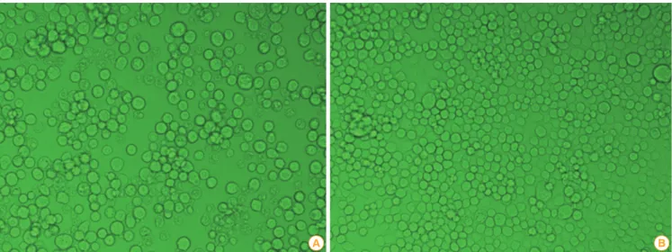
Fig. 2. Cytopathic effect in Sf-9 cells infected with recombinant porcine granulocyte monocyte-colony stimulating factor baculovirus (A, ×200), and normal Sf-9 cells (B, ×200).
A B
Fig. 3. Identification of porcine granulocyte monocyte-colony stimulating factor (GM-CSF) gene from cloned GM-CSF baculovirus. (A) M, 1 kb DNA ladder; lane 1, porcine GM-CSF gene amplified with specific primers; lane 2, normal Sf-9 cell; lane 3, Sf-9 cells inoculated with recombi- nant porcine GM-CSF (reporGM-CSF) baculovirus. The expression of the porcine GM-CSF protein using baculovirus vector system. (B) M, protein marker; lane 1, normal Sf-9 cell; lane 2, supernatant of Sf-9 cells inoculated with reporGM-CSF baculovirus; lane 3, lysate of Sf-9 cell inoculated with reporGM-CSF baculovirus.
1 kbp
M 1 2 3
86 7 bp: Porcine GM-CSF gene in recombinant baculovirus 447 bp
GM-CSF gene
A
M 1 2 3 kDa
24 kDa 240
170130 95 70 56 43 35 28
17
6
B
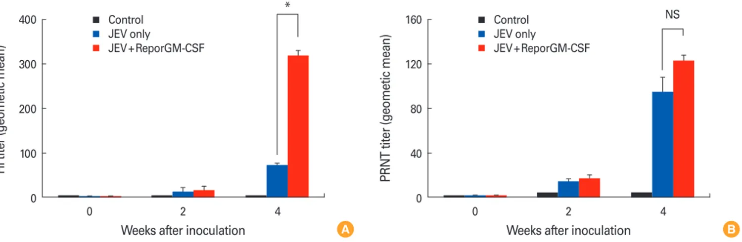
Fig. 4. Antibody responses following vaccination with inactivated Japanese encephalitis virus (JEV) G1 vaccine containing recombinant por- cine granulocyte monocyte-colony stimulating factor (reporGM-CSF) protein or not. The pigs were bled at the day of boosting (pre-boost), and 14 days post-boosting for hemagglutination inhibition (HI) titers (A) and plaque reduction neutralization test (PRNT) titers (B). The HI and PRNT titers were log2 converted and reported as the geometric mean. Data were shown as expressed mean±standard deviation of 8 pigs. NS, not significant. *p < 0.05.
400
300
200
100
0 0 2 4
Weeks after inoculation
HI titer (geometic mean)
Control JEV only
JEV+ReporGM-CSF
* 160
120
80
40
0 0 2 4
Weeks after inoculation
PRNT titer (geometic mean)
Control JEV only
JEV+ReporGM-CSF
NS
A B
(Fig. 3B). The reporGM-CSF baculovirus propagated in Sf-9 cell was added to inactivated JEV genotype 1 vaccine as adju- vants.
Safety and immune response in animals
Mice and guinea pigs inoculated with the vaccines did not any clinical symptoms for 14 days after inoculation. The fat-
tening pigs immunized with two doses of inactivated JEV vac- cines via intramuscular route did not show any symptoms re- lated to encephalitis for period of observation. Virus-neutral- izing antibody is important to protection against JE and over a titer of 1:10 is indicative of protective immunity [8,9]. The anti-JEV antibody titers in pigs inoculated with the inactivat- ed JEV vaccine were compared with those immunized with
Fig. 5. Survival rate of the immunized mice after challenge. After immunization with the inactivated Japanese encephalitis virus (JEV) vaccine with or without recombinant porcine granulocyte monocyte- colony stimulating factor (reporGM-CSF) protein, mice were chal- lenged with 100 LD50 of virulent JEV.
120
100
80
60
40
20
0 0 1 2 3 4 5 6 7 8 9 10 11 12 13 14 15 16
Days after challenge
Survival rates (%)
JEV
JEV+ReporGM-CSF Control
the inactivated JEV vaccine containing reporGM-CSF pro- tein. Fig. 4 showed that HI antibody titers were significantly higher in pigs immunized with the vaccine containing repor- GM-CSF protein than in pigs inoculated with the vaccine alone and ranged from 1:80 to 1:640. The fattening pigs inoc- ulated with the vaccine containing reporGM-CSF protein had JEV neutralizing antibody titers of between 1:16 and 1:512 and mean PRNT titer was also higher in pigs inoculated with the vaccine containing reporGM-CSF protein than in pigs in- oculated with the vaccine alone. Moreover, after the second vaccination at day 14, the HI and PRNT antibody titers were significantly increased in the vaccinated pigs (HI, p<0.05).
These findings indicated that immunization with inactivated JEV vaccine containing reporGM-CSF protein could induce the higher level of HI and PRNT antibody titers than those of immunization with inactivated JEV vaccine only.
Potency of inactivated JEV vaccines in mice
Groups of mice vaccinated with 0.1 dose of inactivated JEV vaccine in the presence or absence of reporGM-CSF protein, were challenged via intracranial route with the low passaged KV1899 strain of JEV at eight days after completion of the vac- cination schedule (Fig. 5). Whilst 90 % of naïve mice succumb- ed to the virus challenge, 87% of mice immunized with the inactivated JEV vaccine survived without showing the char- acteristic signs of JE such as ruffled fur, hunched posture, tre- mors and hind-leg paralysis. Mice immunized with the inac- tivated JEV vaccine containing reporGM-CSF protein showed
53% survival rate.
Discussion
JE continues to be one of the economically significant repro- ductive disorders in the swine industry and has been one of important zoonoses in Asia. JEV G3 was the most major gen- otype in many South and Southeast Asian countries, but gen- otype change G3 into G1 has occurred in Japan, China, and Korea since the 1990s [8,18,19]. In Korea, JEV G1 strain (K9307) was first identified in 1993 and became the dominant circu- lating genotype based on molecular epidemiological analysis of isolated JEVs [7,9,19]. It was reported that mice received an inactivated JEV G3 vaccine had reduced strain-specific neu- tralizing antibody titer against G1 [20]. In accordance with re- placement of JEV G3 by G1, development of JEV vaccine con- taining G1 for swine has been required in Korea.
It has been known that GM-CSF is a glycoprotein involved in the recruitment of antigen presenting cells, differentiation and maturation of dendritic cells [6]. GM-CSF is produced by T cells, epithelial cells, and macrophages in response to a num- ber of inflammatory stimuli such as lipopolysaccharide and tumor necrosis factor-α. Dodecamer assembly of GM-CSF signaling complex on hematopoietic cells predominantly ac- tivates the JAK2/STAT signal pathway [15]. The GM-CSF is one of the most studied cytokines used both therapeutically and diagnostically, as well as in vaccine development and adjuvant technology [14,21]. Mice inoculated with simian immunodeficiency virus VLPs containing GM-CSF increased CD4+ and CD8 T cell response and other study reported that the highest level of foot and mouth disease virus specific hu- moral immune response was induced in mice immunized with recombinant adenovirus expressing both VP1 and GM- CSF [22-24]. Thus, preparation of recombinant GM-CSF pro- tein obtained from each species may be helpful to elicit high immune response. Therefore, we anticipated that reporGM- CSF protein plays a similar role in inducing adequate humor- al immunity in swine. In this study, we cloned porcine GM- CSF gene in baculovirus transfer vector and expressed repor- GM-CSF protein in insect cells. Furthermore, we prepared the inactivated JEV G1 vaccine for swine and evaluated safety and immunogenicity in experimental animals. The results indicated that experimental animals inoculated with two doses of inactivated vaccines did not show any adverse ef- fects such as loss of body weight, death and local reactions, even after second IM injection in fattening pigs.
Humoral immune response is responsible for protecting animals from the JEV challenge [25]. HI and PRNT antibody titers were measured in pigs inoculated with the inactivated JEV vaccine in presence or absence of reporGM-CSF protein.
Given that the antibody responses induced by vaccination appear to play the important protective role [26], pigs inocu- lated with JEV vaccine with or without reporGM-CSF protein generated HI and PRNT antibody titers, suggesting that pigs may be protected from wild JEV infection. A single inocula- tion of inactivated JEV vaccine induced low level of HI and PRNT antibodies raging from 1:10 to 1:20, but boosting sig- nificantly increased circulating HI antibody levels between 1:40 and 1:640. According to our results, it was speculated that reporGM-CSF stimulate more on hemagglutination anti- body response which showed serologically high cross reac- tion within flaviviruses than neutralizing antibody response.
Pigs inoculated with JEV vaccine containing reporGM-CSF protein showed higher HI and PRNT antibody titers, indicat- ing that the reporGM-CSF protein may activate T cells, mac- rophages and endothelial cells in pigs.
Potency of the inactivated JEV G1 vaccine was observed in immunized mice with or without reporGM-CSF protein in accordance with minimum requirements for the inactivated JEV vaccine. Given that the survival rate should be more than 40% in the immunized group [17], the both groups of mice inoculated with the inactivated JEV vaccine with or without reporGM-CSF protein fulfilled the OIE’s requirements.
On the contrary to the above results in pigs, the inactivated JEV vaccine without reporGM-CSF protein was proved to be a better immunogen in protection against virulence JEV chal- lenge than the vaccine containing reporGM-CSF protein. Con- sidering the efficacy test result in mice, GM-CSF still remained the controversy as an effective adjuvant. As to our knowledge, the biological activity of the GM-CSF generally may depend on species specific, due to the low genetic homology among animal species. It was reported that human GM-CSF gene showed approximately 50% homology to murine GM-CSF gene at the deduced amino acid level and recombinant hu- man GM-CSF had no activity in the murine cells in vitro [27].
It was also reported that administration JEV DNA vaccine ex- pressing a GM-CSF gene revealed suppressive effects on the immune response and protective immunity in mice [6]. In this study, JEV vaccine containing porcine GM-CSF protein expressed in insect cell was evaluated the vaccine efficacy in different animal species, mice. Therefore, to evaluate the effi- cacy of the vaccine containing reporGM-CSF protein, it should
be developed other methods such as evaluating the cellular immune response in pigs.
In conclusion, our results demonstrated that two kinds of the inactivated JEV G1 vaccines with or without reporGM- CSF protein was safe in mice, guinea pig, and pigs and in- duced protective immune responses that were capable of protecting pigs against JEV. This result may provide helpful preventive measures against JEV. Our data indicated that in- activated JEV vaccine containing reporGM-CSF protein en- hanced humoral immune responses in pigs. In further study, the effects of the reporGM-CSF protein on the cellular im- mune response should be investigated in pigs.
ORCID
Dong-Kun Yang http://orcid.org/0000000157653043 Jin-Ju Nah http://orcid.org/0000000250210941 Ha-Hyun Kim http://orcid.org/00000001064730035 Jae-Young Song http://orcid.org/0000000295549252
References
1. Weaver SC, Reisen WK. Present and future arboviral threats.
Antiviral Res 2010;85:328-45.
2. Erlanger TE, Weiss S, Keiser J, Utzinger J, Wiedenmayer K.
Past, present, and future of Japanese encephalitis. Emerg Infect Dis 2009;15:1-7.
3. Misra UK, Kalita J, Goel D, Mathur A. Clinical, radiological and neurophysiological spectrum of JEV encephalitis and other non-specific encephalitis during post-monsoon pe- riod in India. Neurol India 2003;51:55-9.
4. Villordo SM, Gamarnik AV. Genome cyclization as strategy for flavivirus RNA replication. Virus Res 2009;139:230-9.
5. Solomon T, Ni H, Beasley DW, Ekkelenkamp M, Cardosa MJ, Barrett AD. Origin and evolution of Japanese enceph- alitis virus in southeast Asia. J Virol 2003;77:3091-8.
6. Chen H, Gao N, Fan D, et al. Suppressive effects on the immune response and protective immunity to a JEV DNA vaccine by co-administration of a GM-CSF-expressing plasmid in mice. PLoS One 2012;7:e34602.
7. Yun SM, Cho JE, Ju YR, et al. Molecular epidemiology of Japanese encephalitis virus circulating in South Korea, 1983-2005. Virol J 2010;7:127.
8. Fan YC, Chen JM, Chen YY, Lin JW, Chiou SS. Reduced neutralizing antibody titer against genotype I virus in swine immunized with a live-attenuated genotype III Japanese
encephalitis virus vaccine. Vet Microbiol 2013;163:248-56.
9. Yang DK, Kim BH, Kweon CH, Kwon JH, Lim SI, Han HR.
Biophysical characterization of Japanese encephalitis vi- rus (KV1899) isolated from pigs in Korea. J Vet Sci 2004;5:
125-30.
10. Appaiahgari MB, Vrati S. IMOJEV((R)): a Yellow fever vi- rus-based novel Japanese encephalitis vaccine. Expert Rev Vaccines 2010;9:1371-84.
11. Li P, Zheng QS, Wang Q, et al. Immune responses of re- combinant adenoviruses expressing immunodominant epitopes against Japanese encephalitis virus. Vaccine 2008;
26:5802-7.
12. Kaur R, Vrati S. Development of a recombinant vaccine against Japanese encephalitis. J Neurovirol 2003;9:421-31.
13. Inumaru S, Kokuho T, Denham S, et al. Expression of bio- logically active recombinant porcine GM-CSF by baculo- virus gene expression system. Immunol Cell Biol 1998;76:
195-201.
14. Taylor DN, Treanor JJ, Sheldon EA, et al. Development of VAX128, a recombinant hemagglutinin (HA) influenza-fla- gellin fusion vaccine with improved safety and immune response. Vaccine 2012;30:5761-9.
15. Broughton SE, Dhagat U, Hercus TR, et al. The GM-CSF/
IL-3/IL-5 cytokine receptor family: from ligand recogni- tion to initiation of signaling. Immunol Rev 2012;250:277- 302.
16. Barteling SJ, Cassim NI. Very fast (and safe) inactivation of foot-and-mouth disease virus and enteroviruses by a com- bination of binary ethyleneimine and formaldehyde. Dev Biol (Basel) 2004;119:449-55.
17. World Health Organisation for Animal Health. Manual of diagnostic tests and vaccines for terrestrial animals (mam- mals, birds and bees). 7th ed. Paris: World Health Organ- isation for Animal Health; 2012. p.188-97.
18. Morita K. Molecular epidemiology of Japanese encepha- litis in East Asia. Vaccine 2009;27:7131-2.
19. Seo HJ, Kim HC, Klein TA, et al. Molecular detection and genotyping of Japanese encephalitis virus in mosquitoes
during a 2010 outbreak in the Republic of Korea. PLoS One 2013;8:e55165.
20. Beasley DW, Li L, Suderman MT, et al. Protection against Japanese encephalitis virus strains representing four gen- otypes by passive transfer of sera raised against Chime- riVax-JE experimental vaccine. Vaccine 2004;22:3722-6.
21. Wang X, Li J, Jiang P, et al. GM-CSF fused with GP3 and GP5 of porcine reproductive and respiratory syndrome virus increased the immune responses and protective ef- ficacy against virulent PRRSV challenge. Virus Res 2009;
143:24-32.
22. Loudon PT, Yager EJ, Lynch DT, et al. GM-CSF increases mucosal and systemic immunogenicity of an H1N1 influ- enza DNA vaccine administered into the epidermis of non- human primates. PLoS One 2010;5:e11021.
23. Skountzou I, Quan FS, Gangadhara S, et al. Incorporation of glycosylphosphatidylinositol-anchored granulocyte- macrophage colony-stimulating factor or CD40 ligand enhances immunogenicity of chimeric simian immuno- deficiency virus-like particles. J Virol 2007;81:1083-94.
24. Zhang C, Wang B, Wang M. GM-CSF and IL-2 as adjuvant enhance the immune effect of protein vaccine against foot- and-mouth disease. Virol J 2011;8:7.
25. Gao N, Chen W, Zheng Q, et al. Co-expression of Japanese encephalitis virus prM-E-NS1 antigen with granulocyte- macrophage colony-stimulating factor enhances humoral and anti-virus immunity after DNA vaccination. Immunol Lett 2010;129:23-31.
26. Lobigs M, Pavy M, Hall RA, et al. An inactivated Vero cell- grown Japanese encephalitis vaccine formulated with Ad- vax, a novel inulin-based adjuvant, induces protective neu- tralizing antibody against homologous and heterologous flaviviruses. J Gen Virol 2010;91(Pt 6):1407-17.
27. Lee F, Yokota T, Otsuka T, et al. Isolation of cDNA for a hu- man granulocyte-macrophage colony-stimulating factor by functional expression in mammalian cells. Proc Natl Acad Sci U S A 1985;82:4360-4.