105
Immune Network
Introduction
Dendritic cells (DCs), as antigen-presenting cells (APCs), play a pivot role in host immune system (1).
After uptaking antigen, DCs migrate from peripheral tissues to lymph nodes, present antigenic peptides to naïve CD4+ T lymphocytes, and thus stimulate anti- gen-specific immune responses. DCs closely connect to the function of other immune cells including natural killer (NK) cells, T, and B lymphocytes and are at a crossroads to modulate a variety of immune functions.
Galectin-3 is an endogenous animal lectin with beta-galactoside-binding affinity. It has been studied in various cells, such as fibroblasts, cancer cells (2,3).
In immune cells, galectin-3 is expressed in T lympho- cytes, macrophages and closely related to prolifer- ation, apoptosis, phagocytosis (4-6). Interestingly, recent data showed that proteomic analysis deter-
mined galectin-3 in the exosome of DCs among dozen proteins (7,8). However, the presence of galectin-3 by other methods and which factor can regulate the expression of galectin-3 have not been studied yet.
This study confirmed the presence of galectin-3 in DCs using Western blot analysis and flow cytometry analysis. And also, it was demonstrated that the expression of galectin-3 was differentially regulated by essential factors for the function of DCs. It thus suggests that galectin-3 in DCs may be involved in a variety of immune responses.
Materials and Methods
Animals and reagents. C57BL/6 mice were purchased from Japan SLC (Shizuoka, Japan) and maintained in the lab animal facility. 7- to 12-week-old female mice were used for experiments. Purified anti-mouse CD8, CD19, Gr-1 monoclonal antibodies (mAbs, BD PharMingen, San Diego, CA) were used for the detection of CD8+ T lymphocytes, B lymphocytes, and granulocytes in bone marrow-derived DCs, res- pectively.
Preparation of DCs. Bone marrow cells were obtained from tibia and femur of mice. DCs were cultured from bone marrow cells using GM-CSF. Briefly, cells
Protein, in Dendritic Cells
Mi-Hyoung Kim and Hong-Gu Joo
Department of Veterinary Medicine, Cheju National University, Jeju, Republic of Korea
ABSTRACT
Background: Dendritic cells (DCs) are the most potent APCs (antigen-presenting cells) and play a critical role in immune responses. Galectin-3 is a biological lectin with a beta-galactoside binding affinity. Recently, proteomic analysis revealed the presence of galectin-3 in the exosome of mature DCs. However, the expression and function of galectin-3 in DCs remains unclear yet. Methods: We used bone marrow-derived DCs of mouse and showed the expression of galectin-3 in DCs by using flow cytometry analysis and Western blot analysis. Results: Galectin-3 was determined as single band of 35 kDa in Western blot analysis. Flow cytometry analysis showed the major growth factor for DCs, granulocyte-macrophage colony stimulating factor (GM-CSF) and maturing agents, anti-CD40 monoclonal antibody (mAb) and lipopolysaccharide (LPS) consistently increased the intracellular expression of galectin-3 in DCs compared to medium alone. In addition, DCs treated with maturing agents did marginally express galectin-3 on their surface. Conclusion: This study suggests that galectin-3 in DCs may be regulated by critical factors for DC function. (Immune Network 2005;5(2):105-109) Key Words: Dendritic cells, galectin-3, viability, proliferaion
Correspondence to: Hong-Gu Joo, Department of Veterinary Medi- cine, Cheju National University, Ara 1-dong, Jeju 690-756, Republic of Korea. (Tel) 82-64-754-3379, (Fax) 82-64-756-3354, (E-mail) [email protected]
This work was supported by Korea Research Foundation Grant (KRF-2003-003-E00243).
were cultured at a concentration of 2×10⁶ cells/ml in 6-well culture plates. RPMI-1640 medium con- taining 5% fetal bovine serum (FBS) and 10 ng/ml mouse GM-CSF (Biosource International, Camarillo, CA) was used. At 6∼10 day of culture, floating cells were over 85% CD11c+ DCs based on flow cytome- try analysis and thus used as DCs for experiments.
Cells were stained with trypan blue solution (Sigma, St. Louis, MO) to check viability.
Chromatin analysis of apoptotic cells. Cells were stained with 2.5μg/ml of Hoechst 33342 fluorochrome and 2.5μg/ml propidium iodide (PI, Sigma), followed by examination on a fluorescence microscope (Olympus optical, Tokyo). Intact blue/white nuclei, condensed/
fragmented pink nuclei, and intact pink nuclei were considered as viable, apoptotic, and necrotic cells, respectively.
Western blot analysis. Cells were dissolved in lysis buffer containing 25μg/ml phenylmethylsulphonyl fluoride, 10μg/ml aprotinin, 10μg/ml leupeptin, 1% Triton X-100, 0.15 M NaCl, 10 mM Tris (pH 7.4) on ice.
After the centrifugation of lysates at 13,000 rpm for 1 min, the supernatants were mixed with 2× sample loading buffer and denatured at 100oC for 5 min.
Protein concentration in the lysates was determined by Bradford protein assay (Bio-Rad, Hercules, CA) and each sample was loaded at the concentration of 25μg/lane in the gel. After electrophoresis, proteins were transferred onto nitrocellulose membrane and probed with 1μg/ml anti-galectin-3 mAb (clone MA1-940, Affinity Bioreagents, Golden, CO) and appropriate secondary antibody. The blot was devel- oped by chemiluminescence reagents (Amersham, Arlington Heights, IL).
Flow cytometry analysis. To block Fc receptors, DCs were incubated with purified anti-mouse CD16/
CD32 mAb (BD PharMingen) at a concentration of 1μg/100μl/106 cells for 15 min at 4oC. For surface staining, cells were incubated with each mAb at a concentration of 1μg/100μl for 30 min at 4oC and washed twice with Hanks’ balanced salt solution (HBSS) containing 5% FBS and 0.1% sodium azide.
For detecting mature DCs, fluorescein isothiocyanate (FITC)-labeled anti-mouse I-Ab mAb, phycoerythrin (PE)-labeled anti-mouse CD11c mAb (BD Phar- Mingen) were used. FITC- or PE-labeled isotype- matched mAb (BD PharMingen) was used as control, respectively. For intracellular staining, cells were fixed in phosphate buffered saline (PBS) containing 1%
formaldehyde for 10 min, permeabilized by adding ice-cold 100% methanol for 30 min on ice, and stained with anti-galectin-3 mAb and secondary anti- body sequentially. After staining, cells were analyzed with FACSCaliber flow cytometer (Becton Dickinson, Mountain View, CA) and CellQuest software.
Results
The culture and characterization of bone marrow-derived DCs.
DCs were cultured from bone marrow cells of tibia and femur by using 10 ng/ml GM-CSF. After 6 days culture, cells were identified as DCs by flow cyto- metry analysis using anti-CD11c mAb. To investigate the expression of galectin-3 in various types of DCs, cells were treated with GM-CSF as a growth factor, anti-CD40 mAb or LPS as maturing agents. The expression level of MHC class II was measured on DCs for detecting the maturation of DCs (data not shown). Since galectin-3 has been correlated to the proliferation and viability of cells, we measured the viability of DCs using trypan blue exclusion test (Fig.
1). GM-CSF, anti-CD40 mAb, or LPS significantly increased the viability of DCs. This data suggest that growth factor or maturing agent may be an essential factor for the viability of DCs.
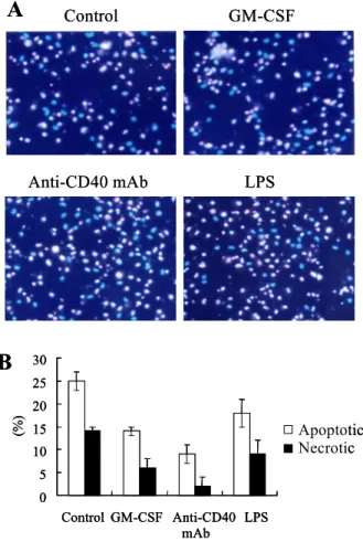
Chromatin analysis revealed the anti-apoptotic effects of anti-CD40 mAb. In chromatin analysis, GM-CSF, anti-CD40 mAb, and LPS significantly decreased the number of DCs containing condensed/fragmented pink nuclei and intact pink nuclei, indicating apop- totic or necrotic cells (Fig. 2A). Apoptotic or necrotic cells were counted and the percentage was calculated (Fig. 2B). Although both anti-CD40 mAb and LPS are maturing agents, anti-CD40 mAb consistently induced higher viability of DCs compared to LPS.
Taken together, chromatin analysis confirmed that GM-CSF, anti-CD40 mAb, or LPS inhibited the cell death of DCs and the maturing agents, anti-CD40 mAb and LPS, may deliver differential signals for viability in DCs.
The identification of galectin-3 in DCs using Western blot
Figure 1. The viability of DCs was enhanced by GM-CSF, anti-CD40 mAb, or LPS. After 6 days culture, DCs were seeded at a concentration of 5×10⁵ cells/ml in 24-well culture plate.
Cells were treated with GM-CSF, anti-CD40 mAb or LPS for 2 days. After washing twice with HBSS, the viability of DCs was measured by using trypan blue exclusion test. Result is a representative of three individual experiments.
0 20 40 60 80 100 120
Control GM-CSF Anti-CD40
mAb LPS
Viability (%)
0 20 40 60 80 100 120
Control GM-CSF Anti-CD40
mAb LPS
Viability (%)
analysis. After 6∼8 days culture, DCs were treated with GM-CSF, anti-CD40 mAb, or LPS for 2 days and the lysates of cells were prepared for Western blot analysis. Fig. 3 revealed that GM-CSF, anti- CD40 mAb, or LPS consistently increased the ex- pression of galectin-3 compared to control. Anti-beta- actin mAb was used for the detection of beta-actin as internal control protein. The molecular weight of galectin-3 in DCs was 35 kDa.
Flow cytometry analysis confirmed the expression of galectin-3 in DCs. DCs were stained by anti-galectin-3 mAb for flow cytometry analysis. The surface or intracellular expression level of galectin-3 was determined (Fig. 4).
Anti-CD40 mAb or LPS marginally increased the expression of galectin-3 on the surface of DCs whereas GM-CSF, anti-CD40 mAb, LPS significantly increased the intracellular expression of galectin-3. It
is suggested that growth factor or maturing agents may increase the expression of galectin-3 in DCs.
Discussion
Galectin-3 has been known to play a critical role in the proliferation of various cells. In this study, we used GM-CSF, a representative growth factor for DCs. GM-CSF consistently increased the expression of galectin-3 in DCs based on Western blot and flow cytometry analysis. Especially, intracellular galectin-3 was significantly enhanced by GM-CSF in flow cytometry analysis. Since GM-CSF is a critical cyto- kine for the proliferation and survival of DCs, galectin-3 seems to be expressed in the growth or generation process of DCs and its intracellular ex- pression closely connected to those processes. Up to date, the relationship between galectin-3 and pro- liferation is not clear yet although the function of intracellular galectin-3 has been studied for many years (9). Previous study demonstrated that intra- cellular galectin-3 in lymphocytes was enhanced by common γ-chain signaling cytokines, interleukin (IL)-2, IL-4, and IL-7, essential cytokines for the proliferation and survival of activated T lymphocytes (4). In addition, intracellular galectin-3 is closely involved in anti-apoptotic activity of cells. Phos- phorylation of galectin-3 and transport from nucleus to cytoplasm were necessary for the anti-apoptotic activity of cancer cells to chemotherapeutic anticancer drugs (10).
Proteomic analysis previously showed the presence of galectin-3 in the exosomes of DCs. Exosome is a subcellular compartment secreted from cells and can be used for cancer immunotherapy (11). Based on this study, growth factor or maturing agents increase the expression of galectin-3 in DCs and thus exosome also may express more galectin-3. The study about galectin-3 function in exosome of DCs treated with various factors should be valuable as a future
Figure 2. GM-CSF, anti-CD40 mAb, or LPS inhibited the apoptosis of DCs. After 6∼8 days culture, cells were seeded at a concentration of 5×10⁴ cells/well in 96-well culture plate. Cells were treated with GM-CSF, anti-CD40 mAb, or LPS for 2 days.
Then, cells were sequentially stained by Hoechst 33342 and propidium iodide as described in Materials & Methods. 200 cells were counted for chromatin analysis. Results are means ± SD from three independent wells and the representative of three individual experiments.
Control GM-CSF
Anti-CD40 mAb LPS
A
Control GM-CSFAnti-CD40 mAb LPS
A
0 5 10 15 20 25 30
Control GM-CSF Anti-CD40
mAb LPS
(%)
Apoptotic Necrotic
B
0 5 10 15 20 25 30
Control GM-CSF Anti-CD40
mAb LPS
(%)
0 5 10 15 20 25 30
Control GM-CSF Anti-CD40
mAb LPS
(%)
Apoptotic Necrotic
B
Figure 3. Western blot analysis confirmed the expression of galectin-3 in DCs. Cell lysates were loaded and blotted on nitrocellulose membrane. Anti-galectin-3 mAb was used for detecting galectin-3 in DCs. Molecular weight of galectin-3 was determined based on the molecular size of pre-stained standards.
Result is a representative of three individual experiments.
Galectin-3
Beta-actin
1 2 3 4
1. Control 2. GM -CSF 3. Anti-CD40 mAb 4. LPS
Galectin-3
Beta-actin
1 2 3 4
1. Control 2. GM -CSF 3. Anti-CD40 mAb 4. LPS
work.
Galectin-3 has been known to be the major non-integrin laminin binding protein and the binding of activated lymphocyte via L-selectin to DCs was mediated by galectin-3 (6,12). Interestingly, our study shows that surface galectin-3 is expressed on the surface of DCs treated with maturing agents. This observation suggests that mature DCs may bind to other immune cells or extracellular matrix protein in a galectin-3 or lactose-dependent manner.
Taken together, galectin-3 is expressed in DCs treated with growth factors or maturing agents. This observation may provide new insights about the expression and function of galectin-3, one of major animal lectins in DCs.
References
1. Mellman I, Steinman RM: Dendritic cells: specialized and regulated antigen processing machines. Cell 106;255-258, 2001
2. Moutsatsos IK, Wade M, Schindler M, Wang JL: Endogenous lectins from cultured cells: nuclear localization of carbo- hydrate-binding protein 35 in proliferating 3T3 fibroblasts.
Proc Natl Acad Sci USA 84;6452-6456, 1987
3. Song YK, Billiar TR, Lee YJ: Role of galectin-3 in breast cancer metastasis: involvement of nitric oxide. Am J Pathol 160;1069-1075, 2002
4. Joo HG, Goedegebuure PS, Sadanaga N, Nagoshi M, Bern- storff W, Eberlein TJ: Expression and function of galectin-3, a beta-galactoside-binding protein in activated T lymphocytes.
J Leuk Biol 69;555-564, 2001
5. Sano H, Hsu DK, Apgar JR, Yu L, Sharma BB, Kuwabara I, Izui S, Liu FT: Critical role of galectin-3 in phagocytosis by macrophages. J Clin Invest 112;389-397, 2003
6. Woo HJ, Shaw LM, Messier JM, Mercurio AM: The major non-integrin laminin binding protein of macrophages is identical to carbohydrate binding protein 35 (Mac-2). J Biol Chem 265;7097-7099, 1990
7. Dietz AB, Bulur PA, Knutson GJ, Matasic R, Vuk-Pavlovic S: Maturation of human monocyte-derived dendritic cells studied by microarray hybridization. Biochem Biophys Res Commun 275;731-738, 2000
8. Thery C, Boussac M, Veron P, Ricciardi-Castagnoli P, Raposo G, Garin J, Amigorena, S: Proteomic analysis of dendritic cell-derived exosomes: a secreted subcellular compartment distinct from apoptotic vesicles. J Immunol 166;7309-7318, 2001
9. Dagher SF, Wang JL, Patterson RJ: Identification of galectin- 3 as a factor in pre-mRNA splicing. Proc Natl Acad Sci USA 92;1213-1217, 1995
10. Takenaka Y, Fukumori T, Yoshii T, Oka N, Inohara H, Kim HR, Bresalier RS, Raz A: Nuclear export of phosphorylated Figure 4. Flow cytometry analysis revealed the differential expression of galectin-3 in DCs. After treating cells as described in Fig.
1, DCs were stained for surface (A) or intracellular (B) galectin-3 using anti-galectin-3 mAb. Results are representative of three experiments.
Galectin-3
Control GM-CSF Anti-CD40 mAb LPS
A
Galectin-3
Control GM-CSF Anti-CD40 mAb LPS
A
Galectin-3
Control GM-CSF Anti-CD40 mAb LPS
B
Galectin-3
Control GM-CSF Anti-CD40 mAb LPS
B
galectin-3 regulates its antiapoptotic activity in response to chemotherapeutic drugs. Mol Cell Biol 24;4395-4406, 2004 11. Zitvogel L, Regnault A, Lozier A, Wolfers J, Flament C,
Tenza D, Ricciardi-Castagnoli P, Raposo G, Amigorena S:
Eradication of established murine tumors using a novel cell-free vaccine: dendritic cell-derived exosomes. Nature
Med 4;594-600, 1998
12. Swarte VV, Mebius RE, Joziasse DH, Van den Eijnden DH, Kraal G: Lymphocyte triggering via L-selectin leads to enhanced galectin-3-mediated binding to dendritic cells. Eur J Immunol 28;2864-2871, 1998