pISSN 2093-596X · eISSN 2093-5978
Article
Effects of Single Vitamin D 3 Injection (200,000 Units) on Serum Fibroblast Growth Factor 23 and Sclerostin Levels in Subjects with Vitamin D Deficiency
Dongdong Zhang1,2, Da Hea Seo3, Han Seok Choi4, Hye-Sun Park3, Yoon-Sok Chung5, Sung-Kil Lim1,3
1Brain Korea 21 PLUS Project for Medical Science, Yonsei University, Seoul, Korea; 2Division of Endocrinology and Metabolism, Department of Internal Medicine, Yantai Affiliated Hospital of Binzhou Medical University, Yantai, China;
3Division of Endocrinology and Endocrine Research Institute, Department of Internal Medicine, Yonsei University College of Medicine, Seoul; 4Division of Endocrinology and Metabolism, Department of Internal Medicine, Dongguk University Ilsan Hospital, Dongguk University College of Medicine, Goyang; 5Division of Endocrinology and Metabolism, Department of Internal Medicine, Ajou University School of Medicine, Suwon, Korea
Background: Vitamin D deficiency remains common in all age groups and affects skeletal and non-skeletal health. Fibroblast growth factor 23 is a bone-derived hormone that regulates phosphate and 1,25-dihydroxyvitamin D homeostasis as a counter regula- tory factor. 1,25-Dihydroxyvitamin D stimulates fibroblast growth factor 23 synthesis in bone, while fibroblast growth factor 23 sup- presses 1,25-dihydroxyvitamin D production in the kidney. The aim of this study was to evaluate the effects of vitamin D3 intramus- cular injection therapy on serum fibroblast growth factor 23 concentrations, and several other parameters associated with bone me- tabolism such as sclerostin, dickkopf-1, and parathyroid hormone.
Methods: A total of 34 subjects with vitamin D deficiency (defined by serum 25-hydroxyvitamin D levels below 20 ng/mL) were randomly assigned to either the vitamin D injection group (200,000 units) or placebo treatment group. Serum calcium, phosphate, urine calcium/creatinine, serum 25-hydroxyvitamin D, fibroblast growth factor 23, sclerostin, parathyroid hormone, and dickkopf-1 levels were serially measured after treatment.
Results: Comparing the vitamin D injection group with the placebo group, no significant changes were observed in serum fibroblast growth factor 23, parathyroid hormone, or dickkopf-1 levels. Serum sclerostin concentrations transiently increased at week 4 in the vitamin D group. However, these elevated levels declined later and there were no statistically significant differences as compared with baseline levels.
Conclusion: Serum fibroblast factor 23, sclerostin, parathyroid hormone, and dickkopf-1 levels were not affected significantly by single intramuscular injection of vitamin D3.
Keywords: Vitamin D3; Injections, intramuscular; Fibroblast growth factor 23; Sclerostin
INTRODUCTION
Vitamin D plays an essential role in bone and mineral metabo-
lism [1]. It is also important in non-skeletal tissues, and its defi- ciency is closely associated with increased risk of cancers, in- fections, autoimmune diseases, cardiovascular diseases, and di-
Received: 1 June 2017, Revised: 9 August 2017, Accepted: 10 October 2017 Corresponding author: Sung-Kil Lim
Division of Endocrinology and Endocrine Research Institute, Department of Internal Medicine, Yonsei University College of Medicine, 50-1 Yonsei-ro, Seodaemun-gu, Seoul 03722, Korea
Tel: +82-2-2228-0878, Fax: +82-2-393-6884, E-mail: [email protected]
Copyright © 2017 Korean Endocrine Society
This is an Open Access article distributed under the terms of the Creative Com- mons Attribution Non-Commercial License (http://creativecommons.org/
licenses/by-nc/4.0/) which permits unrestricted non-commercial use, distribu- tion, and reproduction in any medium, provided the original work is properly cited.
abetes mellitus [2-5]. Despite growing public awareness of the multiple health benefits of vitamin D, epidemiological studies have revealed a very high prevalence of vitamin D deficiency worldwide, especially in Asian countries [6,7].
Vitamin D is mainly produced in the skin when directly ex- posed to sunlight, or obtained from the diet. Active vitamin D maintains calcium and phosphate homeostasis by promoting in- testinal absorption for the bone mineralization process [8].
Aside from its role in the endocrine pathway, active vitamin D is known to affect the differentiation and function of bone cells by targeting key genes involved in bone formation and resorp- tion [9]. Oral supplementation with vitamin D and calcium is common practice in the treatment of vitamin D deficiency. In addition to oral supplementation, vitamin D can also be admin- istered by intramuscular injection. Heikinheimo et al. [10] re- ported that annual intramuscular injection of ergocalciferol vita- min D2 (150,000 to 300,000 IU) resulted in a significant reduc- tion in the incidence of fractures.
To maintain calcium and phosphate homeostasis, active vita- min D works in combination with two other hormones: fibro- blast growth factor 23 (FGF23) and parathyroid hormone (PTH) [11]. 1,25-Dihydroxyvitamin D (1,25(OH)2D) stimulates FGF23 synthesis in bone, while FGF23 suppresses the production of 1,25(OH)2D; thus, acting as a counter-regulatory factor [12].
FGF23 levels are reportedly significantly elevated in patients with chronic kidney disease (CKD), who are at increased risk of mortality mainly from cardiovascular disease [13]. CKD-mineral and bone disorder (CKD-MBD) occurs from the early stages of CKD, and there is a strong association between FGF23 and car- diovascular risks, left ventricular hypertrophy, and vascular cal- cification [13-15]. A study conducted in patients with vitamin D deficiency who were given a high dose vitamin D2 injection (300,000 IU) combined with usual daily oral supplementations of calcium (1.2 g) and vitamin D3 (800 IU) reported that high dose vitamin D increased 1,25(OH)2D and FGF23 concentra- tions [16]. However, there is a paucity of data regarding the ef- fects of single intramuscular injections of 200,000 IU of vitamin D3.
Sclerostin and dickkopf-1 (DKK1) are two important endoge- nous Wnt signaling antagonists, mainly produced in bone [17,18]. Sankaralingam et al. [19] reported that bolus intramus- cular injection of 300,000 IU of vitamin D combined with oral supplementation of vitamin D and calcium increased sclerostin and DKK1 concentrations. In contrast, another study demon- strated that in patients with vitamin D deficiency who were giv- en a monthly intramuscular injection of 300,000 IU of vitamin
D, sclerostin levels decreased considerably after treatment [20].
The aim of our study was to determine the effects of single intramuscular injection of vitamin D3 at 200,000 IU on circulat- ing concentrations of 25-hydroxyvitamin D (25(OH)D), FGF23, sclerostin, and DKK1 in patients with vitamin D deficiency.
METHODS
Study subjects and design
Thirty-four subjects (five males and 29 females) aged 33.2±7.7 years, diagnosed with vitamin D deficiency at Severance Hospi- tal, Yonsei University College of Medicine and Dongguk Uni- versity Ilsan Hospital in Korea were recruited in this study. Vi- tamin D deficiency was diagnosed if serum 25(OH)D concen- trations were below 20 ng/mL. Patients who were taking medi- cations that may affect vitamin D metabolism were excluded from the study.
This study was designed as a randomized, double-blinded, and placebo-controlled clinical trial. Subjects were randomly assigned to the experimental group or the placebo group at a 2:1 ratio. All subjects were given either a single intramuscular injec- tion of vitamin D3 (200,000 IU), or a placebo at the beginning of the study. During this intervention period, consumption of any additional vitamin D supplements and exposure to direct sunlight for more than 10 hours were prohibited. Usual diets were maintained, except diets containing abundant vitamin D.
Laboratory measurements
Fasting blood samples were collected at baseline and weeks 4, 8, 12, and 14 during follow-up visits after vitamin D or placebo injections. Routine biochemical parameters including basic chemistry tests (measuring glucose, calcium, phosphorus, blood urea nitrogen, and creatinine), hematology tests (hemoglobin, hematocrit, and hemoglobin A1c), and urinalyses were conduct- ed via standard methods at each institution, and additional se- rum samples were stored at –70 for subsequent analyses. Serum 25(OH)D and intact PTH concentrations were measured by electrochemiluminescence immunoassay (ECLIA) (Roche Di- agnostics, Mannheim, Germany). The intra- and inter-assay co- efficients of variation (CV) for 25(OH)D measurements were 2.2% to 6.8% and 3.4% to 13.1%, respectively. The intra- and inter-assay CV of intact PTH measurements were 1.1% to 2.0%
and 2.8% to 3.4%, respectively. Serum FGF23 levels were mea- sured using an intact FGF23 (iFGF23) enzyme-linked immuno- sorbent assay (ELISA) kit (Millipore Corporation, Billerica, MA, USA). The intra- and inter-assay CV of iFGF23 measure-
ments were 7.8% to 11.2% and 2.4% to 11.31%, respectively.
Sclerostin and DKK1 concentrations were also measured using human sclerostin and DKK-1 ELISA kits (R&D Systems, Min- neapolis, MN, USA) according to the manufacturer’s instruc- tions. The intra- and inter-assay CV of sclerostin measurements were 1.8% to 2.1% and 8.2% to 10.8%, respectively. The intra- and inter-assay CV of DKK1 measurements were 3.3% to 4.2%
and 4.6% to 7.6%, respectively.
Ethics statement
This study protocol was reviewed and approved by the Institu- tional Review Broad (IRB: 4-2014-0377) at both institutions.
Informed consent was submitted by all subjects when they were enrolled.
Statistical analysis
All statistical analyses were performed using GraphPad Prism version 5.01 (GraphPad Software, CA, USA). Results are pre- sented as mean±standard error (SE). Student paired t test was used to compare the data from different time points with base- line values when the values were normally distributed, and Wil- coxon signed rank test was used when the data were nonpara- metric. An analysis of variance (ANOVA) was used to compare different time points between groups. Dunnett’s test and Bon- ferroni correction were performed for the repeated measures test of one-way ANOVA and two-way ANOVA, respectively. A P value of <0.05 (95% confidence interval) was considered statis- tically significant.
RESULTS
Demographic characteristics and changes in routine biochemical parameters following single vitamin D injection
Demographic information and other baseline characteristics of the subjects are shown in Table 1. There were no statistically significant differences between the vitamin D and placebo groups (P>0.05). Compared with the placebo group, no statisti- cally significant differences were observed in serum calcium, phosphorus, and the ratio of urine calcium to creatinine at weeks 4, 8, and 12 after vitamin D3 injection (Fig. 1). Serum 25(OH)D concentrations increased significantly throughout the study (P<0.001) after vitamin D3 injection (Fig. 2A-C). Moreover, there were no significant differences between the two subgroups receiving vitamin D3 injections which had previously been grouped based on baseline levels of 25(OH)D (Fig. 2D). On the
other hand, serum PTH concentrations at weeks 4, 8, and 12 af- ter vitamin D3 injection in the vitamin D group did not change significantly (P>0.05) (Fig. 3A, D). No statistically significant differences were observed in the vitamin D group compared with the placebo group (P>0.05) (Fig. 3).
Effects of vitamin D on serum FGF23 concentrations To determine whether a single injection of vitamin D3 (200,000 IU) increases circulating FGF23 levels, we measured serum FGF23 concentrations at weeks 4, 8, 12, and 14 after vitamin D3 treatment. The results showed that no significant difference were observed between vitamin D3 treatment group and placebo group (Fig. 4A). Similar results were found in the two sub- groups (Fig. 4B-D). However, subject 35 (placebo group) showed particularly elevated FGF23 levels throughout the study period including at baseline, even though this was in the placebo group without vitamin D3 injection.
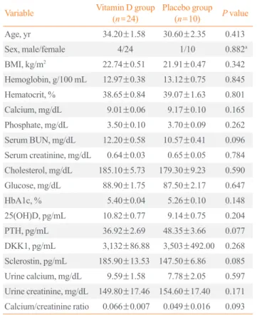
Table 1. Demographic and Other Baseline Characteristics of the Study Subjects
Variable Vitamin D group
(n=24) Placebo group (n=10) P value Age, yr 34.20±1.58 30.60±2.35 0.413 Sex, male/female 4/24 1/10 0.882a BMI, kg/m2 22.74±0.51 21.91±0.47 0.342 Hemoglobin, g/100 mL 12.97±0.38 13.12±0.75 0.845 Hematocrit, % 38.65±0.84 39.07±1.63 0.801 Calcium, mg/dL 9.01±0.06 9.17±0.10 0.165 Phosphate, mg/dL 3.50±0.10 3.70±0.09 0.262 Serum BUN, mg/dL 12.20±0.58 10.57±0.41 0.096 Serum creatinine, mg/dL 0.64±0.03 0.65±0.05 0.784 Cholesterol, mg/dL 185.10±5.73 179.30±9.23 0.590 Glucose, mg/dL 88.90±1.75 87.50±2.17 0.647 HbA1c, % 5.40±0.04 5.26±0.10 0.148 25(OH)D, pg/mL 10.82±0.77 9.14±0.75 0.204 PTH, pg/mL 36.92±2.69 48.35±3.66 0.077 DKK1, pg/mL 3,132±86.88 3,503±492.00 0.268 Sclerostin, pg/mL 185.90±13.53 147.50±6.86 0.085 Urine calcium, mg/dL 9.59±1.58 7.78±2.05 0.597 Urine creatinine, mg/dL 149.80±17.46 154.60±17.40 0.171 Calcium/creatinine ratio 0.066±0.007 0.049±0.016 0.093 Values are expressed as mean±standard error.
BMI, body mass index; BUN, blood urea nitrogen; HbA1c, hemoglobin A1c; 25(OH)D, 25-hydroxyvitamin D; PTH, parathyroid hormone;
DKK1, dickkopf-1.
aChi-square test.
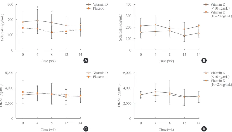
Changes in sclerostin and DKK1 levels
To evaluate whether vitamin D3 injection stimulates the expres- sion of Wnt inhibitors, we measured serum sclerostin and DKK1 concentrations after vitamin D3 injection. In the vitamin D group, serum sclerostin levels were transiently increased at week 4 after vitamin D3 injection. Comparing the vitamin D group with the placebo group, there were significant differences at weeks 4 and
8 (P<0.05) (Fig. 5A). However, the slightly increased levels de- clined thereafter, and were not significantly different when com- pared to baseline (Fig. 5A). Moreover, there were no significant differences in sclerostin levels between the two subgroups with vitamin D3 injection (Fig. 5B). DKK1 levels were not increased in the vitamin D group and there were no significant differences between this and the placebo group (Fig. 5C, D).
10.0
9.5 9.0 8.5 8.0
4.5
4.0 3.5 3.0 2.5
0.20
0.15 0.10 0.05 0
Serum calcium (mg/dL) Serum phosphorus (mg/dL) Urine calcium/creatinine ratio
Time (wk) Time (wk) Time (wk)
0 4 8 12 0 4 8 12 0 4 8 12
Fig. 1. (A) Serum calcium, (B) phosphorus concentrations, and (C) ratio of urine calcium to creatinine in subjects were measured before and after vitamin D3 or placebo treatment.
A B C
Placebo Placebo Placebo
Vitamin D Vitamin D Vitamin D
30
20
10
0
40
30
20
10
0
40
30
20
10
0 30
20
10
0
25(OH)D (ng/mL)25(OH)D (ng/mL) 25(OH)D (ng/mL)25(OH)D (ng/mL)
Time (wk)
Time (wk) Time (wk)
Time (wk) 0 4 8 12 14
0 4 8 12 14 0 4 8 12 14
0 4 8 12 14
Fig. 2. Serum 25-hydroxyvitamin D (25(OH)D) concentrations were increased at different time points after treatment. (A) All subjects, (B) subjects with baseline 25(OH)D below 10 ng/mL, (C) subjects with baseline levels of 10 to 20 ng/mL, and (D) the vitamin D group.
aP<0.05; bP<0.01; cP<0.001.
A
C D
B Vitamin D
Vitamin D
Vitamin D (10–20 ng/mL)
(10–20 ng/mL) (<10 ng/mL)
Vitamin D (<10 ng/mL) Placebo
Placebo Vitamin D
Placebo
c
b
c c
a
c c
c
c c
c
c
80
60
40
20
0
80
60
40
20
0
80
60
40
20
0
60
40
20
0
PTH (pg/mL)PTH (pg/mL) PTH (pg/mL)PTH (pg/mL)
Time (wk)
Time (wk)
Time (wk)
Time (wk) 0 4 8 12
0 4 8 12
0 4 8 12
0 4 8 12
Fig. 3. Serum parathyroid hormone (PTH) levels were detected at the different time points after treatment. The comparison in (A) all sub- jects, (B) the subjects with less 10 ng/mL of baseline 25-hydroxyvitamin D (25(OH)D), (C) the subjects with 10 to 20 ng/mL of baseline 25(OH)D, and (D) the vitamin D group.
A
C
B
D Vitamin D
Vitamin D
Vitamin D (10–20 ng/mL)
(10–20 ng/mL) (<10 ng/mL)
Vitamin D (<10 ng/mL) Placebo
Placebo Vitamin D
Placebo
30
20
10
0
60
40
20
0
18
16
14
12
10 20
15
10
5
0
FGF-23 (pg/mL)FGF-23 (pg/mL) FGF-23 (pg/mL)FGF-23 (pg/mL)
Time (wk)
Time (wk) Time (wk)
Time (wk) 0 4 8 12 14
0 4 8 12 14 0 4 8 12 14
0 4 8 12 14
Fig. 4. Serum fibroblast growth factor 23 (FGF23) levels were measured at the indicated times. (A) All subjects, (B) subjects with baseline 25(OH)D below 10 ng/mL, (C) subjects with baseline levels of 10 to 20 ng/mL, and (D) the vitamin D group.
A
C D
B Vitamin D
Vitamin D
Vitamin D (10–20 ng/mL)
(10–20 ng/mL) (<10 ng/mL)
Vitamin D (<10 ng/mL) Placebo
Placebo
Vitamin D Placebo
DISCUSSION
In the present study, we have shown that serum 25(OH)D levels significantly increased following a single intramuscular injec- tion of vitamin D3 with 200,000 IU, which is consistent with the report [21]. However, the levels of serum PTH, FGF23, and DKK1 did not change during the 14-week follow-up period af- ter vitamin D3 injection. Serum sclerostin levels were slightly increased 4 weeks post-treatment but declined thereafter.
Calcium alone, or combined with oral vitamin D, has been suggested as an inexpensive therapeutic method to prevent os- teoporotic bone loss and fractures. However, this treatment is less effective if the patients’ compliance is poor. Furthermore, therapeutic levels are only reached after a long period. Intra- muscular injection of vitamin D alone or in combination with oral supplementation can maintain increased levels of serum 25(OH)D for at least 6 months [22]. Turner et al. [16] reported that intramuscular injection of vitamin D together with oral cal- cium and vitamin D supplementation, increased 1,25(OH)2D concentrations significantly. Similar findings were also demon- strated recently where 25(OH)D levels were elevated in vitamin D deficient patients who were given a monthly intramuscular
injection of 300,000 IU of vitamin D for 3 consecutive months [20]. Our results are consistent with these findings over the 14 weeks following a single vitamin D3 injection at the dose of 200,000 IU.
Active vitamin D stimulates the production of FGF23 in os- teocytes and osteoblasts [23]. Furthermore, increased FGF23 levels reduce expression of 1α-hydroxylase but increase expres- sion of 24-hydroxylase, which converts 1,25(OH)2D to the les biologically active 24,25(OH)2D, resulting in decreased 1,25(OH)2D production [12]. FGF23 concentrations were also increased after intramuscular injection of vitamin D (300,000 IU) administered in conjunction with usual daily supplementa- tion of vitamin D and calcium [18]. In contrast, Uzum et al. [24]
reported that FGF23 concentrations further declined during vi- tamin D replacement therapy in vitamin D deficient patients who were treated daily with an oral combination of vitamin D and calcium for 6 weeks. In our study, serum FGF23 levels were not significantly elevated after intramuscular injection of vitamin D3 (200,000 IU). In contrast with the previous reports, our study did not provide any evidence that single intramuscular injection of vitamin D3 (200,000 IU) could increase or decrease serum FGF23 levels at all. Participants, medication dosage, and
300
200
100
0
6,000
4,000
2,000
0
400
300
200
100
0
6,000
4,000
2,000
0
Sclerostin (pg/mL)DKK1 (pg/mL) Sclerostin (pg/mL)DKK1 (pg/mL)
Time (wk)
Time (wk)
Time (wk)
Time (wk) 0 4 8 12 14
0 4 8 12 14
0 4 8 12 14
0 4 8 12 14
Fig. 5. (A, B) Serum sclerostin and (C, D) dickkopf-1 (DKK1) concentrations were measured at different time points after treatment.
Mean±SEM are shown. aP<0.05 compared to placebo.
A
C
B
D Vitamin D
Vitamin D
Vitamin D
Vitamin D (<10 ng/mL)
(<10 ng/mL) Placebo
Placebo
Vitamin D
Vitamin D (10–20 ng/mL)
(10–20 ng/mL)
a a
treatment methods were all different in the various studies cited.
Therefore, serum FGF23 levels may be affected by these fac- tors. In this study, one subject in the placebo group showed high FGF23 concentrations at baseline and after-treatment, and was diagnosed with anemia without other abnormal test parameters.
It is known that iron deficiency could elevate C-terminal FGF23 (cFGF23) levels, but not iFGF23 levels [25]. Following intrave- nous iron repletion, cFGF23 levels fell within 24 hours, whereas iFGF23 did not change significantly. Interestingly, culprit iron formulations could uncouple FGF23 production and cleavage, by decreasing cleavage to a greater extent than production, and thereby increase the serum concentration [26]. We used an iFGF23 detection ELISA kit which only measures iFGF23 and no information was available regarding iron supplementation in the subject. Therefore, the reasons behind the high iFGF23 lev- els in this subject are still unknown.
Previous reports have shown that the loading dose in vitamin D supplementation influences PTH levels [27]. The sustained increase in serum PTH may affect bone metabolism negatively by increasing bone turnover, and it is expected that decreased PTH levels after vitamin D3 injection could be beneficial for skeletal bone health. In this study, we did not observe signifi- cant changes in serum PTH, calcium, and phosphate levels. The following may explain our findings: firstly, patients did not have secondary hyperparathyroidism at baseline; secondly, short term intramuscular injection of vitamin D3 at a dose of 200,000 IU may not reveal the PTH suppression effect.
1,25(OH)2D was reported to increase the expression of low density lipoprotein receptor-related protein 5 (LRP5), a Wnt co- receptor that plays a key role in Wnt signaling and bone forma- tion [9]. Hence, we hypothesized that canonical Wnt signaling could be also regulated after vitamin D injection through up or down regulating Wnt inhibitors, such as DKK1 and sclerostin which bind to LRP5/6 and inhibit bone formation. In the previ- ous study, bisphosphonate and denosumab, both used in the treatment of osteoporosis, have been shown to increase scleros- tin levels, and either decrease or have no effect on DKK1 levels [28,29]. The DKK1 response was lower than that of sclerostin, which occurred at 12 months following treatment with deno- sumab [28]. In our study, a slight increase in sclerostin levels was observed at week 4, and there was no significant change in DKK1 levels after vitamin D3 injection. These findings are con- sistent with the previous study where a significant increase in sclerostin was observed after 3 months, but no change was seen in DKK1 after a loading dose of vitamin D injection [19].
The limitations of this study include the fact that only the ef-
fects of intramuscular injection of vitamin D3 (200,000 IU) in subjects with vitamin D deficiency were assessed, while serum 1,25(OH)D and bioavailable 25(OH)D levels were not mea- sured. In addition, FGF23 levels were mostly below the limit of detection of the assay used, and a different assay with better sensitivity should be performed in future. The effects of vitamin D3 injection on FGF23 should be assessed further in patients with CKD, in whom 1,25(OH)2D replacement thereby for sup- pression of PTH increases serum FGF23 concentrations signifi- cantly.
In conclusion, a single vitamin D3 injection (200,000 IU) sig- nificantly increased serum 25(OH)D concentrations, without af- fecting serum FGF23, PTH, and DKK1 levels during short term follow up of 14 weeks, and caused only a slight increase in se- rum sclerostin levels.
CONFLICTS OF INTEREST
No potential conflict of interest relevant to this article was re- ported.
ACKNOWLEDGMENTS
This research was supported by the Brain Korea 21 PLUS Proj- ect for Medical Science of Yonsei University, and supported by Kwangdong pharmaceutical company.
ORCID
Dongdong Zhang https://orcid.org/0000-0002-8803-2546 Da Hea Seo https://orcid.org/0000-0003-2767-0293 Han Seok Choi https://orcid.org/0000-0002-6506-4342 Hye-Sun Park https://orcid.org/0000-0002-9426-7253 Yoon-Sok Chung https://orcid.org/0000-0003-0179-4386 Sung-Kil Lim https://orcid.org/0000-0002-2734-7341
REFERENCES
1. Hofbauer LC, Dunstan CR, Spelsberg TC, Riggs BL, Khos- la S. Osteoprotegerin production by human osteoblast lin- eage cells is stimulated by vitamin D, bone morphogenetic protein-2, and cytokines. Biochem Biophys Res Commun 1998;250:776-81.
2. Lin J, Manson JE, Lee IM, Cook NR, Buring JE, Zhang SM. Intakes of calcium and vitamin D and breast cancer risk in women. Arch Intern Med 2007;167:1050-9.
3. Kamen D, Aranow C. Vitamin D in systemic lupus erythe- matosus. Curr Opin Rheumatol 2008;20:532-7.
4. Judd SE, Tangpricha V. Vitamin D deficiency and risk for cardiovascular disease. Am J Med Sci 2009;338:40-4.
5. Choi HS, Kim KA, Lim CY, Rhee SY, Hwang YC, Kim KM, et al. Low serum vitamin D is associated with high risk of diabetes in Korean adults. J Nutr 2011;141:1524-8.
6. Yu S, Fang H, Han J, Cheng X, Xia L, Li S, et al. The high prevalence of hypovitaminosis D in China: a multicenter vi- tamin D status survey. Medicine (Baltimore) 2015;94:e585.
7. Rizzoli R, Eisman JA, Norquist J, Ljunggren O, Krishnara- jah G, Lim SK, et al. Risk factors for vitamin D inadequacy among women with osteoporosis: an international epidemi- ological study. Int J Clin Pract 2006;60:1013-9.
8. Anderson PH, May BK, Morris HA. Vitamin D metabolism:
new concepts and clinical implications. Clin Biochem Rev 2003;24:13-26.
9. Jurutka PW, Bartik L, Whitfield GK, Mathern DR, Barthel TK, Gurevich M, et al. Vitamin D receptor: key roles in bone mineral pathophysiology, molecular mechanism of ac- tion, and novel nutritional ligands. J Bone Miner Res 2007;
22 Suppl 2:V2-10.
10. Heikinheimo RJ, Inkovaara JA, Harju EJ, Haavisto MV, Kaarela RH, Kataja JM, et al. Annual injection of vitamin D and fractures of aged bones. Calcif Tissue Int 1992;51:105- 10.
11. Shimada T, Hasegawa H, Yamazaki Y, Muto T, Hino R, Takeuchi Y, et al. FGF-23 is a potent regulator of vitamin D metabolism and phosphate homeostasis. J Bone Miner Res 2004;19:429-35.
12. Liu S, Tang W, Zhou J, Stubbs JR, Luo Q, Pi M, et al. Fibro- blast growth factor 23 is a counter-regulatory phosphaturic hormone for vitamin D. J Am Soc Nephrol 2006;17:1305-15.
13. Seiler S, Reichart B, Roth D, Seibert E, Fliser D, Heine GH.
FGF-23 and future cardiovascular events in patients with chronic kidney disease before initiation of dialysis treat- ment. Nephrol Dial Transplant 2010;25:3983-9.
14. Faul C, Amaral AP, Oskouei B, Hu MC, Sloan A, Isakova T, et al. FGF23 induces left ventricular hypertrophy. J Clin In- vest 2011;121:4393-408.
15. Nasrallah MM, El-Shehaby AR, Salem MM, Osman NA, El Sheikh E, Sharaf El Din UA. Fibroblast growth factor-23 (FGF-23) is independently correlated to aortic calcification in haemodialysis patients. Nephrol Dial Transplant 2010;25:
2679-85.
16. Turner C, Dalton N, Inaoui R, Fogelman I, Fraser WD,
Hampson G. Effect of a 300,000-IU loading dose of ergo- calciferol (vitamin D2) on circulating 1,25(OH)2-vitamin D and fibroblast growth factor-23 (FGF-23) in vitamin D in- sufficiency. J Clin Endocrinol Metab 2013;98:550-6.
17. Atkins GJ, Rowe PS, Lim HP, Welldon KJ, Ormsby R, Wi- jenayaka AR, et al. Sclerostin is a locally acting regulator of late-osteoblast/preosteocyte differentiation and regulates mineralization through a MEPE-ASARM-dependent mech- anism. J Bone Miner Res 2011;26:1425-36.
18. Morvan F, Boulukos K, Clement-Lacroix P, Roman Roman S, Suc-Royer I, Vayssiere B, et al. Deletion of a single allele of the Dkk1 gene leads to an increase in bone formation and bone mass. J Bone Miner Res 2006;21:934-45.
19. Sankaralingam A, Roplekar R, Turner C, Dalton RN, Hamp- son G. Changes in dickkopf-1 (DKK1) and sclerostin fol- lowing a loading dose of vitamin D 2 (300,000 IU). J Osteo- poros 2014;2014:682763.
20. Acibucu F, Dokmetas HS, Acibucu DO, Kilicli F, Aydemir M, Cakmak E. Effect of vitamin D treatment on serum sclerostin level. Exp Clin Endocrinol Diabetes 2017;125:634-7.
21. Chung YS, Chung DJ, Kang MI, Kim IJ, Koh JM, Min YK, et al. Vitamin D repletion in Korean postmenopausal women with osteoporosis. Yonsei Med J 2016;57:923-7.
22. Burns J, Paterson CR. Single dose vitamin D treatment for osteomalacia in the elderly. Br Med J (Clin Res Ed) 1985;
290:281-2.
23. Saito H, Maeda A, Ohtomo S, Hirata M, Kusano K, Kato S, et al. Circulating FGF-23 is regulated by 1alpha,25-dihy- droxyvitamin D3 and phosphorus in vivo. J Biol Chem 2005;
280:2543-9.
24. Uzum AK, Salman S, Telci A, Boztepe H, Tanakol R, Alagol F, et al. Effects of vitamin D replacement therapy on serum FGF23 concentrations in vitamin D-deficient women in short term. Eur J Endocrinol 2010;163:825-31.
25. Wolf M, Koch TA, Bregman DB. Effects of iron deficiency anemia and its treatment on fibroblast growth factor 23 and phosphate homeostasis in women. J Bone Miner Res 2013;
28:1793-803.
26. Wolf M, White KE. Coupling fibroblast growth factor 23 production and cleavage: iron deficiency, rickets, and kid- ney disease. Curr Opin Nephrol Hypertens 2014;23:411-9.
27. Romagnoli E, Mascia ML, Cipriani C, Fassino V, Mazzei F, D’Erasmo E, et al. Short and long-term variations in serum calciotropic hormones after a single very large dose of ergo- calciferol (vitamin D2) or cholecalciferol (vitamin D3) in the elderly. J Clin Endocrinol Metab 2008;93:3015-20.
28. Gatti D, Viapiana O, Fracassi E, Idolazzi L, Dartizio C, Po- vino MR, et al. Sclerostin and DKK1 in postmenopausal os- teoporosis treated with denosumab. J Bone Miner Res 2012;
27:2259-63.
29. Gatti D, Viapiana O, Adami S, Idolazzi L, Fracassi E, Rossi- ni M. Bisphosphonate treatment of postmenopausal osteo- porosis is associated with a dose dependent increase in se- rum sclerostin. Bone 2012;50:739-42.