D I A B E T E S & M E T A B O L I S M J O U R N A L
This is an Open Access article distributed under the terms of the Creative Commons Attribution Non-Commercial License (http://creativecommons.org/licenses/by-nc/4.0/) which permits unrestricted non-commercial use, distribution, and reproduction in any medium, provided the original work is properly cited.
Diabetes and Subclinical Coronary Atherosclerosis
Chang Hoon Lee1, Seung-Whan Lee2, Seong-Wook Park2
1Department of Cardiology, Veterans Health Service Medical Center, Seoul,
2Department of Cardiology, Asan Medical Center, University of Ulsan College of Medicine, Seoul, Korea
It is well known that diabetic patients have a high risk of cardiovascular events, and although there has been a tremendous effort to reduce these cardiovascular risks, the incidence of cardiovascular morbidity and mortality in diabetic patients remains high.
Therefore, the early detection of coronary artery disease (CAD) is necessary in those diabetic patients who are at risk of cardio- vascular events. Significant medical and radiological advancements, including coronary computed tomography angiography (CCTA), mean that it is now possible to investigate the characteristics of plaques, instead of solely evaluating the calcium level of the coronary artery. Recently, several studies reported that the prevalence of subclinical coronary atherosclerosis (SCA) is higher than expected, and this could impact on CAD progression in asymptomatic diabetic patients. In addition, several reports suggest the potential benefit of using CCTA for screening for SCA in asymptomatic diabetic patients, which might dramatically decrease the incidence of cardiovascular events. For these reasons, the medical interest in SCA in diabetic patients is increasing. In this ar- ticle, we sought to review the results of studies on CAD in asymptomatic diabetic patients and discuss the clinical significance and possibility of using CCTA to screen for SCA.
Keywords: Atherosclerosis; Computed tomography angiography; Coronary vessels; Diabetes mellitus
Corresponding author: Seung-Whan Lee https://orcid.org/0000-0002-2662-5952 Department of Cardiology, Asan Medical Center, University of Ulsan College of Medicine, 88 Olympic-ro 43-gil, Songpa-gu, Seoul 05505, Korea
E-mail: [email protected]
INTRODUCTION
Cardiovascular (CV) disease is the main cause of death in dia- betic patients [1], with diabetic patients having a 2- to 4-fold higher risk of a CV event than nondiabetic patients. Further- more, diabetic patients can sometimes be asymptomatic, even after progression of CV disease. Therefore, it is necessary to identify those patients with diabetes who are at risk of CV events before the onset of their symptoms. The American Dia- betes Association reviewed the issue of coronary artery disease (CAD) screening in patients with diabetes to identify high-risk subgroups and improve their CV outcome with intensive modification of risk factors, medical surveillance, and revascu- larization [2]. However, the risk assessment may be limited, because nuclear and echocardiography stress tests have a poor prognostic value in diabetic patients compared with the gener- al population with suspected CAD [2-4].
The emergence of high-resolution multi-detector coronary computed tomography angiography (CCTA) has provided an opportunity to evaluate coronary anatomy noninvasively, and to determine the presence and extent of coronary atheroscle- rosis [5]. Several CCTA studies showed that diabetic patients have a higher prevalence of CAD and fewer normal coronary arteries than nondiabetic patients [6,7]. Nevertheless, the re- cent European Society of Cardiology guidelines do not recom- mend CCTA for risk assessment and advise the use of other noninvasive image testing methods in high-risk diabetic pa- tients [8]. By contrast, the American College of Cardiology Foundation/American Heart Association guidelines for detec- tion and risk assessment of CAD suggest that CCTA may be appropriate in high-risk diabetic patients [9]. In this article, we sought to review the impact of subclinical coronary atheroscle- rosis (SCA) on asymptomatic diabetic patients, and to deter- mine the relevance of SCA as an important risk factor in the https://doi.org/10.4093/dmj.2018.0041
pISSN 2233-6079 · eISSN 2233-6087
screening of asymptomatic diabetic patients before they pres- ent with symptoms of CAD.
PREVALENCE OF SUBCLINICAL CORONARY ATHEROSCLEROSIS
The actual prevalence of SCA in a truly representative popula- tion of diabetic patients has not been ascertained. Previous studies exploring the association between CV risk factors and CCTA-determined coronary atherosclerotic burden in type 2 diabetes mellitus (T2DM) patients have shown various preva- lence rates for SCA. A large cohort study (the Multi-Ethnic Study of Atherosclerosis) reported that 63% of diabetic patients had a coronary artery calcium (CAC) score >0, compared with 48% of those subjects without diabetes [7]. In a similar man- ner, a Korean single-center registry study indicated that plaque of any type was more frequently observed in patients with dia- betes than in patients without diabetes (58.4% vs. 51.2%, P=0.001) [6]. In an article focusing on Southern Asians, as- ymptomatic T2DM patients had a higher rate of CAC score >0 (69% vs. 55%, P<0.05) and more obstructive CAD (39% vs.
27%, P<0.05) than Caucasian patients selected according to matched criteria [10]. The differences in prevalence could be explained by the diversity in race. Another study reported that the differences in CAC score associated with diabetes varied with race, with values of 1.37 (95% confidence interval [CI], 1.03 to 1.81) in Caucasians, 1.38 (95% CI, 1.02 to 1.87) in His- panics, 1.58 (95% CI, 1.20 to 2.09) in African Americans, and 2.37 (95% CI, 1.59 to 3.53) in Chinese [11]. However, the rela- tionships between risk factors and CAC showed no significant differences between races. Thus, the racial differences in CAC prevalence among diabetic patients are likely due to unmea- sured risk factors and genetic susceptibility [12]. As many pre- vious studies were confined to Western populations and the prevalence of SCA in diabetic patients differs between coun- tries, there is a need to investigate the actual prevalence of SCA in diabetic patients in each individual country.
THE CHARACTERISTICS OF CORONARY ATHEROSCLEROSIS
The technology behind CCTA has advanced dramatically, with the result that physicians can now investigate the characteris- tics of plaque, instead of only evaluating the calcium level of the coronary artery. Published studies assessing plaque charac-
teristics have shown a relatively high proportion of nonob- structive plaques in diabetes, ranging from 70% to 80% [13,14].
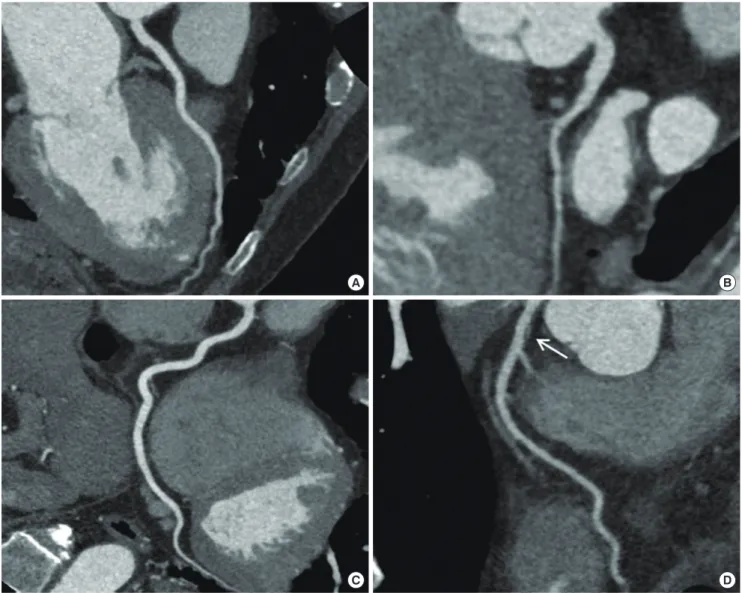
In terms of plaque constitution, previous studies reported that 34% and 41% of plaques were classified as noncalcified plaque, proportions that are higher than those of 21% and 24% in non- diabetic patients [6,13,14]. These noncalcified plaques were as- sociated with a family history of diabetes, and newly diagnosed patients tended to have a large volume of noncalcified plaque, especially patients with asymptomatic diabetes [15,16]. As- ymptomatic diabetic patients therefore appear to have a rela- tively high prevalence of noncalcified plaques, with these being mainly nonobstructive. Although such nonobstructive lesions are not associated with induced ischemia, plaque rupture may occur frequently in nonobstructive plaques (Fig. 1) [17].
Therefore, the detection of an increased nonobstructive plaque burden using CCTA may be clinically important in asymp- tomatic diabetes patients with a familial history of CAD and those patients who have been recently diagnosed with asymp- tomatic diabetes.
An analysis of the locations of CAD lesions revealed that 30.5% of patients had significant CAD, and that 16.5% of pa- tients showed a significant stenosis in the left main or proximal left anterior descending artery on CCTA [13]. Although as- ymptomatic diabetes patients showed a higher CAC score and more significant CAD than nondiabetic subjects, there were no significant differences in high-risk CAD, which was defined as at least 2-vessel CAD with proximal left anterior descending artery involvement, 3-vessel CAD, or left main disease [6].
These characteristics of coronary atherosclerotic plaque in as- ymptomatic CAD in diabetic patients were summarized in Ta- ble 1. According to the studies published so far, high-risk CAD involvement is not related to the presence of diabetes, although the volume of atherosclerosis or plaque invading the coronary arteries, and unstable features of plaque, are associated with di- abetes [6,13,18,19].
PROGRESSION AND PREDICTORS OF SUBCLINICAL CORONARY
ATHEROSCLEROSIS
Diabetic patients are characterized by the presence of an accel- erated progressive atheroma burden with a significantly in- creased incidence of CV events [20]. A recent study reported that asymptomatic patients with newly diagnosed diabetes had plaque features associated with increased vulnerability [16], a
finding that indicates that early-stage diabetic patients consti- tute an important therapeutic target for the prevention of fu- ture CV events. Several studies have investigated the predictors
of atherosclerosis progression and the impact of atherosclero- sis progression on CV events in diabetic patients. These have reported rates of atherosclerosis progression ranging from Table 1. Characteristics of coronary atherosclerotic plaque in asymptomatic coronary artery disease in patients with diabetic mel- litus
Asymptomatic diabetic patients have higher CAC score than nondiabetic patients.
A proportion of nonobstructive plaque is relatively high in asymptomatic diabetes, ranging from 70% to 80%.
Noncalcified plaque is a main constitution associated with a family history of diabetes.
Asymptomatic diabetic patients have more significant (≥50% stenosis) CAD than nondiabetic patients CAC, coronary artery calcium; CAD, coronary artery disease.
Fig. 1. Examples of different coronary computed tomography findings. Curved multiplanar reconstructions of the (A) left anteri- or descending (LAD) coronary artery, (B) left circumflex coronary artery, and (C) right coronary artery of a patient with normal coronary arteries. The curved multiplanar reconstruction of the LAD shows tubular coronary artery disease with a nonobstruc- tive and noncalcified lesion (arrow, D) in a newly diagnosed diabetic patient.
B
D A
C
8.8% to 29.6% during follow-up periods (about 2 years) [21- 23]. The determinants of progression of CAC in diabetic pa- tients were established as age, male gender, blood pressure, smoking, hyperlipidemia, waist-hip ratio, duration of diabetes, presence of retinopathy, statin use, white race, and Framing- ham risk score [21].
There are conflicting opinions over reducing CAC or coro- nary atherosclerosis in diabetic patients by strict glycemic con- trol. While one study did not find a decrease in the progression of CAC by intensive glycemic control [24], other studies re- ported that patients with suboptimal glycemic control had more progression of CAC, and that a poor prognosis was ob- served in those patients where coronary atherosclerosis had al- ready occurred [23,25]. A recent study revealed that, in well- controlled diabetic patients, blood pressure, statin use, and the length of coronary artery lesions were independent factors predicting progression of CAC and atherosclerosis [25]. There- fore, good glycemic control could be important in the progres- sion of CAC or atherosclerosis, and many clinical risk factors could aggravate plaque in asymptomatic diabetic patients.
SCREENING AND THE POTENTIAL BENEFIT OF CORONARY COMPUTED TOMOGRAPHY IN ASYMPTOMATIC DIABETES
Although it is an invasive and complicated technique, fraction- al flow reserve (FFR) is nowadays considered the most accu- rate method for detecting coronary ischemia. The Fractional Flow Reserve vs. Angiography for Multivessel Evaluation (FAME) trial demonstrated FFR to be a clinical and prognostic standard method, because its ability to guide decisions on re- vascularization resulted in better clinical outcomes than angio- graphic-guided revascularization [26]. In the FAME study, 47% of lesions were considered intermediate in stenosis severi- ty, with only one-third of these lesions resulting in coronary ischemia [27]. A prospective clinical head-to-head compara- tive study in suspected CAD patients compared the diagnostic accuracy of CCTA, single-photon emission computed tomog- raphy (SPECT), and positron emission tomography with ex- amination of all coronary arteries by FFR [28]. CCTA exhibit- ed a high sensitivity (90%) and negative predictive value (89%), even in comparison with FFR [28]. SPECT was limited by a high rate of false negatives in cases of balanced ischemia, such as 3-vessel disease or left main disease. This limitation was also identified in a recently published meta-analysis suggesting a
low sensitivity for SPECT in comparison with the FFR refer- ence standard [29]. Therefore, despite a lack of information on the coronary functional state, CCTA is a powerful noninvasive method for ruling out hemodynamically significant CAD.
Nevertheless, the most recent American Diabetic Associa- tion guidelines recommend against screening for CV disease in asymptomatic diabetic patients, because there is a paucity of data suggesting any specific benefits of invasive interventions over medical therapy alone [30]. However, many potential rea- sons for appropriate screening of obstructive CAD may still re- main. First, the identification of asymptomatic CAD by appro- priate screening could result in the provision of precise and ap- propriate treatment strategies for patients, such as suggesting more aggressive lifestyle modification and better medical or invasive interventions, efforts that may dramatically decrease the CV death rate. In addition, effective screening of obstruc- tive CAD could help prevent the various complications associ- ated with CAD, including heart failure and arrhythmias, and eventually the overall cost of managing diabetic patients may be significantly reduced. In patient trials, traditional individual risk factors for predicting CAD are subject to limitations such as overestimation and heterogeneity, and it takes a long time for diseases to present, and hence to evaluate risk factors. For these reasons, the detection of subclinical atherosclerosis or the screening of CAD in asymptomatic diabetic patients is valuable, and options has been suggested for overcoming sev- eral limitations of the traditional risk factors.
In the Detection of Ischemia in Asymptomatic Diabetics (DIAD) trial, no significant differences in cardiac death or non- fatal myocardial infarction were found between the screened and not-screened subjects, because the screening group showed a low rate of CV events that was below the expectation, as well as less coronary revascularizations [31]. One randomized trial has evaluated the use of CCTA in asymptomatic patients. The Effect of Screening for Coronary Artery Disease Using CT An- giography on Mortality and Cardiac Events in High-Risk Pa- tients with Diabetes (FACTOR-64 study) showed that patients with diabetes and large amounts of coronary atherosclerosis did not show reduced rates of CV events after changes in med- ical care, because the overall CV event rate was lower than ex- pected [32]. Another recent study, the Does Coronary Athero- sclerosis Deserve to be Diagnosed Early in Diabetic Patients (DADDY-D) study, enrolled diabetic patients without CAD, who were then randomly assigned to screening for silent myo- cardial ischemia followed by revascularization or to continuing
follow-up [33]. Again, only a small portion (4.6%) of the screened patients received revascularization, and therefore this study failed to demonstrate a significant reduction in CV events, a finding similar to those of previous studies (Table 2).
To date, there are no data demonstrating improvement in clini- cal outcomes with the use of CCTA screening in asymptomatic diabetic patients. Possible reasons for failure of previous stud- ies include a lower manifestation of CV events after optimal management during follow-up and coronary revasculariza- tion, which is not able to reduce future CV events in asymp- tomatic patients. Because of the low CV risk of asymptomatic diabetes, previous studies could not provide evidence demon- strating the effectiveness of screening using CCTA.
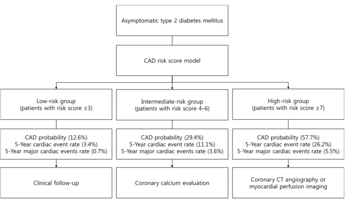
To overcome this, a risk-score model for the assessment of CAD was suggested for use in asymptomatic diabetes. Several
studies show that CCTA is an effective method for risk assess- ment and re-stratification in high-risk asymptomatic patients with diabetes [34-36]. After formulating a risk-score model with variables for significant CAD, the low- (≤3), intermedi- ate- (4 to 6), and high-risk (≥7) groups showed significant dif- ferences in 5-year cardiac event-free survival rate (96.6%±
1.5% vs. 88.9%±1.8% vs. 73.8%±4.1%, respectively; log-rank P for trend <0.001) and the probability of CAD (12.6% vs.
29.4% vs. 57.7%, respectively; P for all <0.001) [36]. On the basis of this CAD risk-score model, the authors proposed that further evaluations for CAD are not recommended for low- risk patients, that CAC scoring may be appropriate as a screen- ing test for intermediate-risk patients, and that CCTA might be considered as a first-line test for high-risk patients (Fig. 2) [36]. Another method suggested for overcoming the limita- Table 2. Descriptions of the studies for screening of asymptomatic coronary artery disease in patients with diabetic mellitus
Variable DIAD (2009) FACTOR-64 (2014) DADDY-D (2015)
No. of patients 1,123 899 520
Design of study Randomized, multicenter Randomized, multicenter Randomized, multicenter
Countries USA and Canada USA Italy
Duration of follow-up, yr 5 4 3.6
Screening method SPECT CCTA ETT
Results of screening
No. of screening arm 561 452 262
Positive screening 15 (83/561) 17 (76/452) 8 (20/262)
CAG related to positive screening 4 (25/561) 8 (36/452) 6 (17/262)
P roportion of patients with positive screening who
performed CAG 30 (25/83) 47 (36/76) 85 (17/20)
Outcomes
Cardiovascular death
Screening 1.4 (8/561) 1.5 (7/452) 0.4 (1/262)
No screening 1.2 (7/562) 1.8 (8/447) 1.9 (5/258)
Nonfatal myocardial infarction
Screening 1.3 (7/561) 1.5 (7/452) 4.2 (11/262)
No screening 1.7 (10/562) 1.8 (8/447) 4.7 (12/258)
Coronary revascularization
Screening 5.5 (31/561) 5.8 (26/452) 4.6 (12/262)
No screening 7.8 (44/562) 3.1 (14/447) 8.5 (22/258)
Values are presented as percentage (number/total number).
DIAD, Detection of Ischemia in Asymptomatic Diabetics; FACTOR-64, Screening for Asymptomatic Obstructive Coronary Artery Disease among High-Risk Diabetic Patients Using CT Angiography, Following Core 64; DADDY-D, Does Coronary Atherosclerosis Deserve to be Di- agnosed Early in Diabetic Patients; SPECT, single-photon emission computed tomography; CCTA, coronary computed tomography angiogra- phy; ETT, exercise tolerance test; CAG, coronary angiography.
tions shown by the screening studies is to use a noninvasive technique for the determination of FFR for selected coronary segments; this can be accomplished through analysis of CCTA [37]. CCTA-derived FFR enables noninvasive assessment of the hemodynamic significance of coronary artery lesions cou- pled with determination of the anatomical severity of a coro- nary stenosis. Prior multicenter trials showed CCTA-derived FFR to have a generally high diagnostic performance [38,39].
Although no study has reported on the diagnostic accuracy of CCTA-derived FFR in asymptomatic diabetic patients, we ex- pect that it will be a good method for risk assessment in high- risk asymptomatic diabetic patients.
CONCLUSIONS
In summary, the prevalence of SCA in diabetic patients is high, and the progression of coronary atherosclerosis leads to the onset of future CV events and is associated with a poor prog- nosis. Although CCTA screening has not yet been demon- strated as improving the outcomes of asymptomatic diabetic patients, it has been shown to be beneficial in predicting future risk, and is promising for screening with an additional tech- nique.
CONFLICTS OF INTEREST
No potential conflict of interest relevant to this article was re- ported.
REFERENCES
1. Gregg EW, Gu Q, Cheng YJ, Narayan KM, Cowie CC. Mortali- ty trends in men and women with diabetes, 1971 to 2000. Ann Intern Med 2007;147:149-55.
2. Bax JJ, Inzucchi SE, Bonow RO, Schuijf JD, Freeman MR, Bar- rett EJ; Global Dialogue Group for the Evaluation of Cardio- vascular Risk in Patients with Diabetes. Cardiac imaging for risk stratification in diabetes. Diabetes Care 2007;30:1295-304.
3. Kamalesh M, Feigenbaum H, Sawada S. Assessing prognosis in patients with diabetes mellitus: the Achilles’ heel of cardiac stress imaging tests? Am J Cardiol 2007;99:1016-9.
4. Elhendy A, Arruda AM, Mahoney DW, Pellikka PA. Prognos- tic stratification of diabetic patients by exercise echocardiogra- phy. J Am Coll Cardiol 2001;37:1551-7.
5. American College of Cardiology Foundation Task Force on Expert Consensus Documents, Mark DB, Berman DS, Budoff MJ, Carr JJ, Gerber TC, Hecht HS, Hlatky MA, Hodgson JM,
Coronary CT angiography or myocardial perfusion imaging Coronary calcium evaluation
Clinical follow-up CAD probability (12.6%) 5-Year cardiac event rate (3.4%) 5-Year major cardiac events rate (0.7%)
High-risk group (patients with risk score ≥7) Intermediate-risk group
(patients with risk score 4–6) Low-risk group
(patients with risk score ≤3)
CAD risk score model Asymptomatic type 2 diabetes mellitus
CAD probability (29.4%) 5-Year cardiac event rate (11.1%) 5-Year major cardiac events rate (3.6%)
CAD probability (57.7%) 5-Year cardiac event rate (26.2%) 5-Year major cardiac events rate (5.5%)
Fig. 2. Proposed algorithm for individualized coronary artery disease (CAD) screening in asymptomatic type 2 diabetes mellitus patients based on a risk-score model. Adapted from Park et al. [36]. CT, computed tomography.
Lauer MS, Miller JM, Morin RL, Mukherjee D, Poon M, Rubin GD, Schwartz RS. ACCF/ACR/AHA/NASCI/SAIP/SCAI/
SCCT 2010 expert consensus document on coronary comput- ed tomographic angiography: a report of the American College of Cardiology Foundation Task Force on Expert Consensus Documents. J Am Coll Cardiol 2010;55:2663-99.
6. Park GM, Lee JH, Lee SW, Yun SC, Kim YH, Cho YR, Gil EH, Kim TS, Kim CJ, Cho JS, Park MW, Her SH, Yang DH, Kang JW, Lim TH, Koh EH, Lee WJ, Kim MS, Lee KU, Kim HK, Choe J, Park JY. Comparison of coronary computed tomo- graphic angiographic findings in asymptomatic subjects with versus without diabetes mellitus. Am J Cardiol 2015;116:372-8.
7. Bild DE, Bluemke DA, Burke GL, Detrano R, Diez Roux AV, Folsom AR, Greenland P, Jacob DR Jr, Kronmal R, Liu K, Nel- son JC, O’Leary D, Saad MF, Shea S, Szklo M, Tracy RP. Multi- Ethnic Study of Atherosclerosis: objectives and design. Am J Epidemiol 2002;156:871-81.
8. Perk J, De Backer G, Gohlke H, Graham I, Reiner Z, Versch- uren M, Albus C, Benlian P, Boysen G, Cifkova R, Deaton C, Ebrahim S, Fisher M, Germano G, Hobbs R, Hoes A, Karaden- iz S, Mezzani A, Prescott E, Ryden L, Scherer M, Syvanne M, Scholte op Reimer WJ, Vrints C, Wood D, Zamorano JL, Zan- nad F; European Association for Cardiovascular Prevention &
Rehabilitation (EACPR); ESC Committee for Practice Guide- lines (CPG). European guidelines on cardiovascular disease prevention in clinical practice (version 2012). The fifth joint task force of the European Society of Cardiology and other so- cieties on cardiovascular disease prevention in clinical practice (constituted by representatives of nine societies and by invited experts). Eur Heart J 2012;33:1635-701.
9. Wolk MJ, Bailey SR, Doherty JU, Douglas PS, Hendel RC, Kramer CM, Min JK, Patel MR, Rosenbaum L, Shaw LJ, Stain- back RF, Allen JM; American College of Cardiology Founda- tion Appropriate Use Criteria Task Force. ACCF/AHA/ASE/
ASNC/HFSA/HRS/SCAI/SCCT/SCMR/STS 2013 multimo- dality appropriate use criteria for the detection and risk assess- ment of stable ischemic heart disease: a report of the American College of Cardiology foundation appropriate use criteria task force, American Heart Association, American Society of Echo- cardiography, American Society of Nuclear Cardiology, Heart Failure Society of America, Heart Rhythm Society, Society for Cardiovascular Angiography and Interventions, Society of Cardiovascular Computed Tomography, Society for Cardio- vascular Magnetic Resonance, and Society of Thoracic Sur- geons. J Am Coll Cardiol 2014;63:380-406.
10. Gobardhan SN, Dimitriu-Leen AC, van Rosendael AR, van Zwet EW, Roos CJ, Oemrawsingh PV, Kharagjitsingh AV, Juke- ma JW, Delgado V, Schalij MJ, Bax JJ, Scholte AJ. Prevalence by computed tomographic angiography of coronary plaques in South Asian and white patients with type 2 diabetes mellitus at low and high risk using four cardiovascular risk scores (UKP- DS, FRS, ASCVD, and JBS3). Am J Cardiol 2017;119:705-11.
11. Bild DE, Detrano R, Peterson D, Guerci A, Liu K, Shahar E, Ouyang P, Jackson S, Saad MF. Ethnic differences in coronary calcification: the Multi-Ethnic Study of Atherosclerosis (MESA).
Circulation 2005;111:1313-20.
12. Wagenknecht LE, Divers J, Bertoni AG, Langefeld CD, Carr JJ, Bowden DW, Elbein SC, Shea S, Lewis CE, Freedman BI. Cor- relates of coronary artery calcified plaque in blacks and whites with type 2 diabetes. Ann Epidemiol 2011;21:34-41.
13. Park GM, Lee SW, Cho YR, Kim CJ, Cho JS, Park MW, Her SH, Ahn JM, Lee JY, Park DW, Kang SJ, Kim YH, Lee CW, Koh EH, Lee WJ, Kim MS, Lee KU, Kang JW, Lim TH, Park SW, Park SJ, Park JY. Coronary computed tomographic angiographic find- ings in asymptomatic patients with type 2 diabetes mellitus.
Am J Cardiol 2014;113:765-71.
14. Scholte AJ, Schuijf JD, Kharagjitsingh AV, Jukema JW, Pun- dziute G, van der Wall EE, Bax JJ. Prevalence of coronary artery disease and plaque morphology assessed by multi-slice com- puted tomography coronary angiography and calcium scoring in asymptomatic patients with type 2 diabetes. Heart 2008;94:
290-5.
15. Park GM, Cho YR, Lee SW, Yun SC, Gil EH, Kim DW, Kim TS, Kim CJ, Cho JS, Park MW, Her SH, Kim YH, Yang DH, Kang JW, Lim TH, Jung CH, Koh EH, Lee WJ, Kim MS, Lee KU, Kim HK, Choe J, Park JY. Family history of diabetes and the risk of subclinical atherosclerosis. Diabetes Metab 2016;42:170- 7.
16. Mrgan M, Funck KL, Gaur S, Ovrehus KA, Dey D, Kusk MW, Nørgaard BL, Gram JB, Olsen MH, Gram J, Sand NPR. High burden of coronary atherosclerosis in patients with a new diag- nosis of type 2 diabetes. Diab Vasc Dis Res 2017;14:468-76.
17. Wackers FJ. Asymptomatic patients with diabetes mellitus should be screened for coronary artery disease. J Nucl Cardiol 2006;13:609-15.
18. Pundziute G, Schuijf JD, Jukema JW, Boersma E, de Roos A, van der Wall EE, Bax JJ. Prognostic value of multislice comput- ed tomography coronary angiography in patients with known or suspected coronary artery disease. J Am Coll Cardiol 2007;
49:62-70.
19. Min JK, Berman DS, Dunning A, Achenbach S, Al-Mallah M, Budoff MJ, Cademartiri F, Callister TQ, Chang HJ, Cheng V, Chinnaiyan K, Chow BJ, Cury R, Delago A, Feuchtner G, Had- amitzky M, Hausleiter J, Kaufmann P, Karlsberg RP, Kim YJ, Leipsic J, Lin FY, Maffei E, Plank F, Raff G, Villines T, Labounty TM, Shaw LJ. All-cause mortality benefit of coronary revascu- larization vs. medical therapy in patients without known coro- nary artery disease undergoing coronary computed tomo- graphic angiography: results from CONFIRM (COronary CT Angiography EvaluatioN For Clinical Outcomes: An InteRna- tional Multicenter Registry). Eur Heart J 2012;33:3088-97.
20. Nicholls SJ, Tuzcu EM, Kalidindi S, Wolski K, Moon KW, Sipa- hi I, Schoenhagen P, Nissen SE. Effect of diabetes on progres- sion of coronary atherosclerosis and arterial remodeling: a pooled analysis of 5 intravascular ultrasound trials. J Am Coll Cardiol 2008;52:255-62.
21. Anand DV, Lim E, Darko D, Bassett P, Hopkins D, Lipkin D, Corder R, Lahiri A. Determinants of progression of coronary artery calcification in type 2 diabetes role of glycemic control and inflammatory/vascular calcification markers. J Am Coll Cardiol 2007;50:2218-25.
22. Kiramijyan S, Ahmadi N, Isma’eel H, Flores F, Shaw LJ, Raggi P, Budoff MJ. Impact of coronary artery calcium progression and statin therapy on clinical outcome in subjects with and without diabetes mellitus. Am J Cardiol 2013;111:356-61.
23. Ndrepepa G, Iijima R, Kufner S, Braun S, Cassese S, Byrne RA, Sorges J, Schulz-Schupke S, Hoppmann P, Fussaro M, Laugwitz KL, Schunkert H, Kastrati A. Association of progression or re- gression of coronary artery atherosclerosis with long-term prognosis. Am Heart J 2016;177:9-16.
24. Saremi A, Moritz TE, Anderson RJ, Abraira C, Duckworth WC, Reaven PD; Veterans Affairs Diabetes Trial (VADT).
Rates and determinants of coronary and abdominal aortic ar- tery calcium progression in the Veterans Affairs Diabetes Trial (VADT). Diabetes Care 2010;33:2642-7.
25. Kataoka Y, Yasuda S, Miyamoto Y, Sase K, Kosuge M, Kimura K, Yoshimasa Y, Miyazaki S; DIANA study investigators. Clini- cal predictors of atheroma progression despite optimal glyce- mic control in early-stage diabetic patients with coronary ar- tery disease: insight from the DIANA study. J Atheroscler Thromb 2014;21:509-18.
26. De Bruyne B, Fearon WF, Pijls NH, Barbato E, Tonino P, Piroth Z, Jagic N, Mobius-Winckler S, Rioufol G, Witt N, Kala P, Mac- Carthy P, Engstrom T, Oldroyd K, Mavromatis K, Manoharan G, Verlee P, Frobert O, Curzen N, Johnson JB, Limacher A,
Nuesch E, Juni P; FAME 2 Trial Investigators. Fractional flow reserve-guided PCI for stable coronary artery disease. N Engl J Med 2014;371:1208-17.
27. Tonino PA, Fearon WF, De Bruyne B, Oldroyd KG, Leesar MA, Ver Lee PN, Maccarthy PA, Van’t Veer M, Pijls NH. Angio- graphic versus functional severity of coronary artery stenoses in the FAME study fractional flow reserve versus angiography in multivessel evaluation. J Am Coll Cardiol 2010;55:2816-21.
28. Danad I, Raijmakers PG, Driessen RS, Leipsic J, Raju R, Naoum C, Knuuti J, Maki M, Underwood RS, Min JK, Elmore K, Stuijfzand WJ, van Royen N, Tulevski II, Somsen AG, Huis- man MC, van Lingen AA, Heymans MW, van de Ven PM, van Kuijk C, Lammertsma AA, van Rossum AC, Knaapen P. Com- parison of coronary CT angiography, SPECT, PET, and hybrid imaging for diagnosis of ischemic heart disease determined by fractional flow reserve. JAMA Cardiol 2017;2:1100-7.
29. Danad I, Szymonifka J, Twisk JWR, Norgaard BL, Zarins CK, Knaapen P, Min JK. Diagnostic performance of cardiac imag- ing methods to diagnose ischaemia-causing coronary artery disease when directly compared with fractional flow reserve as a reference standard: a meta-analysis. Eur Heart J 2017;38:991- 8.
30. Fox CS, Golden SH, Anderson C, Bray GA, Burke LE, de Boer IH, Deedwania P, Eckel RH, Ershow AG, Fradkin J, Inzucchi SE, Kosiborod M, Nelson RG, Patel MJ, Pignone M, Quinn L, Schauer PR, Selvin E, Vafiadis DK; American Heart Associa- tion Diabetes Committee of the Council on Lifestyle and Car- diometabolic Health; Council on Clinical Cardiology, Council on Cardiovascular and Stroke Nursing, Council on Cardiovas- cular Surgery and Anesthesia, Council on Quality of Care and Outcomes Research; American Diabetes Association. Update on prevention of cardiovascular disease in adults with type 2 diabetes mellitus in light of recent evidence: a scientific state- ment from the American Heart Association and the American Diabetes Association. Diabetes Care 2015;38:1777-803.
31. Bansal S, Wackers FJ, Inzucchi SE, Chyun DA, Davey JA, Staib LH, Young LH; DIAD Study Investigators. Five-year outcomes in high-risk participants in the Detection of Ischemia in As- ymptomatic Diabetics (DIAD) study: a post hoc analysis. Dia- betes Care 2011;34:204-9.
32. Muhlestein JB, Lappe DL, Lima JA, Rosen BD, May HT, Knight S, Bluemke DA, Towner SR, Le V, Bair TL, Vavere AL, Ander- son JL. Effect of screening for coronary artery disease using CT angiography on mortality and cardiac events in high-risk pa- tients with diabetes: the FACTOR-64 randomized clinical trial.
JAMA 2014;312:2234-43.
33. Turrini F, Scarlini S, Mannucci C, Messora R, Giovanardi P, Magnavacchi P, Cappelli C, Evandri V, Zanasi A, Romano S, Cavani R, Ghidoni I, Tondi S, Bondi M. Does coronary Ath- erosclerosis Deserve to be Diagnosed earlY in Diabetic pa- tients? The DADDY-D trial. Screening diabetic patients for un- known coronary disease. Eur J Intern Med 2015;26:407-13.
34. Min JK, Labounty TM, Gomez MJ, Achenbach S, Al-Mallah M, Budoff MJ, Cademartiri F, Callister TQ, Chang HJ, Cheng V, Chinnaiyan KM, Chow B, Cury R, Delago A, Dunning A, Feuchtner G, Hadamitzky M, Hausleiter J, Kaufmann P, Kim YJ, Leipsic J, Lin FY, Maffei E, Raff G, Shaw LJ, Villines TC, Berman DS. Incremental prognostic value of coronary com- puted tomographic angiography over coronary artery calcium score for risk prediction of major adverse cardiac events in as- ymptomatic diabetic individuals. Atherosclerosis 2014;232:298- 304.
35. Fujimoto S, Kondo T, Kodama T, Orihara T, Sugiyama J, Kon- do M, Endo A, Fukazawa H, Nagaoka H, Oida A, Ikeda T, Yamazaki J, Takase S, Narula J. Coronary computed tomogra- phy angiography-based coronary risk stratification in subjects presenting with no or atypical symptoms. Circ J 2012;76:2419- 25.
36. Park GM, An H, Lee SW, Cho YR, Gil EH, Her SH, Kim YH,
Lee CW, Koh EH, Lee WJ, Kim MS, Lee KU, Kang JW, Lim TH, Park SW, Park SJ, Park JY. Risk score model for the assess- ment of coronary artery disease in asymptomatic patients with type 2 diabetes. Medicine (Baltimore) 2015;94:e508.
37. Min JK, Taylor CA, Achenbach S, Koo BK, Leipsic J, Norgaard BL, Pijls NJ, De Bruyne B. Noninvasive fractional flow reserve derived from coronary CT angiography: clinical data and sci- entific principles. JACC Cardiovasc Imaging 2015;8:1209-22.
38. Norgaard BL, Leipsic J, Gaur S, Seneviratne S, Ko BS, Ito H, Jensen JM, Mauri L, De Bruyne B, Bezerra H, Osawa K, Mar- wan M, Naber C, Erglis A, Park SJ, Christiansen EH, Kaltoft A, Lassen JF, Botker HE, Achenbach S; NXT Trial Study Group.
Diagnostic performance of noninvasive fractional flow reserve derived from coronary computed tomography angiography in suspected coronary artery disease: the NXT trial (analysis of coronary blood flow using CT angiography: next steps). J Am Coll Cardiol 2014;63:1145-55.
39. Leipsic J, Yang TH, Thompson A, Koo BK, Mancini GB, Taylor C, Budoff MJ, Park HB, Berman DS, Min JK. CT angiography (CTA) and diagnostic performance of noninvasive fractional flow reserve: results from the Determination of Fractional Flow Reserve by Anatomic CTA (DeFACTO) study. AJR Am J Roentgenol 2014;202:989-94.