bloodresearch.or.kr Blood Res 2016;51:204-14.
Letters to the Editor 213
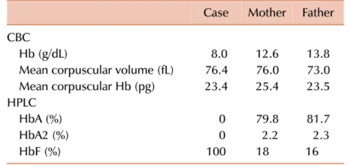
Table 1. Laboratory parameters of the case and parents.
Case Mother Father CBC
Hb (g/dL) 8.0 12.6 13.8
Mean corpuscular volume (fL) 76.4 76.0 73.0 Mean corpuscular Hb (pg) 23.4 25.4 23.5 HPLC
HbA (%) 0 79.8 81.7
HbA2 (%) 0 2.2 2.3
HbF (%) 100 18 16
Abbreviations: CBC, complete blood count; Hb, hemoglobin;
HPLC, high-performance liquid chromatography.
2011;26:162-5.
7. Cho HY, Lee BS, Moon KC, Ha IS, Cheong HI, Choi Y. Complete factor H deficiency-associated atypical hemolytic uremic syn- drome in a neonate. Pediatr Nephrol 2007;22:874-80.
8. Frémeaux-Bacchi V, Miller EC, Liszewski MK, et al. Mutations in complement C3 predispose to development of atypical hemo- lytic uremic syndrome. Blood 2008;112:4948-52.
9. Fan X, Yoshida Y, Honda S, et al. Analysis of genetic and predis- posing factors in Japanese patients with atypical hemolytic ure- mic syndrome. Mol Immunol 2013;54:238-46.
10. Matsukuma E, Imamura A, Iwata Y, et al. Postoperative atypical hemolytic uremic syndrome associated with complement c3 mutation. Case Rep Nephrol 2014;2014:784943.
11. Lhotta K, Janecke AR, Scheiring J, et al. A large family with a gain-of-function mutation of complement C3 predisposing to atypical hemolytic uremic syndrome, microhematuria, hyper- tension and chronic renal failure. Clin J Am Soc Nephrol 2009;4:1356-62.
12. Köse O, Zimmerhackl LB, Jungraithmayr T, Mache C, Nürnberger J. New treatment options for atypical hemolytic uremic syn- drome with the complement inhibitor eculizumab. Semin Thromb Hemost 2010;36:669-72.
13. Al-Akash SI, Almond PS, Savell VH Jr, Gharaybeh SI, Hogue C.
Eculizumab induces long-term remission in recurrent post-transplant HUS associated with C3 gene mutation. Pediatr Nephrol 2011;26:613-9.
Delta beta thalassemia:
a rare hemoglobin variant
TO THE EDITOR: Delta beta ()-thalassemia results from a deletion in both the delta and beta genes on chromosome 11. The gamma genes on the affected chromosome increase their production of gamma globin, thereby increasing the amount of hemoglobin F (HbF). -Thalassemia hetero- zygotes clinically show characteristics of thalassemia minor.
However, homozygous -thalassemia may give a clinical picture of thalassemia intermedia with a mild anemia.
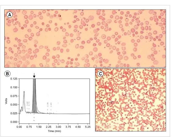
A 12-month-old boy presented to the hematology out- patient department for evaluation of pallor and jaundice that had been for the past 2 months. He had no history of a blood transfusion. His family history was insignificant for congenital anemia. His parents had a consanguineous marriage. Physical examination revealed pallor and palpable spleen 2 cm below the left costal margin. Other examination findings were unremarkable. A complete blood count (CBC) revealed an Hb level of 8.0 g/dL, WBC count of 8.9×10⁹/L, and platelet count of 341×109/L (Table 1). A peripheral blood smear revealed anisopoikilocytosis with hypochromic mi- crocytic red cells, target cells, and basophilic stippling (Fig.
1A). The corrected reticulocyte count was 1.6%. Liver func- tion tests showed raised levels of serum total bilirubin (3.5 mg/dL) and indirect bilirubin (3.0 mg/dL). High-perform-
ance liquid chromatography (HPLC) showed 100% HbF and an absence of HbA and HbA2 (Fig. 1B). The Kleihauer- Bekte test revealed a pancellular pattern (Fig. 1C). Conse- quently, a CBC followed by HPLC was performed for both parents who were apparently healthy and had no history of blood transfusions (Table 1). Kleihauer-Betke tests of both parents showed a heterocellular distribution of HbF.
Hence, the patient was diagnosed with homozygous
-thalassemia, whereas the parents with heterozygous
-thalassemia. Unfortunately, mutational analysis could not be performed because the patient was lost to follow-up.
-Thalassemia results from the deletion of both and
genes. Homozygotes for -thalassemia have 100% HbF and, because of the increased synthesis of HbF, may have thalassemia intermedia rather than thalassemia major [1].
However, the phenotype of heterozygotes resembles that of the -thalassemia trait, but the HbA2 percentage is not increased and is often normal. HbF in such individuals is consistently elevated, varying from 5% to 20%. Peripheral blood film findings are similar to those for the -thalassemia trait, and the distribution of HbF is heterocellular, which is best observed via flow cytometry. It is necessary to dis- tinguish it from hereditary persistence of fetal hemoglobin (HPFH). The two groups of disorders are distinguished by the phenotype of heterozygous individuals. Heterozygotes of -thalassemia mutations have 5% to 20% HbF, which is heterocellularly distributed in red cells, whereas hetero- zygotes of HPFH mutations have 17% to 30% HbF, with a pancellular distribution. In addition, homozygotes of HPFH are asymptomatic, whereas -thalassemic homo- zygotes have thalassemia intermedia-like features [2].
At least nine mutations can result in -thalassemia. This type of thalassemia is observed in many ethnic groups, in- cluding some Mediterranean populations (Italians, Greeks, and Turks). Although the exact diagnosis of -thalassemia requires genetic analysis for mutations, Hb electrophoresis or HPLC findings of markedly elevated HbF may be suggestive. An extensive PubMed search was done to de- termine the incidence of -thalassemia in different parts of the world, but owing to the rarity of this Hb variant,
Blood Res2016;51:204-14. bloodresearch.or.kr
214 Letters to the Editor
Fig. 1. (A) Peripheral blood smear of the patient with anisopoikilo- cytosis and target cells. (B) High- performance liquid chromatography showing 100% HbF indicated by the arrow. (C) Pan-cellular pattern on the Kleihauer-Bekte test.
only a handful of case reports were identified from across the world [3-6].
Huma Mansoori, Sidra Asad, Anila Rashid, Farheen Karim
Department of Haematology, Aga Khan University Hospital, Karachi, Pakistan Correspondence to: Huma Mansoori Department of Haematology, Aga Khan University Hospital, Stadium Road P. O. Box 3500 Karachi 74800, Pakistan E-mail: [email protected]
Received on Jul. 23, 2015; Revised on Nov. 26, 2015; Accepted on Jan. 10, 2016 http://dx.doi.org/10.5045/br.2016.51.3.213
AuthorsÊ Disclosures of Potential Conflicts of Interest No potential conflicts of interest relevant to this article were reported.
REFERENCES
1. Bain BJ. Haemoglobinopathy diagnosis. 2nd ed. Oxford, UK:
Blackwell Publishing Ltd, 2006:116-24.
2. Bollekens JA, Forget BG. Delta beta thalassemia and hereditary persistence of fetal hemoglobin. Hematol Oncol Clin North Am 1991;5:399-422.
3. Khunger JM, Gupta M, Singh R, Kapoor R, Pandey HR.
Haematological characterisation and molecular basis of asian Indian inversion deletions delta Beta thalassemia: a case report.
J Clin Diagn Res 2014;8:FD01-2.
4. Verma S, Bhargava M, Mittal S, Gupta R. Homozygous delta-beta thalassemia in a child: a rare cause of elevated fetal hemoglobin.
Iran J Ped Hematol Oncol 2013;3:222-7.
5. Ramot B, Ben-Bassat I, Gafni D, Zaanoon R. A family with three beta-delta-thalassemia homozygotes. Blood 1970;35:158-65.
6. Silvestroni E, Bianco I, Reitano G. Three cases of homozygous be- ta, delta-thalassemia (or microcythaemia) with high haemoglo- bin F in a Sicilian family. Acta Haematol 1968;40:220-9.