• 검색 결과가 없습니다.

Caspase-8 Potentiates Triglyceride (TG)-Induced Cell Death of THP-1 Macrophages via a Positive Feedback Loop

N/A
N/A
Protected

Academic year: 2021

Share "Caspase-8 Potentiates Triglyceride (TG)-Induced Cell Death of THP-1 Macrophages via a Positive Feedback Loop"

Copied!
7
0
0

로드 중.... (전체 텍스트 보기)

전체 글

(1)

Corresponding author: Yoon Suk Kim

Department of Biomedical Laboratory Science, College of Software and Digital Healthcare Convergence, Yonsei University, 1 Yeonsedae-gil, Heungeop-myeon, Wonju 26493, Korea

E-mail: yoonsukkim@yonsei.ac.kr

ORCID: https://orcid.org/0000-0002-1956-0126

These authors contributed equally to this work.

ORIGINAL ARTICLE

Caspase-8 Potentiates Triglyceride (TG)-Induced Cell Death of THP-1 Macrophages via a Positive Feedback Loop

Byung Chul Jung 1,2,† , Jaewon Lim 2,3,† , Sung Hoon Kim 2,4 , Yoon Suk Kim 2

1

Department of Nutritional Sciences and Toxicology, University of California, Berkeley, United States

2

Department of Biomedical Laboratory Science, College of Software and Digital Healthcare Convergence, Yonsei University, Wonju, Korea

3

Department of Biomedical Laboratory Science, College of Rehabilitation and Health, Daegu Haany University, Gyeongsan, Korea

4

Department of Biomedical Laboratory Science, Korea Nazarene University, Cheonan, Korea

Caspase-8의 양성 피드백 방식을 통한 중성지방-유도 THP-1 대식세포 사멸 증가

정병출 1,2,† , 임재원 2,3,† , 김성훈 2,4 , 김윤석 2

1

캘리포니아대학교 버클리캠퍼스 영양과학 및 독성학과,

2

연세대학교 소프트웨어디지털헬스케어융합대학 임상병리학과,

3

대구한의대학교 재활보건대학

임상병리학과,

4

나사렛대학교 임상병리학과

ARTICLE INFO ABSTRACT

Received February 25, 2021 Revised April 5, 2021 Accepted April 13, 2021

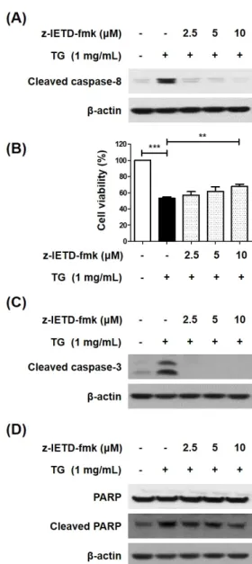
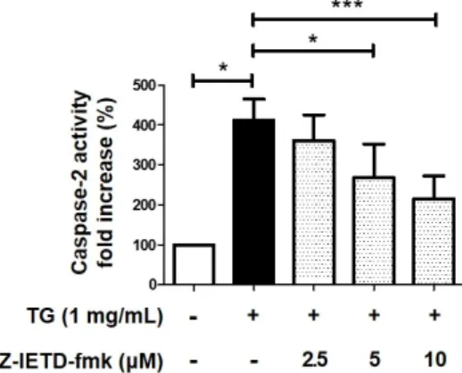
Hypertriglyceridemia is the main risk factor for atherosclerosis. It is reported that triglyceride (TG) induces macrophage cell death, and is involved in the formation of plaques and development of atherosclerosis. We previously reported that TG-induced cell death of macrophages is mediated via pannexin-1 activation, which increases the extracellular ATP and subsequent increase in potassium efflux, thereby activating the caspase-2/caspase-1/apoptotic caspases, including the caspase-8 pathway. Contrarily, some studies have reported that caspase-8 is an upstream molecule of caspase-1 and caspase-2 in several cellular processes. Therefore, this study was undertaken to investigate whether caspase-8 influences its upstream molecules in TG-stimulated macrophage cell death. We first confirmed that caspase-8 induces caspase-3 activation and poly ADP-ribose polymerase (PARP) cleavage in TG-treated macrophages. Next, we determined that the inhibition of caspase-8 results in reduced caspase-1 and -2 activity, which are upstream molecules of caspase-8 in TG-induced cell death of macrophages. We also found that ATP treatment restores the caspase-8 inhibitor-induced caspase-2 activity, thereby implying that caspase-8 affects the upstream molecules responsible for increasing the extracellular ATP levels in TG-induced macrophage cell death. Taken together, these findings indicate that caspase-8 potentiates the TG-induced macrophage cell death by activating its upstream molecules.

Copyright Ⓒ 2021 The Korean Society for Clinical Laboratory Science. All rights reserved.

Key words Caspase-1 Caspase-2 Caspase-8 THP-1 macrophages Triglyceride

INTRODUCTION

Atherosclerosis is a chronic inflammatory disorder, which can be driven by the abnormal recruitment of circulating immune cells such as monocyte [1]. The recruited monocytes are infiltrated and thus differen-

Korean Society for Clinical Laboratory Science

(2)

tiated into macrophages, which has been considered as indispensable immune cells throughout all the stages of atherosclerosis development [2]. In the initial and mid-stage of atherosclerosis, macrophage engulfs excessive lipids such as oxidative low-density lipo- proteins (Oxi-LDL) and triglyceride (TG) and become foam cells [3]. In the advanced stage, macrophage undergoes prominent death resulting in vulnerable plaques which trigger a life-threatening thrombosis.

Recently, we reported that TG activates pannexin-1 to release ATP into extracellular space, which subsequently activates ATP-sensitive potassium channels leading to an increase in potassium efflux. The imbalance of potassium ion triggered caspase-2 activation and subsequent activation of caspase-1 in a sequential manner, which propagates further caspase processing including caspase-8 and eventually results in cell death of THP-1 macrophages [4].

Caspases are the unique family of cysteine proteases executing programmed cell death known as apoptosis [5]. Classically, caspases are categorized by three functional groups; inflammatory caspases (caspase-1, -4, and -5), apoptotic initiator caspases (caspase-2, -8, -9 and -10), apoptotic effector caspases (caspase-3, -6, and -7) [6]. Initiator caspase can be activated by apoptotic stimuli and cleaved effector caspase to procced to apoptosis. Although both caspase-2 and caspase-8 are categorized in the same group, several investigations showed caspase-2 acted as an upstream molecule of caspase-8 [4, 7, 8]. However, another study showed caspase-8 cleaved procaspase-2 in HeLa cells during apoptosis, which implies caspase-8 can act as the upstream molecule of caspase-2 [9]. In addition, it has been reported that caspase-8 plays as an essential factor in caspase-1 activation in response to bacterial infection [10]. Hence, in this study, we investigated whether caspase-8 can be upstream of caspase-1 and -2 in TG-induced macrophage cell death.

MATERIALS AND METHODS

1. Reagents

TG emulsion (Lipofundin MCT/LCT 20%) was purchased from B. Braun Melsungen AG (Melsungen, Hessen, Germany). Lipofundin was used to transform THP-1 macrophages into foam cells as previously described [11]. For convenience, Lipofundin will be referred to as TG emulsion or TG. Ac-YVAD-pNA (caspase-1 substrate) was purchased from Biomal (Plymouth Meeting, PA, USA). Ac-VAVAD-pNA (caspase-2 substrate) and adenosine-5’-triphospate (ATP) were purchased from Sigma-Aldrich (St. Louis, MO, USA).

The caspase-8 specific inhibitor, Z-IETD-fmk was obtained from BioVision (Mountain View, CA, USA).

Primary antibodies used for Western blotting are as follows; caspase-1, -3 and -8, as well as PARP (Cell signaling technology; Denvers, MA, USA).

2. Cell culture

The human monocytic cell line, THP-1 (ATCC, Manassas, VA, USA) was and differentiated into macro- phages as previously described [4]. Briefly, THP-1 cells were cultured in RPMI 1640 supplemented with penicillin-streptomycin (Thermo Fisher Scientific, MA, USA) and 10% (v/v) fetal bovine serum (FBS; Thermo Fisher Scientific) and incubated at 37°C in a humidified atmosphere with 5% CO 2 . To allow the THP-1 cells to differentiate into macrophages, cells were seeded in 6-well plates at a density of 1×10 6 cells/well and incubated with 200 nM of phorbol-myristate-acetate (PMA) for 48 h.

3. Trypan blue dye exclusion assay

To enumerate viable cells, cells were trypsinized and

10 μL of 0.4% trypan blue stain solution was mixed with

10 μL of the trypsinized cell suspension. Non-stained

cells in the resulting mixture were counted using

hemocytometer (Marienfeld, Lauda-Königshofen, Ger-

many).

(3)

4. Western blot analysis

THP-1 cells were washed with PBS dissolved in lysis buffer contained with 1% Triton X-100, protease inhibitor cocktail (Sigma-Aldrich), phosphatase inhibitor cocktail (Roche, Mannheim, Germany), and PBS. Cell lysates clarified and subjected to Western blotting as described previously [12].

5. Measurement of caspase-1 and caspase-2 activities The activity of caspase-1 and -2 was measured as previously described [13]. Briefly, THP-1 cells were harvested and re-suspended in cell lysis buffer supplemented 1% Triton-X 100. Cell lysates were centrifuged at 19,000 g for 10 min at 4°C. The super- natant was collected and the protein concentrations were determined using Lowry protein assay kit (Bio-Rad, Hercules, CA, USA). To determined caspase-1 activity, 90 μg of protein samples were mixed with 200 μM of the Ac-YVAD-pNA substrate in 150 μL of PBS. To determined caspase-2 activity, protein samples were mixed with the Ac-VAVAD-pNA substrate in PBS. After incubation at 37°C for 3 h, the activity was determined by measuring the absorbance at 405 nm.

6. Statistical analysis

Quantified data were statistically evaluated by student’s t-test using GraphPad Prism 5 (GraphPad Software Inc., San Diego, CA, USA). Values are shown as the mean and standard error of the mean (SEM). Each experiment was conducted three times and the date were pooled for analysis. Differences were considered to be statistically significant at *P<0.05, **P<0.01, or

***P<0.001.

RESULTS

1. Caspase-8 induces the activation of caspase-3 and the cleavage of PARP in TG-treated THP-1 macrophages

We previously reported that TG activates caspase-8

and -3 to induce cell death in THP-1 macrophages [14].

Caspase-8 participates in extrinsic apoptotic pathways leading to cell death through the direct cleavage of caspase-3 [15]. Therefore, we investigated the impact of the TG-mediated activation of caspase-8 on caspase-3 activation and subsequent PARP cleavage in THP-1 macrophages. First, we confirmed that the caspase-8 inhibitor Z-IETD-fmk inhibites the activity of caspase-8 in TG-treated THP-1 macrophages (Figure 1A). Furthermore, when macrophages were treated with Z-IETD-fmk, TG-induced cell death was restored in a dose-dependent manner (Figure 1B). Then, we found that the inhibition of caspase-8 by Z-IETD-fmk causes a decrease in the TG-mediated cleavage of caspase-3 and PARP in THP-1 macrophages (Figure 1C and 1D). These results indicate that caspase-8 activates caspase-3, which in turn cleave PARP, leading to cell death in TG-treated THP-1macrophages.

2. Caspase-8 is involved in the activation of caspase-1 in TG-induced macrophage cell death We had previously reported that the caspase-1 induces caspase-8 activation in TG-treated macrophage cell death [14]. Meanwhile, there were some reports showing that caspase-8 is an upstream molecule required in the activation of caspase-1 [16]. Therefore, we examined wheather caspase-8 induces the activation of caspase-1 in TG-treated THP-1 macro- phages. To this end, TG-treated THP-1 macrophages were incubated with the caspase-8 inhibitor, Z-IETD-FMK for 24 h and the activity of caspase-1 was measured. Caspase-1 activity and the cleavage of caspase-1 were decreased in cells treated with Z-IETD- fmk in an inhibitor dose-dependent manner (Figure 2A and 2B). These results imply that caspase-8 is not only activated by caspase-1 but also activates caspase-1 in TG-treated THP-1 macrophages.

3. Caspase-8 is implicated in caspase-2 activation in TG-treated THP-1 macrophages

We have also reported that caspase-2 activates

(4)

Figure 1. Caspase-8 activates effector caspases and cleaves PAPR in TG-accumulated THP-1 macrophages. THP-1 cells were differen- tiated into macrophages by treatment of 200 nM PMA for 48 h. The differentiated THP-1 macrophages were incubated with TG in the absence or presence of the caspase-8 inhibitor, Z-IETD-fmk for additional 24 h. (A) Activation of caspase-8 was detected by Western blotting. β-actin was used as an internal control. (B) Viable cells were enumerated by the trypan blue dye exclusion assay. The number of viable cells in THP-1 macrophages without TG treatment was set as 100%. (C) Activation of caspase-3 and (D) cleaved form of PARP was detected by Western blotting. All data are expressed as the mean±SEM of three independent experiments. P -values were determined with Student’s t-test. ** P <0.01, *** P <0.001.

Figure 2. Caspase-8 is involved in activation of caspase-1 in TG-induced THP-1 cell death. (A) THP-1 macrophages were incubated with TG in the presence of the Z-IETD-fmk for 24 h, after which caspase-1 activity was assessed. The absorbance of THP-1 cells without TG was set to 100%. (B) Cleavage of caspase-1 was detected by Western blotting. All data are expressed as the mean±SEM of three independent experiments. P -values were determined with Student’s t-test. * P <0.05, ** P <0.01.

caspase-1, which in turn induces caspase-8 activation in TG-induced macrophage cell death [14]. On the other hand, some studies have reported that caspase-2 is a substrate of caspase-8 and is activated by it [17, 18].

Therefore, we investigated wheather caspase-8 is

associated with caspase-2 activation in TG-treated THP-1 macrophages. To investigate whether caspase-8 can act as upstream molecule of caspase-2 in TG-stimulated macrophage, Z-IETD-fmk was challenged to TG-treated THP-1 macrophages for 24 h. The result showed that TG-mediated caspase-2 activation was rescued by caspase-8 inhibitor in a dose-dependent manner (Figure 3). These data indicate that the TG-mediated caspase-8 activation is an upstream of caspase-2.

4. Caspase-8 acts as an upstream molecule of pannexin-1 in TG-stimulated cell death of macrophages

We recently showed that TG treatment increases the release of ATP from cells through pannexin-1 channels, which in turn induces potassium efflux, leading to activation of the caspase-2/caspase-1/

apoptotic caspases/PARP pathway [4]. To test whether

caspase-8 contributes to the ATP-dependent activation

(5)

Figure 3. Caspase-8 is implicated in caspase-2 activation in TG-treated THP-1 macrophages. THP-1 macrophages were incubated with TG in the presence of the Z-IETD-fmk for 24 h, and caspase-2 activity was assessed. The absorbance of THP-1 cells without TG was set to 100%. These data are expressed as the mean±SEM of three independent experiments. P -values were determined with Student’s t-test. * P <0.05, *** P <0.001.

Figure 4. Caspase-8 acts as an upstream molecule of ATP- dependent caspase-2 activation in TG-stimulated cell death of macrophages. THP-1 macrophages were incubated with TG in the absence or presence of the Z-IETD-fmk and ATP for 24 h, after which caspase-2 activity was assessed. The absorbance of THP-1 cells without TG treatment was set to 100%. These data are expressed as the mean±SEM of three independent experiments. P -values were determined with Student’s t-test. * P <0.05, *** P <0.001.

of apoptotic pathway involving caspase-2, TG-treated THP-1 macrophages were incubated with Z-IETD-fmk in the absence or presence of ATP for 24 h and caspase-2 activity was measured. Caspase-2 activity was decreased by treatment with caspase-8 inhibitor and restored by the addition of the extracellular ATP (Figure 4). This finding suggested that the mechanism of caspase-8 mediated caspase-2 activation was involved in either increase of extracellular ATP or activation its upstream molecules. Taken together, we suggest that caspase-8, which was previously reported to be activated via pannexin-1/caspase-2/caspase-1 pathway in TG-induced macrophage cell death, can be inversely involved in the activation of its upstream molecules, caspase-2 and caspase-1. Thus, feed forward loop may exist and potentiate TG-induced macrophage cell death.

DISCUSSION

Caspase-8 is one of the essential initiator caspases which play a pivotal role in extrinsic apoptotic signaling via cell surface death-receptor such as Fas [19]. It has been well studied that activated caspase-8 cleaves the effector caspases such as caspase-3, -6, and -7 to activate those caspases, which eventually induce

cell death upon apoptotic stimulation [20]. Here, we showed that TG-mediated activation of caspase-8 can lead to activation of caspase-1 and -2, which contributes to TG mediated apoptosis in the human macrophage cell line.

Recently, our group showed that TG treatment stimulates the secretion of ATP into extracellular space via pannexin-1 activation [4]. The released extracellular ATP promoted upregulation of the ATP sensitive potassium channel and subsequent activation of the caspase cascade. In the present study, we showed that caspase-8 mediated caspase-2 activation, which was involved in increased extracellular ATP. Interestingly, another group also observed that FasL-mediated activation of caspase-8 induces activation of potassium channel and pannexin-1 in human T lymphocyte cell line [21]. In addition, they also reported a similar phenomenon to the present study that ATP release was observed in caspase-8 dependent manner, which in turn promoted caspase cascade to cause cell death.

Meanwhile, it was reported that caspase-8 can cleave

another potassium channel, tandem-pore domain

halothane-inhibited K+ channel 1 (THIK-1), in

response to apoptotic stimuli [22]. During apoptosis,

this THIK-1 cleavage is known to contribute to cell

shrinkage, which is a typical hallmark of apoptosis.

(6)

In conclusion, the current study suggests the additional mechanism of how TG induces apoptosis in the human macrophage cell line, which is associated with caspase-8-mediated increase of extracellular ATP.

Since apoptosis of macrophage contributes to the development of vulnerable plaques in advanced atherogenesis, our results may provide evidence for the role of TG in the atherogenic risk factor.

요 약

고중성지방혈증은 죽상동맥경화증의 주요한 위험 요인 중 하 나이다. 중성지방은 대식세포의 세포 사멸을 유도하여 죽상동 맥경화증 발생에 기여하는 것으로 알려져 있다. 본 연구팀은 앞 선 연구에서 대식세포의 중성지방-유도 세포 사멸이 pannexin-1 활성화에 의한 세포 외 ATP 농도 증가, caspase-2와 caspase-1 활성화, caspase-8을 포함한 apoptotic caspase 활성화 경로 로 일어나는 것을 보고하였다. 한편 다른 연구들에서는 세포 내 다른 여러 기전에서 caspase-8이 caspase-1과 -2의 상위 단 백질이라 보고하고 있다. 따라서 본 연구에서는 caspase-8이 중성지방-유도 대식세포 사멸 과정에서 상위단백지로 영향을 미치는지 여부를 조사하기 위해 수행되었다. 본 연구진은 caspase-8이 중성지방-유도 대식세포 사멸 과정에서 caspase-3 활성화 및 PARP 절단을 유도하였다. 다음으로 중성지방이 처리 된 대식세포에서 caspase-8 억제 시, caspase-8의 상위 단백 질로 보고한 caspase-1 및 -2의 활성이 감소하는 것을 확인하 였다. 또한 ATP 처리 시 caspase-8 억제제 처리에 의해 감소된 caspase-2의 활성이 회복되는 것을 확인하였다. 위의 결과를 통해 caspase-8이 중성지방-유도 대식세포 사멸 과정에서 세 포 외부 ATP 농도 증가에 관여하는 단백질 또는 그 상위 기전에 양성피드백 방식으로 영향을 미쳐 caspase-1과 -2를 활성화하 여 중성지방-유도 대식세포 사멸을 증진시킴을 알 수 있다.

Acknowledgements: This Research was supported by the Korea Nazarene University Research Grants 2021.

Conflict of interest: None

Author’s information (Position): Jung BC 1,2 , Researcher;

Lim J 2,3 , Professor; Kim SH 2,4 , Professor; Kim YS 2 , Professor.

REFERENCES

1. Martinet W, Coornaert I, Puylaert P, De Meyer GRY. Macrophage death as a pharmacological target in atherosclerosis. Front Pharmacol. 2019;10:306. https://doi.org/10.3389/fphar.2019.00306 2. Tabas I. Macrophage apoptosis in atherosclerosis: Consequences

on plaque progression and the role of endoplasmic reticulum stress. Antioxid Redox Signal. 2009;11:2333-2339. https://doi.

org/10.1089/ars.2009.2469

3. Persson J, Nilsson J, Lindholm MW. Cytokine response to lip- oprotein lipid loading in human monocyte-derived macrophages.

Lipids Health Dis. 2006;5:17. https://doi.org/10.1186/1476-511X- 5-17

4. Jung BC, Kim SH, Lim J, Kim YS. Activation of pannexin-1 medi- ates triglyceride-induced macrophage cell death. BMB Rep.

2020;53:588-593. https://doi.org/10.5483/BMBRep.2020.53.11.179 5. Grutter MG. Caspases: Key players in programmed cell death.

Curr Opin Struct Biol. 2000;10:649-655. https://doi.org/10.

1016/s0959-440x(00)00146-9

6. Lavrik IN, Krammer PH. Life and death decisions in the cd95 sys- tem: main pro-and anti-apoptotic modulators. Acta Naturae.

2009;1:80-83. https://doi.org/10.32607/20758251-2009-1-1-80-83 7. Lin CF, Chen CL, Chang WT, Jan MS, Hsu LJ, Wu RH, et al.

Sequential caspase-2 and caspase-8 activation upstream of mi- tochondria during ceramideand etoposide-induced apoptosis. J Biol Chem. 2004;279:40755-40761. https://doi.org/10.1074/jbc.

M404726200

8. Jelinek M, Balusikova K, Kopperova D, Nemcova-Furstova V, Sramek J, Fidlerova J. et al. Caspase-2 is involved in cell death in- duction by taxanes in breast cancer cells. Cancer Cell Int.

2013;13:42. https://doi.org/10.1186/1475-2867-13-42

9. Van de Craen M, Declercq W, Van den brande I, Fiers W, Vandenabeele P. The proteolytic procaspase activation network:

An in vitro analysis. Cell Death Differ. 1999;6:1117-1124. https://

doi.org/10.1038/sj.cdd.4400589

10. Philip NH, Dillon CP, Snyder AG, Fitzgerald P, Wynosky-Dolfi MA, Zwack EE, et al. Caspase-8 mediates caspase-1 processing and innate immune defense in response to bacterial blockade of NF-κB and MAPK signaling. Proc Natl Acad Sci U S A. 2014;111:

7385-7390. https://doi.org/10.1073/pnas.1403252111

11. Aronis A, Madar Z, Tirosh O. Mechanism underlying oxidative stress-mediated lipotoxicity: Exposure of j774.2 macrophages to triacylglycerols facilitates mitochondrial reactive oxygen species production and cellular necrosis. Free Radic Biol Med. 2005;

38:1221-1230. https://doi.org/10.1016/j.freeradbiomed.2005.01.015 12. Jo HS, Kim DS, Ahn EH, Kim DW, Shin MJ, Cho SB, et al.

Protective effects of Tat-NQO1 against oxidative stress-induced HT-22 cell damage, and ischemic injury in animals. BMB Rep.

2016;49:617-622. https://doi.org/10.5483/BMBRep.2016.49.11.117 13. Joo D, Woo JS, Cho KH, Han SH, Min TS, Yang DC, et al. Biphasic

activation of extracellular signal-regulated kinase (ERK) 1/2 in epidermal growth factor (EGF)-stimulated SW480 colorectal cancer cells. BMB Rep. 2016;49:220-225. https://doi.org/10.5483/

BMBRep.2016.49.4.004

14. Lim J, Kim HK, Kim SH, Rhee KJ, Kim YS. Caspase-2 mediates

triglyceride (tg)-induced macrophage cell death. BMB Rep.

(7)

2017;50:510-515. https://doi.org/10.5483/bmbrep.2017.50.10.106 15. Pozzesi N, Fierabracci A, Liberati AM, Martelli MP, Ayroldi E,

Riccardi C, et al. Role of caspase-8 in thymus function. Cell Death Differ. 2014;21:226-233. https://doi.org/10.1038/cdd.2013.

166

16. Gurung P, Anand PK, Malireddi RK, Vande Walle L, Van Opdenbosch N, Dillon CP, et al. Fadd and caspase-8 mediate pri- ming and activation of the canonical and noncanonical nlrp3 inflammasomes. J Immunol. 2014;192:1835-1846. https://doi.

org/10.4049/jimmunol.1302839

17. Fava LL, Bock FJ, Geley S, Villunger A. Caspase-2 at a glance. J Cell Sci. 2012;125:5911-5915. https://doi.org/10.1242/jcs.115105 18. Aksenova VI, Bylino OV, Zhivotovskii BD, Lavrik IN. Caspase-2:

What do we know today?. Mol Biol. 2013;47:165-180. https://doi.

org/10.1134/S0026893313010020

19. Fritsch M, Gunther SD, Schwarzer R, Albert MC, Schorn F, Werthenbach JP, et al. Caspase-8 is the molecular switch for

apoptosis, necroptosis and pyroptosis. Nature. 2019;575:683- 687. https://doi.org/10.1038/s41586-019-1770-6

20. Nhan TQ, Liles WC, Schwartz SM. Role of caspases in death and survival of the plaque macrophage. Arterioscler Thromb Vasc Biol. 2005;25:895-903. https://doi.org/10.1161/01.ATV.0000159519.

07181.33

21. Aguirre A, Shoji KF, Saez JC, Henriquez M, Quest AF. FasL-trig- gered death of Jurkat cells requires caspase 8-induced, ATP-de- pendent cross-talk between Fas and the purinergic receptor P2X(7). J Cell Physiol. 2013;228:485-493. https://doi.org/10.1002/

jcp.24159

22. Sakamaki K, Ishii TM, Sakata T, Takemoto K, Takagi C, Takeuchi A, et al. Dysregulation of a potassium channel, thik-1, targeted by caspase-8 accelerates cell shrinkage. Biochim Biophys Acta.

2016;1863:2766-2783. https://doi.org/10.1016/j.bbamcr.2016.08.

010

수치

Figure  2.  Caspase-8  is  involved  in  activation  of  caspase-1  in  TG-induced THP-1 cell death
Figure  4.  Caspase-8  acts  as  an  upstream  molecule  of  ATP-  dependent  caspase-2  activation  in  TG-stimulated  cell  death  of  macrophages

참조

관련 문서

PI3K inhibition decreased antioxidants/GD-induced apoptosis in A549 cells, and PI3K inhibitor LY294002 had inhibitory effect on antioxidants/GD-induced caspase-3

부산항 그린 모니터링 시스템 부산항 관광정보 시스템 부산항 e-Marketplace 시스템 부산항 배후물류부지 지원시스템.

These results suggest that the bilobalide inhibits the cell proliferation and induces the apoptotic cell death in FaDu human pharyngeal squamous cell carcinoma via both

The combination treatment of PBA and 6-gingerol induced the cleavage of caspase-3, caspase-4, PARP, GRP78, p-eIF2α, and CHOP in DU145 cells and Western blot analysis for

[r]

이 를 위해 cell proliferation assay (MTT), cytochrome C ELISA assay, caspase―3 enzyme activity assay, fluorescence microscopy에 의한 apoptosis 관찰,

저탄소원의 전력 생산량을 기존 탄소중립 공표안보다 확대해야 하며 이를 위해 재생 에너지, 원자력 활용과 수소 생산 확대를 언급함.. ■ 보고서에서는 2050

Glutamate excitotoxicity induced by excessive activation of NMDA receptor causes various damage to cells, which leads to cell death.. In previous studies, increased ROS