측두골 전산화단층촬영에서 상반고리관 결손의 빈도와 상부 골벽의 두께 측정
부산대학교 의학전문대학원 부산대학교병원 이비인후과학교실,1 부산대학교 의학전문대학원 양산부산대학교병원 이비인후과학교실2
최성원
1· 박성환1· 공수근1· 오세준1· 이일우2· 고의경1
Prevalence of Superior Semicircular Canal Dehiscence and
· 오세준1· 이일우2· 고의경1
Prevalence of Superior Semicircular Canal Dehiscence and
· 고의경1
Prevalence of Superior Semicircular Canal Dehiscence and
It’s Overlying Bone Thickness in Computed Tomography
Sung-Won Choi, MD
1, Sung-Hwan Park, MD
1, Soo-Keun Kong, MD
1, Se-Joon Oh, MD
1, Il-Woo Lee, MD
2and Eui-Kyung Goh, MD
11
Department of Otorhinolaryngology-Head and Neck Surgery, Pusan National University School of Medicine, Pusan National University Hospital, Busan; and
2
Department of Otorhinolaryngology-Head and Neck Surgery, Pusan National University School of Medicine, Pusan National University Yangsan Hospital, Yangsan, Korea
- ABSTRACT -
Background:Superior semicircular canal dehiscence syndrome (SCDS) is diagnosed by clinical symptoms and
signs. Computed tomography (CT) has been used to confirm the diagnosis. A consecutive series of temporal bone CT scans was reviewed to define the prevalence of a dehiscent-appearing superior semicircular canal in temporal bone and measured to know the thickness of bone overlying the superior semicircular canal. These datas support to make a diagnosis of SCDS. Methods:Retrospective review of CT scans of the temporal bone performed at our institution between January 2006 and December 2008. Prevalence figures for dehiscent-appearing superior semicircular canal were determined by consensus. An additional 50 control subjects who underwent temporal bone multi-detector row CT for other reasons, were prospectively selected to measure the thickness of bone over- lying the superior semicircular canal. Results:A dehiscent-appearing superior semicircular canal was seen in 30 ears (1.5%) of studies, at the middle fossa floor (n=22) and where the superior petrosal sinus was in contact with the canal (n=8). The thickness of bone overlying the superior canal in controls measured 1.02±0.42 mm (mean±SD). And the thickness of the bone overlying the superior canal on one side correlated with that on the other side in controls (r=0.62, p<0.0001). Conclusions:Determination of Superior canal dehiscence (SCD) should be based on the coronal CT scans that correlated with clinical symptoms. (J Clinical Otolaryngol 2012;23:206-211)
KEY WORDS
:Temporal boneㆍComputed tomographyㆍSuperior canal dehiscence syndrome.논문접수일 :2012년 9월 14일 / 논문수정일 :2012년 9월 28일 / 심사완료일 :2012년 10월 23일
교신저자 :고의경, 602-739 부산광역시 서구 아미동 1가 10 부산대학교 의학전문대학원 부산대학교병원 이비인후과학교실 전화 :(051) 240-7332·전송:(051) 246-8668·E-mail:[email protected]
서 론
상반고리관 결손증후군(Superior semicircular canal dehiscence syndrome, SCDS)은 강한 소리 자극이나 내 이 또는 두개골내 압력 변화에 의해 어지럼증과 이충만 감(ear fullness), 경도 난청 등의 증상이 나타나는 비교적 드문 질환이다. 정상인의 경우 난원창을 통하여 와우로 전달된 소리 자극은 정원창으로 배출되어 전정계로는 거 의 전달되지 않는다.1) 이는 골의 결손 부위가 내이의 제3 의 창으로 작용하여 반고리관이 소리와 압력 자극에 비정 상적으로 반응하기 때문이다.2) 반고리관 결손증후군 환 자 중 드물게 전정증상 없이 난청만을 호소하는 경우도 있다.3)
Minor 등은 1998년 병적으로 소리자극에 의해 어지럼 증이 유발되는 Tullio 현상(Tullio phenomenon)4)이 상반 고리관 결손에서 발생하는 것을 보고 하였고,1) 이후 전음 성난청, 이충만감 등의 다양한 증상들이 상반고리관 결 손증후군에 의해 발생될 수 있음이 보고되었다.2,5)
상반고리관 결손증후군의 진단은 앞서 기술한 독특한 임상증상 및 징후가 있고, 전정유발근전위에서 이상을 보 였을 때 의심을 하고, 그 중 고해상도 측두골 전산화단층 촬영을 통하여 상반고리관 결손을 확진할 수 있다.5,6)
본 연구에서는 여러 가지 이유로 촬영한 측두골 전산화 단층 촬영을 통한 상반고리관 결손의 발생 빈도를 조사 하고, 상반고리관 상부 골벽의 두께를 측정하였다. 이과 영역에서 전정증상, 전음성 난청, 이충만감, 자가강청 등 의 증상이 있으나 특별한 원인을 알 수 없는 경우, 상반 고리관 결손증후군의 가능성은 어느 정도인지를 파악하 여, 임상 진단에 기초적 자료로 삼고자 하였다.
대상 및 방법
상반고리관 결손의 빈도
2006년 1월부터 2008년 12월까지 본원에서 만성 중이 염과 난청 등 다른 질환을 진단하기 위해 측두골 전산화 단층촬영을 시행한 18세 이상의 성인을 대상으로 어지럼 증을 호소 하지 않는 1,000명, 2,000귀를 후향적으로 조사 하였다. 남성은 454명, 여성은 546명이었으며, 평균 연령
49.1세(18~83세)이었다.
영상 촬영은 전산화단층촬영기(LightSpeed ; GE Heal- thcare Qx/i)를 이용하여, 4 section multi-detector, axial 0.63 mm, helical 1.25 mm, kVp, 120 ; mA 200, 0.8 sec 의 조건에서 촬영하였다.
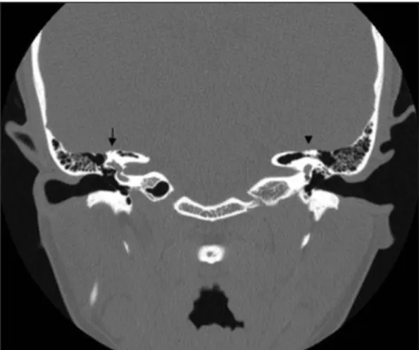
각각의 상반고리관(superior semicircular canal, SCC) 은 상부 골벽(overlying bone of SCC)의 결손 유무에 따 라 정상 또는 결손으로 분류하였고, 상반고리관 결손이 있을 경우 부위에 따라 중두개와저부(floor of the mid- dle cranial fossa)와 상추체정맥동(superior petrosal si- nus)으로 분류하였다(Fig. 1, 2). 영상은 3명의 이비인후
Fig. 2. Coronal computed tomography of the bilateral temporal bone shows bony defect of the superior semi- circular canal. right (arrow), left (arrow head).
Fig. 1. Coronal computed tomography of the left tem- poral bone shows bony defect of the superior semicircu- lar canal (arrow).
과 의사에 의해 독립적으로 실시되었고, 3명 모두 결손이 있다고 한 경우를 결손으로 분류하였다.
상반고리관 상부 골벽 두께 측정
측두골 전산화단층촬영상의 상반고리관 상부 골벽의 두께 및 양측 두께의 상관 관계를 알아 보기 위하여 재구 성된 고해상도 측두골 전산화단층촬영을 시행한 50명의 환자(100귀)를 대상으로 두께를 측정하였다. 남성 26명, 여성 24명이며, 평균 연령 48.4세(18~70세)이었다.
영상은 전산화단층촬영기(Siemens ; Definition 64 sec- tion multi-detector)를 이용하여, axial 0.6 mm, FOV 160 mm, kVp, 120 ; mA 336, 1.0 sec의 조건에서 촬영하였다.
상반고리관과 평행한 면(the plane of Po¨schl)으로 재구 성하여, 상반고리관 전체 고리와 주위를 둘러싸는 뼈를 한 단면에서 볼 수 있게 하였다(Fig. 3).
뼈의 두께의 측정은 PACS(Picture Archiving and Communications System)를 이용하여 상반고리관과 평 행한 면(the plane of Po¨schl)을 대상으로 상반고리관의 전각(anterior crus), 후각(posterior crus) 및 구형낭(sac- cule)이 한 단면에 모두 나타난 면에서 상반고리관 상부 골벽의 가장 얇은 두께를 CT 이미지 확대 배율 500%, window level 350 HU, window width 3000 HU의 동일 조건하에서 한 사람의 의사가 계측하였다. 전자 캘리퍼 (electronic caliper)를 이용하여 마우스 클릭으로 1/100 mm까지 계측할 수 있었다.
컴퓨터 통계분석 프로그램(SPSS 12.0 k for Windows ; SPSS. Inc., Chicago, IL)에서 χ2 test를 사용하여 통계학 적으로 검증을 하였으며, 유의 수준 p<0.05인 경우 유의 한 것으로 평가하였다. 또한 상관계수를 측정하여 양측 상반고리관 상부 골벽의 두께 사이에 어느 정도 선형 상 관관계를 가지는 지를 조사하였다.
결 과
상반고리관 결손의 빈도 및 분류
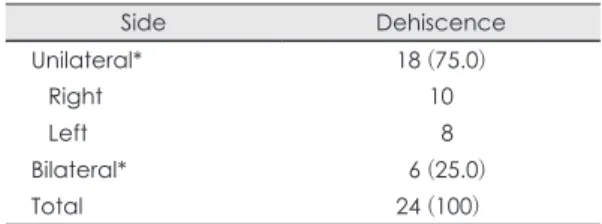
1,000명, 2,000귀에서 총 24명(30귀)에서 방사선학적 으로 상반고리관 결손을 보여 결손의 빈도는 1.5%이었 다. 양측이 6명, 편측이 18명으로 카이제곱검정에서 편측 이 유의하게 많았다(p<0.05)(Table 1). 우측 결손이 16 귀, 좌측 결손은 14귀이었으며, 총 30귀 중 22귀(73.3%) 에서 중두개와저부(floor of the middle cranial fossa)에 결손 부위가 있었으며, 8귀에서 상추체정맥동(superior Table 1. Dehiscence of overlying bone of superior semi- circular canal in CT scan case (%)
Side Dehiscence
Unilateral* 18 (75.0)
Right 10
Left 8
Bilateral* 6 (25.0)
Total 24 (100)
* : p<0.05
Fig. 3. Plane of Pöschl. A : Angle of reformation demonstrated on transverse scout image. B : Intact superior semicircu- lar canal and thickness of bone overlying the superior semicircular canal.
A B
petrosal sinus)에 결손 부위가 있었다(Table 2). 상추체정 맥동보다 중두개와저부로 결손 부위가 유의하게 많았다 (p<0.05). 상반고리관 결손을 보인 24명 중 상반고리관 결손증후군을 의심하고 영상을 촬영한 경우는 없었으며 이번 후향적 조사에서 상반고리관 결손증후군의 증상이 있었는지에 대한 직접적인 질문은 평가하지 않았다.
상반고리관 상부 골벽 두께 측정
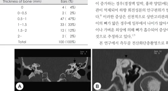
정상인 50명, 100귀에서 재구성한 고해상도 측두골 전 산화단층촬영을 상반고리관 상부 골벽의 두께를 측정하 기 위해 시행하였다(Fig. 3). 상반고리관을 덮고 있는 뼈 의 두께는 평균 1.02±0.42 mm이었고, 결손이 있는 경우 가 4귀(4%)이었고, 0.5~1 mm가 47귀(47%)로 가장 많았
다(Table 3). 80%에서 0.5~1.5 mm 사이에 분포하였다.
측두골 전산화 단층촬영의 관상면에서 보이는 중두개 와의 상반고리관 결손은 재구성한 고해상도 측두골 전 산화단층촬영에서도 관찰되었고(Fig. 4), 축면에서 보이 는 상추체정맥동으로의 결손도 재구성한 고해상도 측두 골 전산화단층촬영에서도 관찰되었다(Fig. 5).
양측을 비교하기 위해 우측 상반고리관 상부 골벽의 두 께를 좌측과 비교하여 도표화하였다(Fig. 6). 상관관계 분석 결과는 뚜렷한 양적 선형 관계가 나타났다(r=0.62, p<0.0001).
고 찰
Minor 등1)이 소리나 압력에 의해 유발되는 현훈이 발 생하는 상반고리관 결손 증후군을 처음 보고한 이후 많 은 연구가 있었으며, 다양한 증상 및 증후들이 동반될 수 있음이 보고되었다.2,5,7) 상반고리관 결손을 의심할 수 있 는 대표적인 증상으로는 소리나 압력 등의 자극에 의해 발생하는 현훈이 있고, 그 외이도 이충만감이나 바닥의 진동이 그대로 울려 들릴 때 의심할 수 있다. 내이의 압 력이 중두개와의 뇌압보다 높아지는 경우(기침, 코풀기, valsalva법 등)에는 상반고리관이 흥분되어 상향 회전성 분의 안구편위가 발생하게 되며, 반대로 내이보다 뇌압 이 증가하는 경우(경정맥 압박, 흉곽 양압)에는 상반고리 관이 억제되어 하향 회전성분의 안구편위가 발생하게 된 다.8) 이러한 증상은 선천적으로 상반고리관과 두개강 사 이의 뼈가 얇은 경우에 일부에서 나이가 많아지면서 뇌압 이나 가벼운 외상에 의해 뼈가 흡수되어 증상이 발생하는 것으로 추정하고 있다.1,7)
본 연구에서 측두골 전산화단층촬영으로 확인된 상반
Fig. 4. A : Coronal reformatted image from thin-section transverse CT examination shows bony covering of superior semicircular canal is intact on right but absent on left (arrow). B : Same patient Plane of Pöschl also shows dehiscent at the floor of the middle cranial fossa (arrow head).
A B
Table 2. Site of dehiscence of the superior canal between the bony superior canal and the upper surface of the tem- poral bone (middle cranial fossa) or through of the supe- rior petrosal sinus
Site of dehiscence Ears (%)
Middle Cranial Fossa* 22 (73.3)
Superior Petrosal Sinus* 8 (26.7)
Total 30 (100)
* : p<0.05
Table 3. The thickness of the bone overlying superior ca- nal in plane of Pöschl
Thickness of bone (mm) Ears (%)
0 4 ( 4%)
0-0.5 2 ( 2%)
0.5-1 47 ( 47%)
1-1.5 33 ( 33%)
1.5-2 12 ( 12%)
2- 2 ( 2%)
Total 100 (100%)
고리관 결손의 빈도는 1.5%였다. 이번 결과는 Carey 등 이 측두골의 조직학적 연구에서 보고한 상반고리관 결손 유병율인 0.5%보다 높게 나타났다.7)
Carey 등은 1,000개의 측두골을 상반고리관의 평면과 수직이 되게 절단한 조직학적 연구에서 상반고리관의 완 전 결손은 0.5%에서 발견되었고, 가장 얇은 부분의 두께 가 0.1 mm를 넘지 않는 경우도 1.4%였다고 보고하였다.7) 실제로 주관적인 증상을 동반하여 ‘상반고리관 결손증후 군’으로 진단에 이르는 경우는 그리 많지는 않은 것 같다.
고해상도 측두골 전산화단층촬영은 반고리관 결손이 의심되는 경우 반드시 필요한 검사로 상반고리관 결손의 진단에 민감도 100%, 특이도 99%, 양성예측도 93%, 음 성예측도 100%를 보인다.9)
Robert 등은 후향적 측두골 전산화단층촬영을 분석하 여 442귀(223명) 중 39귀(9%)의 높은 상반고리관결손율 을 보고하였다.6) 이에 대하여 Carey 등은 상반고리관과 중두개저 사이의 골 두께가 일반인의 1.3%에서는 고해상 도 전산화단층촬영에서도 골 결손이 있는 것으로 해석될 정도인 0.1 mm 이하로 관찰되어 전산환단층촬영에만 진 단을 의존할 경우 과다한 위양성을 보일 수 있음을 지적 하였다.7) 이번 연구에서도 상반고리관과 평행인 평면(the plane of Po¨schl)으로 재구성한 100귀중 4귀(4%)에서 골 이 결손된 것처럼 나타나 이들은 위양성일 가능성이 높다.
따라서, Weber 검사상 병변쪽으로의 편위, 전음성 난청 과 소리에 대한 특징적 안구 운동의 유발 유무 및 전정유 발근전위검사상의 비정상적으로 낮은 역치 등의 소견이
관찰될 경우에 전산화단층촬영으로 확진하는 것이 필요 하다고 한다.
Barton 등은 측두골 전산화단층촬영의 관상 영상(co- ronal image)과 상반고리관과 평행인 평면(the plane of Po¨schl)으로 재구성한 영상과의 유의한 방사선학적인 진 단의 차이가 없다고 보고하였다.10) 또한 Loke 등은 싱가 포르인의 481명 중 10명(2%)에서 상반고리관결손을 보 고하였으며, 측두골 전산화단층촬영과 비교하여 Po¨schl plane에서 얻어지는 장점은 없다고 하였다.11)
Hirvonen 등은 상반고리관을 덮고 있는 뼈의 결손이 나 얇음이 종종 양측으로 나타났고, 일측에 상반고리관 결손이 있을 때, 반대측의 상반고리관을 덮고 있는 뼈가 얇은 경향이 있다고 하였다.12) 이러한 결과는 상반고리관 결손이 발생학적인 문제라는 점을 지지해준다. 본 연구
Fig. 5. A : Axial image from thin-section transverse CT examination shows left SSCD along posterior groove formed by superior petrosal sinus (arrow). B : Same patient Plane of Pöschl also shows dehiscent at the superior petrosal sinus (ar- row head).
A B
2.5 2 1.5 1 0.5
Bone thickness on the left 0
(mm
)
0 0.5 1 1.5 2 2.5 Bone thickness on the right (mm)
Fig. 6. The thickness of the bone overlying the right and left superior canals is plotted for each control subject and a regression line is fitted (r=0.62; p<0.0001).
에서도 30귀, 1.5%에서 상반고리관 결손이 있었는데, 이 중 6명(25%)에서 양측 상반고리관 결손이 있었으며, 상 반고리관 상부 골벽의 양측 사이에는 뚜렷한 양적 선형 관계가 나타났다(r=0.62, p<0.0001).
상반고리관 결손증후군의 진단은 자세한 문진과 적절 한 신체검사를 통한 임상적 의심이 중요하다. 흔히 임상 증상 및 증후, 전정유발근전위 그리고 고해상도 전산화단 층촬영 등으로 하게 되며,5) 소리 및 압력에 의해 유발되 는 안진의 관찰이 중요한 역할을 하게 된다.
이과 영역에서 다른 여러 질환과 유사한 임상양상을 보 이기 때문에 이들 질환과의 감별 진단이 중요하다. 전음 성 난청은 이경화증에서, 자가강청은 개방성 이관(patu- lous Eustachian tube)에서 흔히 나타날 수 있으며, 압력 이나 큰 소리에 의한 전정증상은 외림프누공으로 오인되 기 쉽다. 이러한 증상으로 내원한 환자에서 원인을 알 수 없을 때, 본 연구 결과를 토대로 하여, 상반고리관 결손증 후군의 가능성을 염두에 두고, 측두골 전산화단층촬영을 시행하는 것이 의미 있는 것으로 생각된다.
상반고리관 결손 증후군은 강한 소리 자극이나 압력 변 화에 의해 어지럼증, 귀막힘 등이 나타나는 질환으로 어 지럼증 및 난청을 호소하는 환자에서 감별 진단이 필요 하다. 이번 연구는 상반고리관 결손증후군의 진단에 참 고 자료로 이용하고자 방사선학적인 빈도 및 상반고리 관 상부 골벽의 두께에 대하여 조사하였다.
본 연구에서 상반고리관의 방사선학적인 결손은 측두 골 전산화단층촬영결과 1.5%였으며, 상반고리관 상부 골 벽의 두께는 평균 1.02±0.42 mm이었고, 상반고리관 상 부 골벽 양측 사이에는 뚜렷한 양적 선형 관계가 나타났 다(r=0.62, p<0.0001).
상반고리관의 방사선학적인 결손 빈도는 조직학적인 조사7)에서의 완전 결손 빈도 0.5%보다 많았지만, 가장 얇 은 부분의 두께가 고해상도 전산화단층촬영에서도 골 결 손이 있는 것으로 해석될 정도인 0.1 mm를 넘지 않는 경 우 1.4%를 포함하면, 이과 영역에서 다른 여러 질환과 유 사한 양상을 보이는 전정증상, 전음성 난청, 이충만감, 자 가강청 등의 증상을 호소하는 환자에서 원인을 알 수 없 을 때, 상반고리관 결손증후군의 가능성을 염두에 두고
측두골 전산화단층촬영을 시행하는 것이 의미 있는 것으 로 생각되어진다. 하지만 과다한 위양성을 보일 수 있기 때문에 전산화단층촬영에만 진단을 의존하는 것은 신중 해야 하며, 임상적인 증상과 연계한 전산화단층촬영의 판 독이 필요하다고 생각된다
중심 단어:측두골·전산화단층촬영·상반고리관 결손 증후군.
REFERENCES
1) Minor LB, Solomon D, Zinreich JS, Zee DS. Sound- and/
or pressure-induced vertigo due to bone dehiscence of the superior semicircular canal. Arch Otolaryngol Head Neck Surg 1998;124(3):249-58.
2) Minor LB. Clinical manifestations of superior semicircu- lar canal dehiscence. Laryngoscope 2005;115(10):1717-27.
3) da Cunha Ferreira S, de Melo Tavares de Lima MA. Supe- rior canal dehiscence syndrome. Braz J Otorhinolaryngol 2006;72(3):414-8.
4) Watson SR, Halmagyi GM, Colebatch JG. Vestibular hy- persensitivity to sound (tullio phenomenon): structural and functional assessment. Neurology 2000;54(3):722-8.
5) Zhou G, Gopen Q, Poe DS. Clinical and diagnostic char- acterization of canal dehiscence syndrome: a great oto- logic mimicker. Otol Neurotol 2007;28(7):920-6.
6) Williamson RA, Vrabec JT, Coker NJ, Sandlin M. Coro- nal computed tomography prevalence of superior semicir- cular canal dehiscence. Otolaryngol Head Neck Surg 2003;
129(5):481-9.
7) Carey JP, Minor LB, Nager GT. Dehiscence or thinning of bone overlying the superior semicircular canal in a tempo- ral bone survey. Arch Otolaryngol Head Neck Surg 2000;
126(2):137-47.
8) Baloh RW. Superior semicircular canal dehiscence synd- rome: leaks and squeaks can make you dizzy. Neurology 2004;62(5):684-5.
9) Belden CJ, Weg N, Minor LB, Zinreich SJ. CT evaluation of bone dehiscence of the superior semicircular canal as a cause of sound- and/or pressure-induced vertigo. Radiolo- gy 2003;226(2):337-43.
10) Branstetter BF,4th, Harrigal C, Escott EJ, Hirsch BE. Su- perior semicircular canal dehiscence: oblique reformatted CT images for diagnosis. Radiology 2006;238(3):938-42.
11) Loke SC, Goh JP. Incidence of semicircular canal dehis- cence in singapore. Br J Radiol 2009;82(977):371-3.
12) Hirvonen TP, Weg N, Zinreich SJ, Minor LB. High-reso- lution CT findings suggest a developmental abnormality underlying superior canal dehiscence syndrome. Acta Oto- laryngol 2003;123(4):477-81.