INTRODUCTION
Cancer is a major health problem and one of the main causes of mortality worldwide [1]. At present, breast cancer is the most common cancer in women around the world [2]. The common therapies for cancer are surgery and chemotherapy.
Chemotherapeutical agents greatly damage cell DNA, affecting normal cells as well as cancer cells, and cause side effects such as digestive problems, leukopenia, and hair loss. Furthermore, drug resistance limits the effectiveness of chemotherapy [3].
Approximately 60% of the drugs approved for cancer treat- ment are derived from natural sources. Vincristine, etoposide, irinotecan, taxane, and camptothecin are examples of plant-
derived compounds [4]. One of the important compounds in cancer treatment is chitin, (C8H13O5N)n. It was first derived from a fungus and is the first known polysaccharide [5]. Potent acids can dissociate chitin, a water-insoluble polymer, into ace- tic acid and chitosan. Chitosan is a nontoxic polymer produced by the deacetylation of chitin. Chitin is the main component of arthropod exoskeletons, tendons, and respiratory tract lining, and cell walls of the majority of fungi. Crustacean shells are a source of chitin (20%–30%). Crab shell also contains lipidic pigments such as carotenoids (astaxanthin, astatine, and can- thaxanthin), lutein, and β-carotene [6].
Jeon and Kim [7] found that oligomers of chitosan exhibit antitumor activities in vitro and in vivo. Low-molecular- weight chitosan has been shown to have remarkable antimeta- static effects against lung cancer in mice [8]. Maeda and Kimura [9] investigated the antitumor effects of low-molecu- lar-weight chitosan and reported a decline in tumor growth and final tumor weight in mice with sarcomas. In recent years, the viability of cancer cells has remarkably decreased in some cell lines, regardless of whether the chito-oligosaccharide (COS) derivatives were positively or negatively charged [10].
Carotenoids are yellow-orange pigments that are found in all
Antiproliferatory Effects of Crab Shell Extract on Breast Cancer Cell Line (MCF7)
Leila Rezakhani, Zahra Rashidi, Pegah Mirzapur, Mozafar Khazaei
Fertility and Infertility Research Center, Kermanshah University of Medical Sciences, Kermanshah, Iran ORIGINAL ARTICLE
Purpose: Breast cancer is the most common type of cancer in women. Despite various pharmacological developments, the identification of new therapies is still required for treating breast cancer. Crab is often recommended as a traditional medicine for cancer. This study aimed to determine the in vitro effect of a hy- droalcoholic crab shell extract on a breast cancer cell line. Meth- ods: In this experimental study, MCF7 breast cancer cell line was used. Crab shell was powdered and a hydroalcoholic (70° etha- nol) extract was prepared. Five concentrations (100, 200, 400, 800, and 1,000 µg/mL) were added to the cells for three periods, 24, 48, and 72 hours. The viability of the cells were evaluated using trypan blue and 3-(4, 5-dimethylthiazol-2-yl)-2,5-diphenyl- tetrazolium bromide assays. Cell apoptosis was determined us- ing the terminal deoxynucleotidyl transferase dUTP nick end la-
beling method. Nitric oxide (NO) level was assessed using the Griess method. Data were analyzed using analysis of variance, and p<0.05 was considered significant. Results: Cell viability decreased depending on dose and time, and was significantly different in the groups that were treated with 400, 800, and 1,000 µg/mL doses compared to that in the control group (p<0.001).
Increasing the dose significantly increased apoptosis (p<0.001).
NO secretion from MCF7 cells significantly decreased in re- sponse to different concentrations of the extract in a dose- and time-dependent manner (p<0.050). Conclusion: The crab shell extract inhibited the proliferation of MCF7 cells by increasing apoptosis and decreasing NO production.
Key Words: Apoptosis, Breast neoplasms, Cell survival
Correspondence to: Mozafar Khazaei
Fertility and Infertility Research Center, Medical Faculty, University Avenue, Shirodi Blv., Kermanshah, Iran
Tel: +98-83-34281563, Fax: +98-83-34281563 E-mail: [email protected]
This article originated from a M.Sc. thesis (project No: 91379) and supported by Kermanshah University of Medical Sciences, Iran.
Received: May 2, 2014 Accepted: July 18, 2014
Cancer
higher plants and some animals. A diet containing carotenoids can decrease the risk of cancer in various tissues. Clinical stud- ies have shown that some carotenoids have powerful antitumor activities both in vitro and in vivo [11]. The anticancer activities of different natural carotenoids as well as β-carotene have been documented. Carotenoids (α-carotene and lutein), zeaxanthin, lycopene, astaxanthin, crocetin, and β-carotene can prevent cancer [12]. Sachindra et al. [13] examined the quantitative and qualitative distribution of carotenoids in the meat and shell of the major marine crab (Charybdis cruciata) and fresh- water crab (Potamon). The total carotenoid content in both species was low. Astaxanthin and its esters, and zeaxanthin were the major carotenoids found in the fresh-water crab ex- tract.
Selenium is an essential part of the diet of organisms, includ- ing humans, and acts as a protective factor against cancer. Al- though the anticancer activity of selenium is not completely known, it has been suggested that various mechanisms, such as antioxidant protection by selenoenzymes, inhibit the develop- ment of tumor cells, regulate and modulate the cell cycle, in- hibit apoptosis, and repair DNA. Recent studies have indicated that selenium not only has the potential to prevent cancer but could also be used to treat cancer and enhance the effective- ness of cancer therapy in combination with other anticancer drugs or radiation [14]. In cell culture models, selenium com- pounds have been shown to inhibit the development of cancer cells by reducing cell proliferation via cell cycle control or an increase in apoptosis [15].
Considering the current prevalence of breast cancer and the necessity to identify and develop new pharmaceutical com- pounds, especially those with a natural origin, as well as the primary information obtained from traditional medicine, this study was aimed to determining the effect of hydroalcoholic crab shell extract on the viability, apoptosis, and nitric oxide (NO) secretion of MCF7 cell line.
METHODS
The MCF7 cell line was treated with hydroalcoholic crab shell extract (100, 200, 400, 800, and 1,000 µg/mL doses) for three periods, 24, 48, and 72 hours. Each experiment was re- peated 3 to 5 times and the means of the data were used.
Extraction
Fresh-water crab was prepared and identified in terms of sex and species (Potamon persicum) by a zoologist (from Razi University, Kermanshah, Iran). The crab shell was powdered, and 5 g of powder was dissolved in 150 mL of 70% ethanol for 48 hours in darkness. It was subsequently filtered through filter
paper and dried to evaporate the alcohol; thereafter, the final powder was weighed (0.59 g). The solvent of the final powder was a serum-free cell culture medium (RPMI 1640), and the prepared concentrations were passed through a 0.22-µm filter before use.
Cell viability assessment using trypan blue
A total of 3×10⁴ MCF7 cells were cultured in each well of a 24-well culture dish. Subsequently, 750 µL of RPMI 1640 (containing 10% fetal bovine serum) was added to each well and the dishes were kept in a CO2 incubator for 24 hours. The supernatants were then removed and 1 mL of serum-free RPMI 1640 medium containing 0 (control), 100, 200, 400, 800, and 1,000 µg/mL concentrations of crab shell extract was added to each well. After 24 hours, the supernatant of each well was removed and frozen at -20°C for NO measurement.
The cells were then detached using trypsin (0.25%, 100 µL), stained with trypan blue, and counted using a Neubauer cham- ber, and then, their viability was calculated. Cell viability was similarly calculated for wells incubated for 48 and 72 hours.
3-(4,5-Dimethylthiazol-2-yl)-2,5-diphenyltetrazolium bromide (MTT) assay
The basis of this test is that the mitochondrial enzyme suc- cinate dehydrogenase in viable cells is capable of reducing the tetrazolium MTT salt to insoluble formazan, which is purple in color; the greater the number of viable cells, the greater the intensity of the stain and vice versa. To perform the test, 15×103 MCF7 cells were loaded into a 96-well plate and 200 µL of RPMI medium containing 7% serum was added. After 24-hour incubation, 200 µL of various concentrations of the extract (0, 100, 200, 400, 800, and 1,000 µg/mL) dissolved in serum-free RPMI 1640 medium was added to the wells. The cells were separately incubated with different doses for 24, 48, and 72 hours.
After incubation, the MTT test was performed in which the supernatant from each well was removed and 500 µL of MTT solution (5 mg/mL) was added to each well and incubated for 3 hours. The supernatant from each well was then removed and 100 μL of dimethyl sulfoxide was added to dissolve the formazan crystals at room temperature for 30 minutes. The optical density of each well was measured using an enzyme- linked immunosorbent assay (ELISA) reader (STAT FAX 2100; Awareness Technology, Westport, USA) at 570 and 630 nm. The viability of the cells for each concentration was calcu- lated using the following formula [16]:
Cell viability (%)=[A570, 630(sample)/A (control)]×100
NO assay
NO was measured using the Griess staining method. For each sample, 400 µL of thawed supernatant was deproteinized by adding 6 mg of zinc sulfate. The samples were centrifuged at 4°C and 12,000 g for 12 minutes. Separately, 100 µL of 0, 6.25, 12.5, 25, 50, 100, and 200 µM sodium nitrite was poured into standard wells. We then added 100 µL of the deprotein- ized samples to each well, and 100 µL of vanadium chloride, 50 µL of sulfanilamide, and 50 µL of N-(1-naphthyl) ethylenedi- amine dihydrochloride were added to all the wells (standard and samples). All wells were incubated for 15 minutes and were assessed using an ELISA Reader at wavelengths of 570 and 630 nm [17].
Terminal deoxynucleotidyl transferase dUTP nick end labeling (TUNEL) assay
Apoptosis in the MCF7 cells was analyzed using the TUNEL method. This method is used to determine DNA fragmenta- tion during apoptosis cell death. MCF7 cancer cells were cul- tured in 96-well culture plates with 15×103 cells in each well.
Subsequently, 200 µL of RPMI 1640 medium was added to each well and incubated for 24 hours. The supernatant was then removed and 200 µL of each dose of crab shell extract was added to the cells and incubated for 72 hours. The cells were then fixed in 4% paraformaldehyde for 1 hour. The permeabil- ity of the cell membranes was increased by adding 0.2% Triton x-100 solution (Sigma, St. Louis, USA) for 2 minutes on ice.
The cells were then incubated with TUNEL mixture solution at 37°C in dark conditions for 1 hour. Finally, differential stain- ing of the cells was performed using propidium iodide (5 µg/
mL; PI) and the cells were analyzed using a fluorescence mi- croscope (Eclipse TS100; Nikon, Tokyo, Japan) after being washed with phosphate-buffered saline three times [18]. The apoptotic index of the cells was calculated as the percentage of apoptotic cells relative to the total cell number.
Apoptotic index (%)=(number of apoptotic cells/total number of cells)×100
Crab shell components
To determine the extract contents, it was subjected to gas chromatography-mass spectrometry (GC-MS). The type of GC device used was an Agilent 6890 with a HP-5MS column with a length of 30 m, internal diameter of 0.25 mm, and thickness of 0.25 µm. The column temperature plan consisted of a primary temperature of 50°C, with a 5-minute retention period and a thermal gradient of 3°C every minute. The tem- perature was then increased to 180°C with a velocity of 10°C per minute, and then to 260°C with a 4-minute retention peri-
od. Helium was used as the carrier gas with a 1-mL/min flow speed. The total analysis was completed in 45 minutes. The re- sults obtained from the analysis were compared using the data present in the machine. Biochemical compounds (75) were obtained from the analysis, which was related to retention time and extraction of compounds from the extract (Table 1).
Moreover, atomic absorption spectrometry (VGA 77; Varian Inc., Mulgrave, Australia) was used to determine the amount of selenium in the extract.
Data analysis
Data were analyzed using one-way analysis of variance with SPSS version 16.0 (SPSS Inc., Chicago, USA) software, and p<0.05 was considered significant.
RESULTS
MCF7 viability
Comparison of the mean MCF7 cell viabilities analyzed us- ing the trypan blue method after 24, 48, and 72 hours indicated a significant difference between the groups treated with crab shell extract (400, 800, and 1,000 µg/mL) compared to the con- trol group (p<0.001). Increasing the dose significantly de- creased the cell viability (Figure 1A, B).
Comparison of the mean MCF7 cell viabilities analyzed us- ing the MTT assay after 24, 48, and 72 hours revealed a signif- icant difference between the groups treated with crab shell ex- tract (400, 800, and 1,000 µg/mL) in comparison with the control group. Cell viability was again significantly decreased by increasing the dose (Figure 2).
NO levels
NO concentration was evaluated using the Griess method.
The effect of different concentrations of crab shell extract on MCF7 cells after 24, 48, and 72 hours indicated a dose- and time-dependent decrease in NO secretion. The difference compared to the control group was significant with the 800 and 1,000 µg/mL doses (p<0.050) (Figure 3).
Table 1. Biochemical compounds from the analysis with gas chroma- tography-mass spectrometry
The most compounds Eicosane (CAS) Pyrido[2,3-d]pyrimidine N-(2-Acetylcyclopentylidene)cyc Hexadecanoic acid
9-Octadecenoic acid (Z)- (CAS)
CAS=chemical abstracts service; cyc=cyclohexylamine.
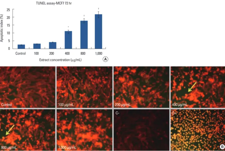
MCF7 apoptosis
The apoptosis index of MCF7 cells treated with crab shell extract showed an increase from control to experimental groups in order of concentration, exhibiting a significant dif- ference with 400 (p<0.050), and 800 and 1,000 µg/mL (p<
0.001) doses (Figure 4A, B).
Extract compounds
Using GC-MS, the presence of chitosan, COS, and carot- enoids such as astaxanthin in the extract was confirmed. Fur- thermore, atomic absorption spectrometry measured the amount of selenium as 12.5 ppm (VGA 77 Varian).
DISCUSSION
In this study, the effect of crab shell hydroalcoholic extract on a breast cancer cell line (MCF7) was evaluated. Five doses (100, 200, 400, 800, and 1,000 µg/mL) of the extract were pre- pared and their effects on the cells were analyzed over three time periods of 24, 48, and 72 hours. MTT assay and trypan blue methods were used to determine cell viability. A decreas- ing trend was observed in cell viability along with a rising trend in apoptosis, with significant differences with 400, 800, and 1,000 µg/mL doses compared with the control group.
The NO level in the MCF7 culture medium of extract-treat- ed cells significantly decreased depending on dose and time at 800 and 1,000 µg/mL doses after 48 and 72 hours. Generally, Figure 1. (A) MCF7 viability (%) after 24, 48, and 72-hour exposed to different doses of crab shell extract. Significant difference between the experi- ments and control groups were seen.
*p<0.05; †p<0.001. (B) MCF7 cells in control and different doses of the extract at 72 hours by invert microscope (×100).
Extract concentration (µg/mL) Trypan blue-MCF7
Viability (%)
100 90 80 70 60
Control 100 200 400 800 1,000
24 hr 48 hr 72 hr
* *
†
† †
†
†
†
†
†
†
†
A
Control 100 μg/mL
200 μg/mL 400 μg/mL 800 μg/mL 1,000 μg/mL
B
Figure 2. MCF7 cell line viability (%) in different crab shell extracts and different culture periods. There is significant difference between the groups.
*p<0.05; †p<0.001.
Extract concentration (µg/mL) MTT assay-MCF7
Viability (%)
100 80 60 40 20 0
Control 100 200 400 800 1,000
24 hr 48 hr 72 hr
†
†
†
†
†
† †
*
†
Figure 3. Nitric oxide (NO) levels in different groups and different culture periods. Significant difference between groups.
*p<0.05.
Extract concentration (µg/mL) NO assay-MCF7
NO concentration (µM)
80 70 60 50 40 30 20
Control 100 200 400 800 1,000
*
*
* *
24 hr 48 hr 72 hr
24, 48, and 72-hour periods [19] and 100–1,000 µg/mL doses [20] are used to analyze the effects of natural compounds on cancer cell lines. The antiproliferative properties of fresh-water crab shell extract can be attributed to the presence of carot- enoids, chitin derivatives (COS and chitosan), and selenium, which induce apoptosis and decrease NO secretion.
Chitin is the second natural polymer frequently found in the exoskeleton of crustaceans and cell wall of fungi. Chitosan and COS are its main derivatives, for which the antiproliferative properties have been reported in different cancer cell lines in various studies [9-11]. COS extracted from fungi has also been reported to inhibit the development of a liver carcinoma cell line (HepG2) at five concentrations and over three periods [19], which is similar to the findings of the present study in terms of inhibition of MCF7 cell proliferation at different doses and times.
The effect of raw and cooked crab shell extract and its deriv- ative carotenoids on a liver-tumor cell line (AH109A) has pre-
viously been evaluated and the inhibitory effect observed has been attributed to the antioxidant effect of the extract [21].
However, in the present study, the inhibitory effect of crab shell extract on a breast cancer cell line was associated with NO de- crease and induction of apoptosis. The inhibitory effect on MCF7 cells was due to decreasing NO secretion, as reported by Yin et al. [22], although decreasing cyclooxygenase 2 and cytokine expression are additional factors to consider.
A decrease in NO secretion and dose- and time-dependent decrease in the viability of a liver carcinoma cell line (HepG2) and subsequent larynx carcinoma (Hep2) have been attributed to saffron extract and its carotenoids [23]; this finding is in line with the results of the present study. The effect of one of the components of selenium, methyl selenic acid, on the viability of MCF7 cancer cells assessed using the MTT assay showed inhibition of cell development by 50% over three periods of 24, 48, and 72 hours and an increase in cell apoptosis after 48 hours [19]. As in the present study, increasing the dose and
Extract concentration (µg/mL) TUNEL assay-MCF7 72 hr
Apoptotic index (%)
25 20 15 10 5 0
Control 100 200 400 800 1,000
*
Figure 4. (A) MCF7 apoptotic index (%) after 72-hour with different amounts of crab shell extract. Significant difference between groups. *p<0.05;
†p<0.001. (B) Identification of apoptosis in MCF7 cell line following terminal deoxynucleotidyl transferase dUTP nick end labeling (TUNEL) staining and counterstaining with propidium iodide after 72 hours of exposure crab shell extract. TUNEL positive cells (apoptotic nuclei) identified by a distinct bright yellow stained chromatin (arrows). C+: The positive control was incubated with ethanol 10% for 10 minutes, all cells are seen as bright yellow.
C-: The positive control was incubated with label solution without enzyme solution. No TUNEL positive cells were observed (×200).
†
†
A
B 400 μg/mL
200 μg/mL 100 μg/mL
1,000 μg/mL 800 μg/mL
Control
C- C+
time caused cell growth to decline but viability did not fall be- low 50%, which could be because raw extract was used in the present study unlike the previous study that used distinct and pure compounds. Furthermore, similar apoptotic effects of selenocysteine on MCF7 cells and of sodium selenite on a prostate cancer cell line (JCA1, DU145) have been reported in the studies of Chen and Wong [18] and Gasparian et al. [24], respectively.
Moreover, an antioxidant and antiproliferative effect on a human bladder cancer cell line (T24) has been reported after 48 hours, depending on chitosan concentration [25]. The effect of one of the compounds derived from chitin, astaxanthin, on a human esophagus cancer cell line (TE-4) over 24, 48, and 72 hours was investigated, and dose-dependent inhibition was observed [26]. Furthermore, astaxanthin derived from the green alga Haematococcus pluvialis exerted an inhibitory effect on a large intestine cancer cell line (HCT-116) via apoptosis in- duction [27]. The findings obtained in the present study are similar to those obtained previously, and confirm the findings regarding different cancer cell lines. Our study had limitations:
we did not evaluate the effect of crab sell extract on normal breast epithelial cells. Moreover, using MCF10A cell as normal breast epithelial cell line, can’t substitute normal cell character- istic completely. MCF10A cell line has a near-diploid karyo- type with modest genetic modifications [28].
In conclusion, crab shell extract inhibited the proliferation of a breast cancer cell line in a dose- and time-dependent manner. It seems that the extract exerts its effect through a re- duction in NO production and induction of apoptosis. Chitin derivatives, carotenoids, and selenium are important factors to consider in the process of growth inhibition.
CONFLICT OF INTEREST
The authors declare that they have no competing interests.
REFERENCES
1. Rani D, Somasundaram VH, Nair S, Koyakutty M. Advances in cancer nanomedicine. J Indian Inst Sci 2012;92:187-218.
2. Curado MP. Breast cancer in the world: incidence and mortality. Salud Publica Mex 2011;53:372-84.
3. Kateb B, Chiu K, Black KL, Yamamoto V, Khalsa B, Ljubimova JY, et al.
Nanoplatforms for constructing new approaches to cancer treatment, imaging, and drug delivery: what should be the policy? Neuroimage 2011;54 Suppl 1:S106-24.
4. Grever MC. Cancer drug discovery and development. In: De Vita VH, Rosenberg SA, editors. Cancer: Principles and Practice of Oncology.
Philadelphia: Lippincott-Raven; 2001. p.328-39.
5. Domard A, Domard M. Chitosan: structure-properties relationship and
biomedical applications. In: Dumitriu S, editor. Polymeric Biomaterials.
2nd ed. New York: Marcel Dekker Inc.; 2002. p.187-212.
6. Acosta N, Jiménez C, Borau V, Heras A. Extraction and characterization of chitin from crustaceans. Biomass Bioenergy 1993;5:145-53.
7. Jeon YJ, Kim SK. Antitumor activity of chitosan oligosaccharides pro- duced in ultrafiltration membrane reactor system. J Microbiol Biotech- nol 2002;12:503-7.
8. Tsukada K, Matsumoto T, Aizawa K, Tokoro A, Naruse R, Suzuki S, et al.
Antimetastatic and growth-inhibitory effects of N-acetylchitohexaose in mice bearing Lewis lung carcinoma. Jpn J Cancer Res 1990;81:259-65.
9. Maeda Y, Kimura Y. Antitumor effects of various low-molecular-weight chitosans are due to increased natural killer activity of intestinal intraepithelial lymphocytes in sarcoma 180-bearing mice. J Nutr 2004;134:945-50.
10. Huang R, Mendis E, Rajapakse N, Kim SK. Strong electronic charge as an important factor for anticancer activity of chitooligosaccharides (COS). Life Sci 2006;78:2399-408.
11. Tanaka T, Shnimizu M, Moriwaki H. Cancer chemoprevention by ca- rotenoids. Molecules 2012;17:3202-42.
12. Nishino H, Murakosh M, Ii T, Takemura M, Kuchide M, Kanazawa M, et al. Carotenoids in cancer chemoprevention. Cancer Metastasis Rev 2002;21:257-64.
13. Sachindra NM, Bhaskar N, Mahendrakar NS. Carotenoids in crabs from marine and fresh waters of India. Lebenson Wiss Technol 2005;38:221-5.
14. Brozmanová J, Mániková D, Vlčková V, Chovanec M. Selenium: a dou- ble-edged sword for defense and offence in cancer. Arch Toxicol 2010;84:919-38.
15. Whanger PD. Selenium and its relationship to cancer: an update. Br J Nutr 2004;91:11-28.
16. Ghosh K, Chandra K, Ojha AK, Sarkar S, Islam SS. Structural identifi- cation and cytotoxic activity of a polysaccharide from the fruits of Lage- naria siceraria (Lau). Carbohydr Res 2009;344:693-8.
17. Miranda KM, Espey MG, Wink DA. A rapid, simple spectrophoto- metric method for simultaneous detection of nitrate and nitrite. Nitric Oxide 2001;5:62-71.
18. Chen T, Wong YS. Selenocystine induces caspase-independent apop- tosis in MCF-7 human breast carcinoma cells with involvement of p53 phosphorylation and reactive oxygen species generation. Int J Biochem Cell Biol 2009;41:666-76.
19. Shah YM, Kaul A, Dong Y, Ip C, Rowan BG. Attenuation of estrogen receptor alpha (ERalpha) signaling by selenium in breast cancer cells via downregulation of ERalpha gene expression. Breast Cancer Res Treat 2005;92:239-50.
20. Shen KT, Chen MH, Chan HY, Jeng JH, Wang YJ. Inhibitory effects of chitooligosaccharides on tumor growth and metastasis. Food Chem Toxicol 2009;47:1864-71.
21. Nakahara S, Miura Y, Yagasaki K. Inhibitory action of carotenoids in crab shell on the invasion of hepatoma cells co-cultured with mesothelial cells. In: European Society of Animal Cell Technology, Japanese Associa- tion for Animal Cell Technology, editors. Animal Cell Technology:
Challenges for the 21st Century. Boston: Kluwer; 2002. p.409-13.
22. Yin MB, Li ZR, Tóth K, Cao S, Durrani FA, Hapke G, et al. Potentiation of irinotecan sensitivity by Se-methylselenocysteine in an in vivo tumor model is associated with downregulation of cyclooxygenase-2, inducible
nitric oxide synthase, and hypoxia-inducible factor 1alpha expression, resulting in reduced angiogenesis. Oncogene 2006;25:2509-19.
23. Parizadeh MR, Gharib FG, Abbaspour AR, Afshar JT, Ghayour-Mobar- han M. Effects of aqueous saffron extract on nitric oxide production by two human carcinoma cell lines: hepatocellular carcinoma (HepG2) and laryngeal carcinoma (Hep2). Avicenna J Phytomed 2011;1:43-50.
24. Gasparian AV, Yao YJ, Lü J, Yemelyanov AY, Lyakh LA, Slaga TJ, et al.
Selenium compounds inhibit I kappa B kinase (IKK) and nuclear fac- tor-kappa B (NF-kappa B) in prostate cancer cells. Mol Cancer Ther 2002;1:1079-87.
25. Kuppusamy S, Karuppaiah J. Antioxidant and cytotoxic efficacy of chi-
tosan on bladder cancer. Asian Pac J Trop Dis 2012;2 Suppl 2:S769-73.
26. Lim SA, Lee JY, Jung WH, Lim EH, Joo MK, Lee BJ, et al. Anticancer ef- fects of astaxanthin and alpha-tocopherol in esophageal cancer cell lines. Korean J Helicobacter Up Gastrointest Res 2011;11:170-5.
27. Palozza P, Torelli C, Boninsegna A, Simone R, Catalano A, Mele MC, et al. Growth-inhibitory effects of the astaxanthin-rich alga Haematococ- cus pluvialis in human colon cancer cells. Cancer Lett 2009;283:108-17.
28. Imbalzano KM, Tatarkova I, Imbalzano AN, Nickerson JA. Increasingly transformed MCF-10A cells have a progressively tumor-like phenotype in three-dimensional basement membrane culture. Cancer Cell Int 2009;9:7.