158
A 69-year-old woman presented to the emergency depart- ment with palpitations and dizziness of half an hour duration.
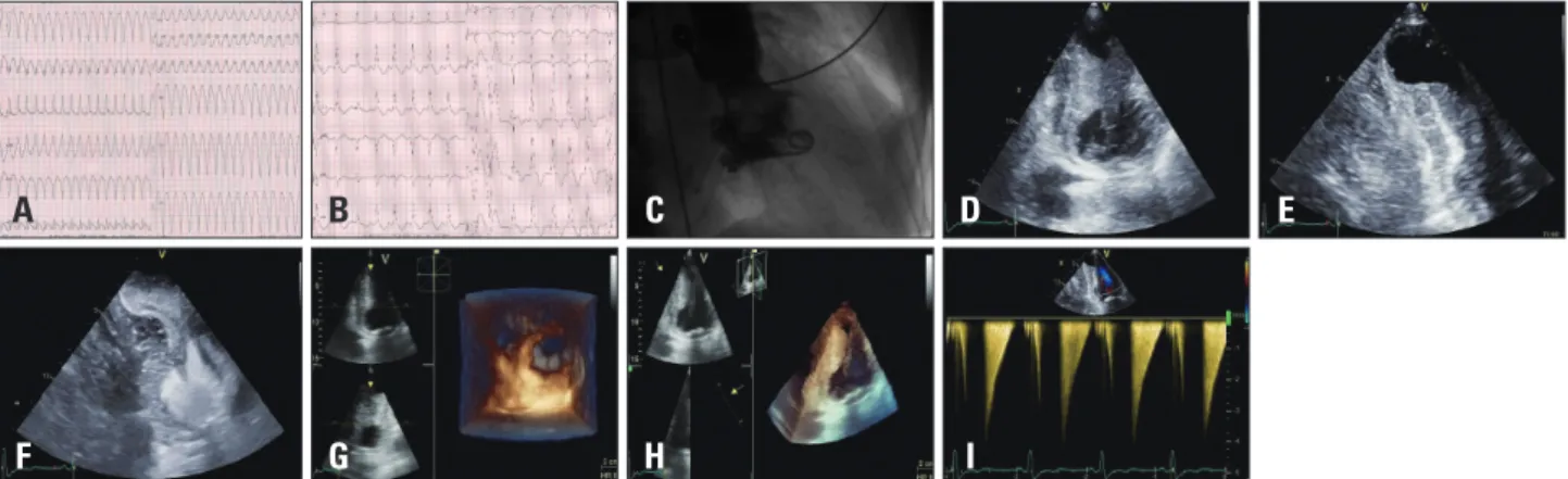
The patient’s history included dyslipidemia under atorvas- tatin. The patient was hemodynamically stable. The 12-lead surface electrocardiogram demonstrated sustained ventricular tachycardia (VT) with a left ventricular origin and north-west axis at 200 beats/min (Fig. 1A). Cardioversion with intrave- nous procainamide administration revealed sinus rhythm with left-axis deviation and deep T-wave inversion in the anterior leads (Fig. 1B). Two-dimensional (Fig. 1D and E), contrast (Fig. 1F), and 3-dimensional (Fig. 1G and H) echocardiogra-
phy revealed mid-ventricular hypertrophy with an apical an- eurysm, and an intraventricular flow velocity of 4 m/s (Fig.
1I). Coronary arteriography demonstrated normal coronary ar- teries, while left ventriculography (Fig. 1C) revealed mid-ven- tricular obliteration with an abrupt drop in intraventricular pressure from 280 mmHg to 160 mmHg, measured with the pig-tail catheter.
Mid-ventricular obstructive hypertrophy cardiomyopathy comprises a rare subtype of hypertrophic cardiomyopathies (HCM), accounting for only 1% of cases.1) It is characterized by the presence of a pressure gradient between the apical and
pISSN 1975-4612/ eISSN 2005-9655 Copyright © 2014 Korean Society of Echocardiography www.kse-jcu.org http://dx.doi.org/10.4250/jcu.2014.22.3.158
Mid-Ventricular Hypertrophic Obstructive Cardiomyopathy Complicated by an Apical Aneurysm, Presenting as Ventricular
Tachycardia
Emmanouil Petrou, MD, Stamatis Kyrzopoulos, MD, Eftychia Sbarouni, MD, Dimitris Tsiapras, MD, and Vassilis Voudris, MD
Division of Cardiology, Onassis Cardiac Surgery Center, Athens, Greece
KEY WORDS: Hypertrophic obstructive cardiomyopathy · Apical aneurysm · Ventricular tachycardia.
• Received: June 9, 2014 • Revised: July 4, 2014 • Accepted: August 20, 2014
• Address for Correspondence: Emmanouil Petrou, Division of Cardiology, Onassis Cardiac Surgery Center, 356 Syggrou Ave., Kallithea, Athens 17674, Greece Tel: +30-694-511-2509, Fax: +30-210-275-1028, E-mail: [email protected]
• This is an Open Access article distributed under the terms of the Creative Commons Attribution Non-Commercial License (http://creativecommons.org/licenses/by-nc/3.0) which permits unrestricted non-commercial use, distribution, and reproduction in any medium, provided the original work is properly cited.
IMAGES IN CARDIOVASCULAR ULTRASOUND J Cardiovasc Ultrasound 2014;22(3):158-159
Fig. 1. A: Presenting electrocardiogram showing ventricular tachycardia at 200 beats/min. B: Electrocardiogram revealing sinus rhythm with deep T-wave inversion in the anterior leads after intravenous procainamide administration. C: Left ventriculography demonstrating mid-ventricular obliteration. D and E: Two-dimensional echocardiography. F: Contrast echocardiography. G and H: Three-dimensional echocardiography, showing left mid-ventricular hypertrophy with an apical aneurysm. I: Intraventricular flow velocity of 4 m/s.
F A
G B
H C
I
D E
Hypertrophic Obstructive Cardiomyopathy | Emmanouil Petrou, et al.
159 basal chambers of the left ventricle (LV). The mid-cavity ob-
struction is the result of the mid-systolic muscular apposition of the septum and LV free wall producing distinct proximal and distal chambers, resembling an “hourglass” shape.2) Fur- thermore, LV apical aneurysms are present in up to 2% of pa- tients with HCM, and are associated with intramural throm- bus and sustained monomorphic VT.3)4)
Our patient received an implantable cardioverter-defibrilla- tor and was discharged with explicit instructions and medica- tion. This case demonstrates multiple complications and pe- culiarities of HCM.
References
1. Maron BJ. Hypertrophic cardiomyopathy: a systematic review. JAMA 2002;287:1308-20.
2. Efthimiadis GK, Pliakos C, Pagourelias ED, Parcharidou DG, Spanos G, Paraskevaidis S, Styliadis IH, Parcharidis G. Hypertrophic cardiomyopathy with midventricular obstruction and apical aneurysm for- mation in a single family: case report. Cardiovasc Ultrasound 2009;7:26.
3. Maron MS, Finley JJ, Bos JM, Hauser TH, Manning WJ, Haas TS, Lesser JR, Udelson JE, Ackerman MJ, Maron BJ. Prevalence, clinical significance, and natural history of left ventricular apical aneurysms in hy- pertrophic cardiomyopathy. Circulation 2008;118:1541-9.
4. Holloway CJ, Betts TR, Neubauer S, Myerson SG. Hypertrophic car- diomyopathy complicated by large apical aneurysm and thrombus, presenting as ventricular tachycardia. J Am Coll Cardiol 2010;56:1961.