Journal of Bacteriology and Virology 2008. Vol. 38, No. 1 p.19 – 27
임상가검물에서 분리된 Vancomycin 내성 Enterococcus faecium의 항균제 내성과 Multilocus Sequence Type 분석
경북대학교 의과대학 미생물학교실, 가톨릭 의과대학 내과학교실1 오재영·허성호1·설성용·이유철·이제철·김정민·조동택*
Antimicrobial Resistance and Multilocus Sequence Typing of Vancomycin-Resistant Enterococcus faecium Isolated from Clinical Specimens
Jae Young Oh, Sung Ho Her1, Sung Yong Seol, Yoo Chul Lee, Je Chul Lee, Jungmin Kim and Dong Taek Cho*
Department of Microbiology, Kyungpook National University School of Medicine, Deagu 700-422, Republic of Korea,
1Department of Cardiology in Internal Medicine, Catholic University of Korea College of Medicine, Seoul, 137-040, Republic of Korea
Received : February 18, 2008 Accepted : March 20, 2008
A total of 58 vancomycin-resistant E. faecium (VREF) was isolated from 3 hospitals located in Daegu, Korea. The VREF isolates were evaluated for the antimicrobial susceptibility pattern and resistance determinants against vancomcin, aminoglycosides, and macrolides. The multilocus sequence types (MLST) were determined to characterize the clonal diversity of the VREF isolates. The VREF isolates were highly resistance to teicoplanin, erythromycin, ciprofloxacin, gentamicin, and streptomycin, whereas quinupristin-dalfopristin and linezolid were the most susceptible drugs. All isolates carried the vanA gene. The aac6'-aph2" (n=53) and aadE (n=27) genes were detected in the high-level aminoglycoside resistant (HLAR) isolates. The aac6'-aph2" gene was located in the conjugally transferable plasmids.
The ermB and ermA genes were detected in the 54 and 3 VREF isolates, respectively. The VREF isolates showed 11 different sequence types (ST). The VREF isolates belonging to ST192 was the most prevalent (n=19), but detected in one hospital, whereas the isolates belonging to ST203 (n=11) were detected in 3 hospitals. These results suggest that the VREF isolates resistant to aminoglycosides and erythromycin are originated from different clones and specific VREF clones are spread in the study hospitals.
Key Words: Vancomycin-resistant E. faecium, aac6'-aph2", MLST, ST192
서 론
건강인의 97%는 장관내에 장알균 (enterococci)을 보균
하고 있지만, 요로감염, 창상감염, 균혈증 등의 장알균 감염은 만성정신질환자, 면역결핍환자, 장기간 입원환자 에서 주로 발생한다 (21). 장알균은 원내감염의 중요 원 인균으로 우리나라의 한 대학병원 조사에서는 장알균 감염이 전체 원내감염률의 약 14%를 차지하는 것으로 보고하였으며 (2), 균종별로는 Enterococcus faecalis와 Enterococcus faecium이 주종을 이룬다.
장알균 감염의 치료를 위해서 1980년대에 beta-lactam 계, cephalosporin계, aminoglycosides 등이 광범위하게 사용
*교신저자: 조동택. 700-422, 대구광역시 중구 동인동2가 101번지, 경북대학교 의과대학 미생물학교실
Phone: +82-53-420-4841, Fax: +82-53-427-5664, e-mail: [email protected]
**이 논문은 2003년도 보건복지부 지원에 의하여 연구되었음 (03-PJ1-PG1-CH03-0002).
19
되면서 이들 항균제에 내성을 가지는 장알균 감염이 심 각한 문제로 대두되었다 (6,8,11,15,20,27,29,32,33). 1990년 대 이후 우리나라에서 분리되는 E. faecalis와 E. faecium 의 aminoglycosides 고도내성률은 45% 이상을 차지하였고, macrolide 내성유전자들도 plasmid나 transposon (Tn917)을 통해 전파되면서 사람이나 동물에서 분리된 streptococcus 균종과 장알균 등에 널리 확산되었다 (22). Vancomycin 내 성 장알균 (vancomycin-resistant enterococci; VRE)은 1988년 에 유럽에서 첫 번째로 보고된 이후 (3), 2004년 KONSAR (Korean Nationwide Surveillance of Antimicrobial Resistance) 의 보고에서는 국내병원에서 분리된 E. faecium의 vanco- mycin 내성률이 16%에 이른다고 보고하였다 (1).
장알균의 vancomycin 내성은 내성유전자를 다른 균 으로부터 획득하는 획득내성과 내인성 내성에 기인한다 (21). Vancomycin에 저농도 내성 (2~16 µg/ml)을 나타내 는 내인성 내성균은 E. gallinarum, E. casseliflavus, E.
falvescens가 있으며, 이들 균종은 vanC를 보유하고 있다 (4). 다른 세균으로부터 내성유전자를 전달받는 획득내성 은 E. faecium, E. faecalis, E. avium, E. durans 등의 균종들 이 대표적이며, 그 중 우리나라에서는 E. faecium이 병원 환경에서 가장 분리빈도가 높고, vanA 유전자가 삽입된 이동인자 (transposon)들이 획득내성에 관여한다 (16). 이 들은 vancomycin (>128 µg/ml)과 teicoplanin (>16 µg/ml)에 고도내성을 나타내는 VanA 표현형을 나타낸다. vanB 유 전자를 보유한 균주들은 vancomycin에 저농도 내성 (16~
64 µg/ml)과 teicoplanin에 감수성 (≤1 µg/ml)을 보이는 VanB 표현형을 나타낸다. 최근 vanD를 갖는 소수의 E.
faecium이 보고되었는데, 이것은 vancomycin에 중등도 내 성 (64~128 µg/ml)과 teicoplanin에도 중등도 내성 (4~8 µg/ml)을 나타내며, 새로운 vanE를 갖는 E. faecalis도 보 고되었다 (17).
최근 각 균종의 계통발생이나 유행균주 (epidemic strain) 들의 유래를 추적하기 위해 pulsed-field gel electrophoresis (PFGE)나 PCR 등의 band-based typing 방법의 단점을 보 완할 수 있는 multilocus sequence typing (MLST) 방법이 고안되어 장알균 뿐만 아니라 다른 그람양성균 및 그람 음성균에서도 많이 사용되고 있다. MLST 방법은 염색체 DNA에서 7개의 housekeeping 유전자의 염기서열을 결정 하고 MLST database에 입력한 후, allelic profile들을 조합 하여 sequence type (ST)을 결정하는 방법이다 (13). MLST 결과는 분리균들의 ST나 clonal complex (CC)를 확인할
수 있기 때문에 실험자와 실험실간의 오차 없이 동일 균 종들의 역학적 기원 및 진화적 배경들을 추정할 수 있는 방법이다.
본 연구는 대구지역의 3개 병원에서 분리한 vancomycin- resistant Enterococcus faecium (VREF) 균주들을 대상으로 항균제 내성과 내성기전을 규명하고, MLST 방법으로 분 리균주들의 역학조사를 실시하여 임상가검물에서 분리되 는 VREF 균주들의 항균제 내성의 유전적 특성과 분리 균주들의 역학적 특성을 규명하였다.
재료 및 방법
1. 균주 수집 및 동정
대구지역에 위치한 3개 병원의 환자검체로부터 VREF 로 동정된 58주를 연구에 사용하였다. 병원환자들로부터 분리된 분리균주들은 2001년도에 경북대병원 (A) 15주, 계명대학교 동산의료원 (B) 11주 및 영남대병원 (C) 2주 와 2004년과 2005년에 경북대병원에서 분리된 11주와 19주이다.
2. 항균제 감수성 검사
VREF 균주에 대한 항균제 감수성 검사와 최소발육 저지농도 (minimum inhibitory concentration; MIC) 측정은 Clinical and Laboratory Standards Institute (CLSI) (10)의 기준 에 따라 우무희석법으로 시행하였다. 항균제 감수성 검 사에 사용된 항균제들은 vancomycin (Sigma Chemical Co., St. Louis, MO, USA), teicoplanin (Sigma Chemical Co), erythromycin (Sigma Chemical Co.), ciprofloxacin (Fluka, Buchs, Switzerland), nitrofurantoin (Sigma Chemical Co.), quinupristin-dalfopristin (Aventis Pharmaceuticals, Greenville, NC, USA), linezolid (Zyvox INJ bag, Pharmacia Korea)와 aminoglycosides에 고도내성을 위한 검사로 gentamicin (DUCHEFA, 500 µg/ml)과 streptomycin (Sigma Chemical Co., 2000 µg/ml)을 포함하여 총 9종을 실험에 사용하였 다. 표준균주로는 E. faecalis ATCC29212와 E. faecalis ATCC51299를 사용하였다.
3. 항균제 내성유전자의 검출
VREF 균주들의 항균제 감수성 검사와 내성유형을 확 인한 뒤, gentamicin과 streptomycin에 고도내성을 나타내 는 균주들은 고도내성 유전인자로 알려진 aac6'-aph2"와
aadE 유전자를 PCR법으로 확인하였고, macrolide계 항균 제인 erythromycin에 내성인 균주들은 ermA, ermB, ermC 유전자들의 primer를 설계하여 PCR법으로 검출하였다 (Table 1) (8,25).
4. 접합 실험
전달성 항균제 내성유전자들을 확인하기 위하여 접합 실험을 시행하였다. 실험방법은 filter mating을 이용하였 으며 수여균은 E. faecium UW64/3을 사용하였다. 수여균 은 독일의 코흐 연구소 (Koch Institute, Germany)에 있는 Dr. Guido Werner로부터 분양받았으며, 공여균은 전체 VREF 균주들을 실험에 사용하였다. 접합 실험은 Claudia 등 (24)의 방법에 따랐다. 공여균과 수여균을 Brain Heart Infusion (BHI, Becton, Dickinson and Company, Franklin Lakes, NJ, USA) 배지에 접종하고 37℃ 배양기에서 하 룻밤 정치배양하고 집락을 5 ml의 BHI 액체배지에 접종 하고, 37℃ 항온기에서 하룻밤 진탕배양하였다. 공여균 과 수여균을 새로운 BHI 액체배지에 1:100으로 희석하 고 균액의 혼탁도 (OD600)가 0.3이 되도록 2~3시간 정도 37℃ 배양기에서 배양하였다. 배양된 공여균과 수여균을 10:1로 혼합하고 BHI 배지 위에 멸균된 0.45 µm pore size 의 cellulose nitrate filter (Millipore, Bedford, MA, USA)에 혼 합액 100 µl를 떨어뜨려 필터 전체에 퍼지게 배지를 위 아래로 조심스럽게 돌려주고 37℃ 배양기에서 하룻밤 배양하였다. 멸균된 핀셋을 가지고 배지 위에 부착된 필터를 제거하고 4 ml의 BHI 액체배지가 들어있는 시험 관에 필터를 넣고 30초간 진탕하였다. 피전달접합균주 (transconjugant)의 선택배지는 50 µg/ml vancomycin, 50 µg/
ml ripampin (Sigma Chemical Co.) 그리고 25 µg/ml의 fusidic acid (Sigma Chemical Co.)가 들어있는 BHI 배지를 이용하였고, 37℃ 배양기에서 하룻밤 배양하여 생성된 집 락을 실험에 이용하였다.
5. Pulsed-field gel electrophoresis (PFGE)
전체 VREF 분리균주에서 피전달균주로 항균제 내성 유전자가 전달된 VREF 분리균주와 그들의 피전달접합 균주들을 대상으로 PFGE (30)를 시행하였다. BHI 용액에 서 18시간 배양한 균액을 원심분리하고 균 침사를 Tris buffer (10 mM Tris, pH 7.6; 1 M NaCl, pH 7.6)에 희석하여 OD600 값이 1.2가 되도록 하였다. 정량된 균부유액 200 µl 를 Tris buffer에 2% InCert agarose (FMC, Rockland, ME,
USA)를 56℃에서 녹인 후, 동량을 부유액과 혼합하고 plug를 만들기 위하여 SDS-PAGE용 comb을 변형한 제조 장치에 마이크로피펫을 가지고 comb의 well에 agarose를 채웠다. 액체 상태의 agarose에서 고형화된 plug를 2 ml의 용균용액 II [1 M NaCl; 10 mM Tris-HCl (pH 7.6); 100 mM EDTA (pH 7.5); 1 mg/ml lysozyme; 7 µg/ ml mutanolysin (Sigma-Aldrich Co, USA)]가 들어있는 24 well plate (Corning, NY, USA)에서 37℃ 수조에서 24시간 보관하고, plug를 2 ml의 proteinase K가 들어있는 용액 III (0.5 M EDTA, pH 9.5, 1% sarcosine; 0.5 mg/ml proteinase K)에 다시 넣고 56℃에서 24시간 보관하였다. 24 well plate에 담겨있는 plug를 2 ml의 TE buffer로 30분씩 3회 세척하였다. Plug 는 TE buffer에 들어있는 채로 실온에서 식히고 제한효 소 처리를 위하여 5 mm 두께로 잘라낸 후, 100 µl의 제 한효소 buffer와 20 U의 SmaI 제한효소 (Roche Diganostics GmbH, Mannheim, Germany)를 첨가하고 37℃ 항온기에 서 20시간 반응시켰다. 전기영동은 CHEF DR III system (Bio-Rad, Richmond, CA, USA)으로 0.5 × TBE buffer에서 1.0% agarose gel (PFGE certified; Bio-Rad)에 영동하였으며, lambda 48.5 kb DNA ladder (Bio-Rad)를 size marker로 사용 하였다. 전기영동은 6 V/cm으로 18시간 동안 진행하였고 pulse 시간은 10~35초 간격으로 주었다. 전기영동이 끝 난 agarose gel을 0.5 mg/ml ethidium bromide 염색액에서 30분간 염색하고 1시간 동안 증류수에서 세척하였다. 염 색이 끝난 gel을 UV transilluminator (SL-20 DNA Image Visualizer, Seoulin Scientific Co., Ltd., Korea)에서 디지털카 메라 (C4000, Olympus, Tokyo, Japan)가 부착된 사진촬영 장치 프로그램 (IMT, Vancouver, BC, Canada)을 이용하여 사진을 얻은 후, PFGE gel은 Southern hybridization에 사용 하였다.
6. Southern Hybridization
접합 실험에 의해 aminoglycoside 고도내성이 전달된 aac6'-aph2" 유전자의 위치를 확인하기 위하여 Southern hybridization을 시행하였다. 5주의 VREF 분리균주와 이들 로부터 항균제 내성유전자를 전달받은 피전달접합균주 를 대상으로 PFGE를 시행하였고, capillary 방법 (28)에 의해 양이온을 띠는 nylon membrane (Hybond N+, Roche Diagnostics GmbH)에 DNA를 흡착시켰다. Hybridization을 위해서 DIG DNA labeling과 detection kit (Roche Diagnostics GmbH)를 이용하여 aac6'-aph2" 유전자 probe를 membrane
에 교잡시킨 후, 발색과정을 통하여 각각의 DNA 상에서 유전자의 위치를 확인하였다.
7. Multilocus sequence typing
MLST는 http://efaecium.mlst.net/misc/info.asp에 제시된 방법에 따라 시행하였다. 실험에 사용된 7개의 house- keeping 유전자들에 대한 PCR primer는 Table 1에 제시 하였다. PCR 반응 총액은 50 µl로 하였으며, 1.25 U의 TaKaRa Ex TaqTM, 2 × buffer (25 mM TAPS, 50 mM KCl, 2 mM MgCl2, 1 mM 2-mercaptoethanol), 0.4 mM의 dNTPs mixture, 10 pM의 primer 1 µl와 순수 분리된 염색체 DNA 10 ng을 넣어 반응액을 구성하였다. PCR 반응조건은 94℃
에서 3분간 첫 번째 변성을 시키고 35번 회전수로 94℃
30초, 52℃ 30초, 72℃ 30초로 반응하였고, 마지막 합성 반응은 72℃에서 5분으로 실행하였다. PCR 산물은 1.2%
의 agarose gel에서 50 V로 1시간 영동하였다. ST를 확인 하기 위하여 7개의 유전자에 대한 PCR 산물들을 (주) 바이오닉스 (Seoul, Korea)에 의뢰하여 염기서열을 분석 하였고, 각각의 분석된 염기서열들은 MLST database 분 석을 통해 최종적으로 E. faecium에 대한 ST를 확인하 였다.
결 과
1. 항균제 내성과 내성유전자
VREF 균주들은 aminoglycosides와 macrolide계 항균제 Table 1. Oligonucleotide primers used in this study
Primer Oligonucleotide sequence (5' → 3') Product size (bp) Target gene GenBank accession no.
acph-F GAGCGATAAGGGCATACCAAAAATC 504 aac6'-aph2" AY969045
acph-R CCGTGCATTTGTCTTAAAAAACTGG
aadE-F ACTGGCTTAATCAATTTGGG 596 aadE AB247327
aadE-R GCCTTTCCGCCACCTCACCG
ermA-F ACATAAGGAGGTTTCAAT 946 ermA X03216
ermA-R TTAGTGAAACAATTTGTA
ermB-F ACAGACGAAACTGGCTAAAAT 524 ermB AF516335
ermB-R CGTGTTTCATTGCTTGAT
ermC-F TTGTCAACCCATTTCATAACG 500 ermC DQ088624
ermC-R TTTGAAATCGGCTCAGGA
adk-F TATGAACCTCATTTTAATGGG 437 adk AF443299
adk-R GTTGACTGCCAAACGATTTT
atpA-F CGGTTCATACGGAATGGCACA 556 atpA AF443320
atpA-R AAGTTCACGATAAGCCACGG
ddl-F GAGACATTGAATATGCCTTATG 465 ddl AF443323
ddl-R AAAAAGAAATCGCACCG
gdh-F GGCGCACTAAAAGATATGGT 530 gdh AF443323
gdh-R CCAAGATTGGGCAACTTCGTCCCA
gyd-F CAAACTGCTTAGCTCCAATGGC 395 gyd AF443344
gyd-R CATTTCGTTGTCATACCAAGC
purK-F GCAGATTGGCACATTGAAAGT 492 purK AF443355
purK-R TACATAAATCCCGCCTGTTTY
pstS-F TTGAGCCAAGTCGAAGC 583 pstS AF443384
pstS-R CGTGATCACGTTCTACTTCC
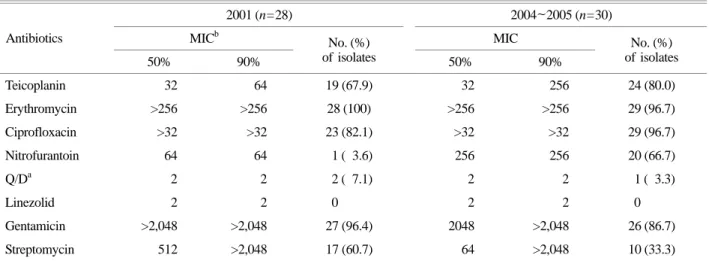
에 고도내성을 가지고 있었다. Gentamicin에 대한 MIC는 연도별 분리균주들에서 큰 차이가 없었지만, streptomycin 에 대한 내성빈도는 2001년 분리균주들에 비해 2004년과 2005년에 분리된 균주들에서 크게 감소하였다. 반면에 2001년 분리균주들은 nitrofurantoin에 대부분 감수성이었 지만, 2004년과 2005년 분리균주에서는 65% 이상이 내성 으로 나타나 통계학적 유의성을 보였다 (p<0.0001, Chi- square test). Macrolide계 항균제인 erythromycin에는 1주를 제외한 57주에서 MIC가 >256 µg/ml 이상으로 나타나 높 은 내성률을 보였다. Streptogramin계 항균제인 quinupristin- dalfopristin에는 3주가 내성이었고, 현재 치료제로 사용되 고 있는 linezolid에 내성인 균주는 없었다 (Table 2).
분리된 대부분의 VREF 균주들이 aminoglycosides와 erythromycin에 고도내성을 나타내어 내성유전자들을 조 사하였다. 고도내성 gentamicin 내성유전자인 aac6'-aph2"
는 전체 분리균주 중 53주 (91.4%)에서, streptomycin 내성
유전자인 aadE는 27주 (46.6%)에서 발견되었다. Erythro- mycin 내성 ermB와 ermA 유전자는 각각 54주 (93.1%)와 3주 (5.2%)에서 발견되었다 (Table 3).
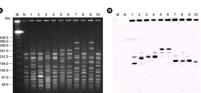
2. 접합에 의한 gentamicin 내성유전자의 분자적 특성 전체균주를 대상으로 접합을 시행한 뒤 항균제 내성 전달 여부를 검색한 결과, 51주는 vancomycin 내성이 전 달되었고, 31주는 erythromycin 내성이 전달되었으며, 5주 는 gentamicin 내성이 수여균으로 전달되는 것을 확인하 였다. 또한 분리균주들과 접합 실험에 의해 gentamicin 내성이 전달된 5주의 피전달접합균주들을 대상으로 내 성유전자의 유전적 위치를 규명하였다 (Fig. 1). Southern hybridization을 시행한 결과, 분리균주들과 피전달접합균 주들은 모두 aac6'-aph2" 유전자가 염색체상에 위치하고 있었다 (Fig. 1B).
3. Multilocus sequence typing (MLST) 분석
분리된 VREF 균주들의 MLST를 시행한 결과, 모두 11개의 ST가 나타났다 (Table 4). 클론의 다양성을 확인하 기 위해 11개의 ST들을 eBURST program으로 분류한 결과 complex 78, 17, 18 그룹에 속하였으며, 이들은 모두 CC78에 속하였다. 11개의 ST에서 ST192 (19주), ST203 (11주), ST17 (9주), 그리고 ST78 (8주)이 흔한 유형이었다.
가장 흔한 ST192는 1개 병원에서 분리된 균주에서만 나 타났고, ST203은 3개 병원으로부터 분리한 균주에서 나 타났다. 또한 ST18, ST64, ST206, ST117, ST132, ST233은 대부분 2001년 분리균주에서만 나타났고 이후의 분리균 Table 2. Antimicrobial susceptibility of vancomycin-resistant E. faecium isolates
2001 (n=28) 2004~2005 (n=30)
MICb MIC
Antibiotics
50% 90%
No. (%)
of isolates 50% 90%
No. (%) of isolates
Teicoplanin 32 64 19 (67.9) 32 256 24 (80.0)
Erythromycin >256 >256 28 (100) >256 >256 29 (96.7) Ciprofloxacin >32 >32 23 (82.1) >32 >32 29 (96.7)
Nitrofurantoin 64 64 1 ( 3.6) 256 256 20 (66.7)
Q/Da 2 2 2 ( 7.1) 2 2 1 ( 3.3)
Linezolid 2 2 0 2 2 0
Gentamicin >2,048 >2,048 27 (96.4) 2048 >2,048 26 (86.7)
Streptomycin 512 >2,048 17 (60.7) 64 >2,048 10 (33.3)
a Q/D, Quinupristin-Dalfopristin, b MIC, minimum inhibitory concentration.
Table 3. Distribution of antimicrobial resistant genes of vancomycin- resistant E. faecium isolates
No. (%) of is olates
HLARa Erythromycin Isolation year
(No. of isolates)
aac6'-aph2" aadE ermA ermB 2001 (n=28) 27 (46.6) 17 (29.3) 3 (5.2) 25 (43.1) 2004~2005
(n=30) 26 (44.8) 10 (17.3) 0 (0.0) 29 (50.0) Total (n=58) 53 (91.4) 27 (46.6) 3 (5.2) 54 (93.1)
a HLAR, high-level aminoglycoside resistance
에서는 나타나지 않았다 (Table 4) (Fig. 2).
고 찰
58주의 VREF 균주 중 57주는 vancomycin에 대한 MIC 가 >256 µg/ml로 나타나 vancomycin 고도내성균주이었으 며, 나머지 1주의 vancomycin MIC가 64 µg/ml인 중등도 내성균주이었다. 또한 vancomycin과 teicoplanin에 모두
내성인 VanA phenotype-vanA genotype 균주 (25)는 57주가 있었으며, 1주는 vancomycin에 내성이지만 teicoplanin에 는 감수성인 VanB phenotype-vanA genotype 균주이었다.
vanA 유전자는 전달성 전위인자인 transposon Tn1546에 의해 다른 균주로 전이될 수 있으며 (9,12,18), VanB 표 현형을 나타내는 균주들이 최근에 일본, 대만 및 한국에 서도 보고되고 있는 실정이므로 VREF 균주들이 보유하 고 있는 vancomycin 내성유전자의 검색과 이들이 나타내 Table 4. Distribution of sequence types of vancomycin-resistant E. faecium isolates
No. of isolates
2001 2004 to 2005
ST (allelic profile)
Hospital A (n=15) Hospital B (n=11) Hospital C (n=2) Hospital A (n=30)
Total (n=58)
192 (15-1-1-1-1-7-1) 6 0 0 13 19
203 (15-1-1-1-1-20-1) 2 5 1 3 11
17 (1-1-1-1-1-1-1) 2 2 0 5 9
78 (15-1-1-1-1-1-1) 0 0 0 8 8
18 (7-1-1-1-5-1-1) 1 1 0 0 2
205 (3-1-1-1-1-1-1) 0 0 1 1 2
64 (7-1-1-1-1-1-1) 1 1 0 0 2
206 (1-3-1-1-1-1-7) 1 1 0 0 2
117 (9-1-1-1-1-1-1) 1 0 0 0 1
132 (7-1-1-1-12-1-1) 1 0 0 0 1
233 (1-1-1-1-1-20-1) 0 1 0 0 1
A B
Figure 1. (A) CHEF electrophoresis of SmaI-digested genomic DNAs from the VREF isolates and their transconjugants. (B) Corresponding Southern blot hybridized with a aac6'-aph2" gene probe. Lane M, Lambda ladder; lane N, recipient strain UW64/3 as a aac6'-aph2" negative control; lane 1, 01DEM01; lane 2, pDEM01W; lane 3, 01YEM01; lane 4, pYEM01W; lane 5, 04KEM10; lane 6, pKEM10W; lane 7, 04KEM13; lane 8, pKEM13W; lane 9, 05KEM26; lane 10, pKEM26W.
는 표현형 검사에 세심한 주의가 필요하다.
전반적인 항균제 내성률은 nitrofurantoin을 제외하고는 2001년 분리균주와 2004년과 2005년 분리균주에서 크게 달라진 점은 없었다. 최근 연구결과에서 요로감염치료제 로 사용되는 nitrofurantoin은 장알균에서 높은 내성빈도를 가지지 않는 것으로 알려져 있지만 (23), 본 연구에서는 2004년과 2005년 분리주들에서 내성률이 크게 증가하여 VREF의 분리빈도가 높은 병원에서의 nitrofurantoin의 사 용은 신중한 선택이 필요할 것으로 생각된다. 그람양성 균의 중증감염 치료제로 사용되고 있는 linezolid에 내 성을 나타내는 linezolid 내성 enterococci (LRE)는 외국에 서는 2000년대 초부터 발견되고 있지만 (14), 본 연구에 서는 발견되지 않았다. 그러나 linezolid의 MIC가 4 µg/ml 인 중등도 내성균주 2주가 2001년도에 분리되었으므로 linezolid의 무분별한 오, 남용의 예방과 LRE 출현에 대 한 꾸준한 검색이 필요하다.
접합 실험의 결과에서 gentamicin 내성이 전달된 5주 중 4주는 공여균과 피전달접합균주들에서 내성유전자가 염색체상의 동일위치에서 관찰되는 것으로 보아 피전달 균주로 전달된 내성유전자는 분리균의 내성유전자가 위 치한 동일 염색체상에 삽입되는 것으로 생각된다. 우리 나라에서 분리되는 VRE 균주들에서는 vancomycin 내성 과 더불어 ampicillin과 aminoglycosides 내성이 흔히 함께 동반되며 (19), Zervos 등 (35)은 gentamicin 내성유전자들 이 Tn924, Tn5281, Tn4031, Tn4001과 같은 전위유전자들과 함께 염색체의 transposon에 위치하는 것을 보고하였다.
본 연구에서도 vancomycin 내성과 함께 gentamicin 내 성 aac6'-aph2" 유전자가 함께 피전달접합균주로 전달 되는 것이 확인되어, gentamicin 내성유전자가 단독으로 수평전달되기 보다는 vancomycin 내성유전자가 포함된 transposon이 전달될 때 부수적으로 gentamicin 내성유 전자가 전달되는 것으로 생각된다. Oancea 등 (24)은 E.
faecium 균종의 접합 실험에서 피전달균주로 전달된 항 균제 내성 중 erythromycin과 tetracycline은 고빈도로 내성 이 전달되지만, gentamicin 내성은 거의 전달되지 않음을 보고하였다. 본 연구에서도 gentamicin 내성이 낮은 빈도 로 피전달접합균주로 전달되는 것을 확인하였다. 따라서 58주의 균주 중 53주가 gentamicin 내성균주라는 사실은 내성유전자의 수평전달보다는 gentamicin 내성을 가진 클 론의 확산이 gentamicin 내성의 더 큰 원인으로 생각할 수 있다.
MLST 분석결과에 의하여, 3개 병원에서 분리된 VREF 균주들의 ST들에 대한 변화를 크게 세 가지로 구분할 수 있었다. 첫 번째는 1개의 병원에서 5년간 지속적으로 나타나는 토착형 (ST192), 두 번째는 3개의 병원에 모두 나타나는 일반형 (ST203), 그리고 세 번째는 잠시 출현 하였다가 사라지는 소멸형 (ST64, ST206, ST117, ST132, ST233)이었다. 이러한 유전학적 분포를 기준으로 대구지 역에서 분리되는 VREF 균주들은 CC78에 속하는 ST192, ST203, ST78 등이 가장 일반적이고 우세한 것을 확인할 수 있었고, MLST 연구결과에서 CC78에 속하는 VREF 균주들은 2005년 한국의 다른 병원에서 분리된 VREF 균 Figure 2. Clustering analysis of 11 STs. The dot with dashed circle indicates each ST.
주의 MLST 결과와 마찬가지로 80% 이상이 CC78에 속 하였다 (16). 2006년에는 브라질에서 분리된 균주들은 대 부분 ST114와 ST222, ST224, ST163에 속하는 세계적으로 알려진 complex 그룹에 포함되지 않는 완전히 다른 독립 적인 그룹의 균주들이 분리되기도 하였다 (5). 그러나 최 근까지 유럽지역과 미국 등에서 MLST 분석에 의해 동 정된 임상유래 분리균주들은 대부분 Complex 17에 속하 는 그룹이고, 이들 균주들은 다약제 내성으로 인한 치료 의 어려움과 감염원으로 추정되는 병인인자들의 빈도가 증가하여 원내감염의 원인균으로 대두되고 있는 추세이 다 (34).
본 연구는 최근 임상가검물에서 분리빈도가 높은 VREF 균주들의 내성과 내성유전자들의 특성을 조사하 였으며, MLST를 통하여 역학적 특성을 규명하였다. 연 구결과 vancomycin 내성균주들은 aminoglycoside계 및 macrolide계 항균제에 고도내성을 동시에 가지는 균주들 이 흔하게 나타났으며, gentamicin 내성유전자인 aac6'- aph2"는 vanA 유전자가 포함된 transposon과 밀접하게 연 관되어 있어서 vanA 유전자의 수평전달 때 부수적으로 동반되어 전달됨을 확인하였다. MLST에 의한 VREF 균 주들의 역학조사는 지역적인 범위의 역학조사뿐만 아니 라 클론의 기원과 전파 양상을 분석하는데도 유용하게 사용될 수 있음을 확인하였다.
참 고 문 헌
1)최원석, 서유빈, 조유미, 김정연, 기세윤, 정혜원, 송준 영, 정희진, 송기준, 김우주: Vancomycin 내성 장구균에 의한 세균뇨의 역학과 임상적 중요성. 감염과화학요법 38: 242-249, 2006.
2)홍기숙, 강은숙, 이미애: Vancomycin 내성 Enterococci의 빈도 조사 및 중합효소연쇄반응을 이용한 유전자형의 분석. 대한임상병리학회지 18: 372-378, 1998.
3) Agerso Y, Pedersen AG, Aarestrup FM: Identification of Tn5397-like and Tn916-like transposons and diversity of the tetracycline resistance gene tet (M) in enterococci from humans, pigs and poultry. J Antimicrob Chemother 57: 832-839, 2006.
4) Biavasco F, Miele A, Vignaroli C, Manso E, Lupidi R, Varaldo PE: Genotypic characterization of a nosocomial outbreak of VanA Enterococcus faecalis. Microb Drug Resist 2: 231-237, 1996.
5) Camargo IL, Gilmore MS, Darini AL: Multilocus sequence
typing and analysis of putative virulence factors in vancomycin- resistant and vancomycin-sensitive Enterococcus faecium isolates from Brazil. Clin Microbiol Infect 12: 1123-1130, 2006.
6) Chang SC, Chen YC, Luh KT, Hsieh WC: Macrolides resistance of common bacteria isolated from Taiwan. Diagn Microbiol Infect Dis 23: 147-154, 1995.
7) del Campo R, Tenorio C, Rubio C, Castillo J, Torres C, Gomez-Lus R: Aminoglycoside-modifying enzymes in high- level streptomycin and gentamicin resistant Enterococcus spp.
in Spain. Int J Antimicrob Agents 15: 221-226, 2000.
8) del Campo R, Ruiz-Garbajosa P, Sanchez-Moreno MP, Baquero F, Torres C, Canton R, Coque TM: Antimicrobial resistance in recent fecal enterococci from healthy volunteers and food handlers in Spain: genes and phenotypes. Microb Drug Resist 9: 47-60, 2003.
9) Eom JS, Hwang IS, Hwang BY, Lee JG, Lee YJ, Cheong HJ, Park YH, Park SC, Kim WJ: Emergence of vanA Genotype Vancomycin-Resistant Enterococci with Low or Moderate Levels of Teicoplanin Resistance in Korea. J Clin Microbiol 42: 1785-1786, 2004.
10) Gordon KA, Jones RN: Susceptibility patterns of orally administered antimicrobials among urinary tract infection pathogens from hospitalized patients in North America:
comparison report to Europe and Latin America. Results from the SENTRY Antimicrobial Surveillance Program (2000).
Diagn Microbiol Infect Dis 45: 295-301, 2003.
11) Harada T, Tsuji N, Otsuki K, Murase T: Detection of the esp gene in high-level gentamicin resistant Enterococcus faecalis strains from pet animals in Japan. Vet Microbiol 106:
139-143, 2005.
12) Hashimoto Y, Tanimoto K, Ozawa Y, Murata T, Ike Y:
Amino acid substitutions in the VanS sensor of the VanA-type vancomycin-resistant Enterococcus strains result in high-level vancomycin resistance and low-level teicoplanin resistance.
FEMS Microbiol Lett 185: 247-254, 2000.
13) Homan WL, Tribe D, Poznanski S, Li M, Hogg G, Spalburg E, Van Embden JD, Willems RJ: Multilocus sequence typing scheme for Enterococcus faecium. J Clin Microbiol 40: 1963 -1971, 2002.
14) Kainer MA, Devasia RA, Jones TF, Simmons BP, Melton K, Chow S, Broyles J, Moore KL, Craig AS, Schaffner W:
Response to emerging infection leading to outbreak of linezolid-resistant enterococci. Emerg Infect Dis 13: 1024-1030,
2007.
15) Klare I, Heier H, Claus H, Witte W: Environmental strains of Enterococcus faecium with inducible high-level resistance to glycopeptides. FEMS Microbiol Lett 106: 23-29, 1993.
16) Ko KS, Baek JY, Lee JY, Oh WS, Peck KR, Lee N, Lee WG, Lee K, Song JH: Molecular characterization of vancomycin-resistant Enterococcus faecium isolates from Korea. J Clin Microbiol 43: 2303-2306, 2005.
17) Kolar M, Pantucek R, Vagnerova I, Cekanova L, Kesselova M, Sauer P, Koukalova D, Kolar V, Ruzi kova M, Dosar J:
Prevalence of vancomycin-resistant enterococci in hospital and community environment. Klin Mikrobiol Infekc Lek 11:
47-50, 2005.
18) Leavis H, Top J, Shankar N, Borgen K, Bonten M, van Embden J, Willems RJ: A novel putative Enterococcal pathogenicity island linked to the esp virulence gene of Enter- ococcus faecium and associated with epidemicity. J Bacteriol 186: 672-682, 2004.
19) Lee WG, Huh JY, Cho SR, Lim YA: Reduction in glyco- peptide resistance in vancomycin-resistant enterococci as a result of vanA cluster rearrangements. Antimicrob Agents Chemother 48: 1379-1381, 2004.
20) Lim JA, Kwon AR, Kim SK, Chong Y, Lee K, Choi EC:
Prevalence of resistance to macrolide, lincosamide and strepto- garmin antibiotics in Gram-positive cocci isolated in a Korean hospital. J Antimicrob Chemother 49: 489-495, 2002.
21) Murray BE: The life and times of the enterococcus. Clin Microbiol Rev 3: 46-65, 1990.
22) Murray BE: Diversity among multidrug-resistant enterococci.
Emerg Infect Dis 4: 37-47, 1998.
23) Nichol KA, Sill M, Laing NM, Johnson JL, Hoban DJ, Zhanel GG: Molecular epidemiology of urinary tract isolates of vancomycin-resistant Enterococcus faecium from North America. Int J Antimicrob Agents 27: 392-396, 2006.
24) Oancea C, Klare I, Witte W, Werner G: Conjugative transfer of the virulence gene, esp, among isolates of Enterococcus faecium and Enterococcus faecalis. J Antimicrob Chemother 54: 232-235, 2004.
25) Oh JY, An S, Jin JS, Lee YC, Cho DT, Lee JC: Phenotypic and genotypic differences of the vancomycin-resistant Enter- ococcus faecium isolates from humans and poultry in Korea. J
Microbiol 45: 466-472, 2007.
26) Poeta P, Costa D, Rodrigues J, Torres C: Antimicrobial resistance and the mechanisms implicated in faecal enterococci from healthy humans, poultry and pets in Portugal. Int J Antimicrob Agents 27: 131-137, 2006.
27) Rice LB, Carias L, Rudin S, Vael C, Goossens H, Konstabel C, Klare I, Nallapareddy SR, Huang W, Murray BE: A potential virulence gene, hylsEfm, predominates in Enterococcus faecium of clinical origin. J Infect Dis 187: 508-512, 2003.
28) Sambrook J, Fritsch EF, Maniatis T: Molecular cloning: a laboratory manual, 10.1~10.52, 2nd ed. Cold Spring Harbor Laboratory, Cold Spring Harbor NY., 1989.
29) Sanchez ML, Flint KK, Jones RN: Occurrence of macrolide- lincosamide-streptogramin resistance among staphylsococcal clinical isolates at a university medical center. Is false susceptibility to new macrolides and clindamycin a contem- porary clinical and in vitro testing problem? Diagn Microbiol Infect Dis 16: 205-213, 1993.
30) Shankar N, Lockatell CV, Baghdayan AS, Drachenberg C, Gilmore MS, Johnson DE: Role of Enterococcus faecalis surface protein Esp in the pathogenesis of ascending urinary tract infection. Infect Immun 69: 4366-4372, 2001.
31) Tunger A, Aydemir S, Uluer S, Cilli F: In vitro activity of linezolid & quinupristin/dalfopristin against Gram-positive cocci. Indian J Med Res 120: 546-552, 2004.
32) Uttley AH, Collins CH, Naidoo J, George RC: Vancomycin- resistant enterococci. Lancet 1: 57-58, 1998.
33) van den Braak N, van Belkum A, van Keulen M, Vliegen- thart J, Verbrugh HA, Endtz HP: Molecular characterization of vancomycin-resistant enterococci from hospitalized patients and poultry products in the Netherlands. J Clin Microbiol 36:
1927-1932, 1998.
34) Willems RJ, Top J, van Santen M, Robinson DA, Coque TM, Baquero F, Grundmann H, Bonten MJ. Global spread of vancomycin-resistant Enterococcus faecium from distinct nosocomial genetic complex. Emerg Infect Dis 11: 821-828, 2005.
35) Zervos MJ, Mikesell TS, Schaberg DR: Heterogeneity of plasmids determining high-level resistance to gentamicin in clinical isolates of Streptococcus faecalis. Antimicrob Agents Chemother 30: 78-81, 1986.