Surgical Approaches for Symptomatic Cerebral Cavernous Malformations of the Thalamus and Brainstem
Dale Ding1, Robert M. Starke2, R. Webster Crowley3, Kenneth C. Liu1,4
1Department of Neurosurgery, University of Virginia, Charlottesville, VA, USA
2Department of Neurological Surgery, University of Miami, Miami, FL, USA
3Department of Neurological Surgery, Rush University, Chicago, IL, USA
4Department of Radiology and Medical Imaging, University of Virginia, Charlottesville, VA, USA
Objective : Surgical resection of thalamic and brainstem cerebral cav- ernous malformations (CCMs) is associated with significant operative mor- bidity, but it may be outweighed, in some cases, by the neurological damage from recurrent hemorrhage in these eloquent areas. The goals of this retrospective cohort study are to describe the technical nuances of surgical approaches and determine the postoperative outcomes for CCMs of the thalamus and brainstem.
Materials and Methods : We reviewed an institutional database of patients harboring thalamic or brainstem CCMs, who underwent surgical resection from 2010 to 2014. The baseline and follow-up neuroimaging and clinical findings of each patient and the operative details of each case were evaluated.
Results : A total of eight patients, including two with thalamic and six with brainstem CCMs, were included in the study cohort. All patients had progressive neurological deterioration from recurrent CCM hemorrhage, and the median modified Rankin Scale (mRS) at presentation was 3. The median CCM maximum diameter and volume were 1.7 cm and 1.8 cm3, respectively. The thalamic CCMs were resected using the anterior trans- callosal transchoroidal and supracerebellar infratentorial approaches each in one case (13%). The brainstem CCMs were resected using the retro- sigmoid and suboccipital trans-cerebellomedullary fissure approaches each in three cases (38%). After a median follow-up of 11.5 months, all pa- tients were neurologically stable or improved, with a median mRS of 2.
The rate of functional independence (mRS 0-2) was 63%.
Conclusion : Microneurosurgical techniques and approaches can be safely and effectively employed for the management of thalamic and brainstem CCMs in appropriately selected patients.
J Cerebrovasc Endovasc Neurosurg.
2017 March;19(1):19-35 Received : 21 April 2015 Revised : 4 January 2017 Accepted : 18 February 2017 Correspondence to Dale Ding
Department of Neurosurgery, University of Virginia, P.O. Box 800212, Charlottesville, VA 22908, USA
Tel : 1-434-924-2203 Fax : 1-434-982-5753
E-mail : [email protected] ORCID : http://orcid.org/0000-0002-2627-446X
This is an Open Access article distributed under the terms of the Creative Commons Attribution Non- Commercial License (http://creativecommons.org/li- censes/by-nc/3.0) which permits unrestricted non- commercial use, distribution, and reproduction in any medium, provided the original work is properly cited.
Keywords Brainstem, Cerebral cavernous malformation, Intracranial hemorrhages, Microsurgery, Thalamus, Vascular malformations
INTRODUCTION
Cerebral cavernous malformations (CCM) are com- monly localized to deep regions of the brain, such as
the thalamus and brainstem. Thalamic and brainstem CCMs account for approximately one-third of CCMs.41)42) The annual hemorrhage risk of these deep-seated CCMs has been reported, in some series, to be sig-
nificantly higher than superficially located CCMs.57) Specifically, the hemorrhage risk of brainstem CCMs with a history of clinically overt hemorrhage may be as high as 20% per year.2) Due to the increased pro- pensity for symptomatic CCMs to hemorrhage and the exquisitely sensitive nature of the thalamus and brainstem, intervention should be considered for ap- propriately selected lesions.24)38)
However, thalamic and brainstem CCMs present a significant surgical challenge, and are associated with relatively high rates of operative morbidity and mortality.41)58) While radiosurgery has been employed as a minimally invasive therapy for surgically unfavorable CCMs, the efficacy of this approach is controversial.
Thus, surgery remains the gold standard of treatment for symptomatic CCMs which necessitate intervention.
The aims of this retrospective cohort study are to (1) evaluate the technical aspects of common surgical ap- proaches to CCMs of the thalamus and brainstem us- ing specific case examples and (2) determine the post- operative outcomes for patients with thalamic and brainstem CCMs in the modern era.
MATERIALS AND METHODS
Patient selection
We performed a retrospective evaluation of an in- stitutional review board (IRB) approved database of CCM patients who underwent surgical management by the senior author (K.C.L.) at the University of Virginia from July 2010 to December 2014. The in- clusion criteria were (1) CCM localization in the thala- mus or brainstem and (2) sufficient data regarding baseline neurological status, intraoperative details, and postoperative outcomes. Patients with vascular malformations other than CCMs, such as cerebral ar- teriovenous malformations (AVM) and dural arterio- venous fistulas, were excluded.
Baseline data and follow-up
The following baseline data were extracted from di- rected chart review: (1) patient attributes, (2) CCM
characteristics, and (3) surgical details. The patient at- tributes were gender, age, presenting symptoms, and modified Rankin Scale (mRS) score at presentation.
The CCM characteristics were size (maximum diame- ter and volume) and location. The surgical details were approach taken to access the lesion, use of supple- mentary localization techniques (e.g. electrophysiologic monitoring of neurological function, stereotactic neu- ronavigation), administration of intraoperative medi- cations, and technical aspects of the surgical procedure, including patient positioning, skin incision, craniotomy or craniectomy, dural opening, intradural dissection, and CCM identification and resection.
Follow-up was classified as perioperative or longitudinal.
Perioperative follow-up comprised all events from the end of surgery to hospital discharge, including post- operative complications, duration of hospital stay, dis- charge destination, and mRS at discharge. Longitudinal follow-up comprised all events from hospital dis- charge to the most recent clinical follow-up, including neuroimaging documentation of the absence of pres- ence of residual CCM, mRS at last clinical follow-up, and duration of radiologic and clinical follow-up. The necessity for additional procedures after CCM resection was also noted for each patient.
RESULTS
Patient cohort and preoperative evaluation
A total of eight patients, two with thalamic CCMs (25%) and six with brainstem CCMs (75%), were in- cluded in the study cohort. The affected brainstem re- gions were the pons and medulla each in two patients (25%), and the midbrain and pontomesencephalic junc- tion each in one patient (13%). Thin-slice (1 mm thick- ness) magnetic resonance imaging (MRI), T1-weighted with and without contrast and T2-weighted sequences, was performed for each patient. Based on the location of the lesion, the neurosurgeon (K.C.L.) decided on the surgical approach. Consideration was given to the accessibility of the lesion, closest pial border, oper- ative corridor afforded by the approach, and morbid-
A B C
D E F
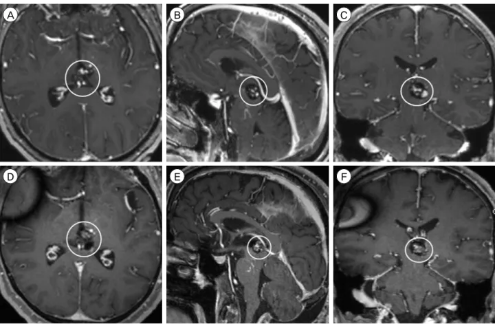
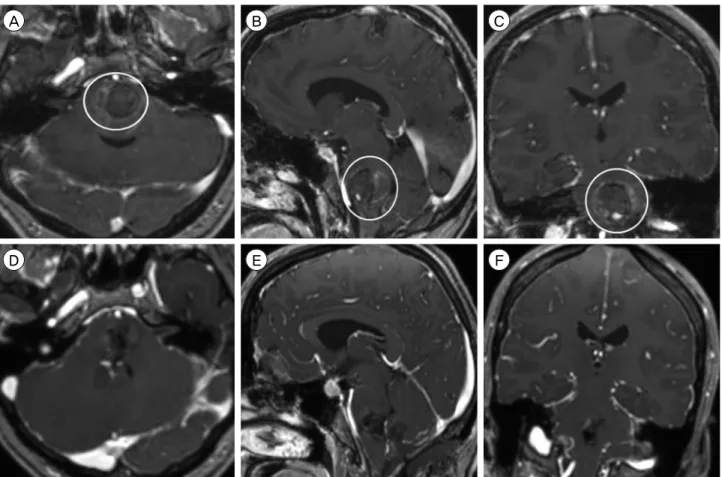
Fig. 1. Preoperative MRI, T1-weighted sequence with contrast, (A) axial, (B) sagittal, and (C) coronal views, shows a 2.3 × 2.0 × 1.9 cm CCM in the right anterior medial thalamus, with medial extension into the third ventricle and inferior extension into the cerebral peduncle (circle). Postoperative MRI (follow-up interval 5 months) after resection via an anterior transcallosal transchoroidal approach, T1-weighted sequence without contrast, (D) axial, (E) sagittal, and (F) coronal views, shows dense hemosiderin staining along the right lateral aspect of the surgical cavity, which is suspicious for a small amount of residual CCM (circle). MRI = magnetic resonance imaging; CCM = cerebral cavernous malformation.
ity of the approach. All patients presented with pro- gressively worsening symptoms from mass effect after recurrent CCM hemorrhage. The median mRS at pre- sentation was 3 (range 3 to 4). The median CCM max- imum diameter and volume were 1.7 cm (range 1.1 to 2.8 cm) and 1.8 cm3 (range 0.6 to 8.7 cm3), respectively.
General neurosurgical considerations
All surgical procedures were performed under gen- eral anesthesia, with the patient's head affixed in a three-point fixation Mayfield Skull Clamp System (Integra, Plainsboro, NJ, USA). After appropriate pa- tient positioning for the planned surgical approach, the StealthStation (Medtronic, Minneapolis, MN, USA) frameless stereotactic neuronavigation system was reg- istered with the patient's neuroimaging. Intraoperative electrophysiologic neurological monitoring of motor and somatosensory evoked potentials (MEP and SSEP,
respectively) were utilized in all cases, and brainstem evoked potentials (BSEPs) were used for all brainstem CCMs. Near the time of skin incision and prior to du- ral opening, mannitol 1 gm/kg and dexamethasone 10 mg were administered, and hyperventilation to a PCO2 of 25 mmHg was performed to reduce brain swelling and intracranial pressure.
The operating microscope was routinely used for all intradural dissection, and both gross intraoperative findings and neuronavigation were used to identify a CCM's location. Afterwards, a combination of hemosi- derin staining of the pial surface, when present, and surface anatomical landmarks were used, in con- junction with neuromonitoring, to determine the pial entry point for accessing the lesion. The dura was closed in a watertight fashion when possible, other- wise a dural substitute graft was sutured to the native
dura to cover the dural defect. When a craniotomy was performed, the bone flap was plated back to the skull with titanium plates and screws. When a cra- niectomy was performed, a titanium mesh plate was placed over the cranial defect. Closure was performed in a standard layered fashion.
Anterior transcallosal transchoroidal approach for anterior thalamic CCMs
A 69 year-old female presented with several months of progressively worsening altered mental status and ataxia (mRS score 3 at presentation). MRI showed a 2.3 cm CCM in the right anterior medial thalamus (Fig. 1). We proceeded with surgical resection of the lesion via an anterior transcallosal transchoroidal approach. The patient was placed in a supine position, and a bicoronal skin incision was made. A left para- sagittal craniotomy was performed, two-thirds ante- rior to the coronal suture. The dura covering the me- dial left frontal lobe was reflected medially over the superior sagittal sinus. We proceeded into the inter- hemispheric fissure to expose the dorsal surface of the corpus callosum.
A callosotomy was performed, located 1.5 cm poste- rior to the genu of the corpus callosum and measur- ing approximately 3 cm in length, in order to enter the lateral ventricle. We determined that we were in the right lateral ventricle, but the right foramen of Monro was obliterated by mass effect from the thala- mic CCM. A septostomy was performed to facilitate cerebrospinal fluid (CSF) egress into the contralateral ventricle. After the anterior septal vein was coagu- lated and divided, the choroidal fissure of the right lateral ventricle, lateral to the fornix and medial to the choroid plexus, was opened in order to access the third ventricle. The CCM did not present to the epen- dymal surface and, thus, could not be readily visualized. The ependyma over the anterior medial thalamus was opened, and the CCM was immediately encountered. Due to the large size of the lesion, it was resected in a piecemeal fashion with standard microsurgical technique.
An external ventricle drain (EVD) was left in the third ventricle for 48 hours postoperatively. The pa- tient had an uncomplicated postoperative course and was discharged from the hospital to an inpatient re- habilitation facility (mRS score 3 at discharge) on POD 10. Follow-up MRI, performed 5 months after surgery, showed a small residual CCM. At 11 months clinical follow-up, there was no evidence of postoperative in- tracranial hemorrhage, although the patient was neu- rologically unchanged, with a mRS score of 3.
Supracerebellar infratentorial approach for posteri- or thalamic CCMs
A 50 year-old male presented with two months of progressively worsening headache, diplopia, and ataxia (mRS score 3 at presentation). MRI showed a 1.7 cm CCM in left posterior medial thalamus and midbrain (Fig. 2). We proceeded with surgical resection of the lesion via a supracerebellar infratentorial approach.
The patient was placed in a prone position, with capi- tal flexion, and a straight incision was made in the midline from the vertex to the C4 spinous process. A median suboccipital craniotomy was performed in or- der to expose the torcula and suboccipital dura. The suboccipital dura was reflected superiorly over the torcula and bilateral transverse sinuses. The occipital sinus was ligated, and the cerebellar hemispheres and vermis were retracted inferiorly in order to proceed through the supracerebellar infratentorial corridor.
Two precentral cerebellar veins were divided to im- prove visualization of the deep anatomy.
At the tentorial incisura, the splenium of the corpus callosum and confluence of the venous structures at the vein of Galen were identified. The quadrigeminal cistern was opened to release CSF and promote brain relaxation. The arachnoid adhesions tethering the cer- ebellar vermis to the deep venous complex were re- leased, and dissection was continued under the left basal vein of Rosenthal, lateral to the divided pre- central cerebellar vein. The tectum, perforator arteries, and a large developmental venous anomaly were not- ed, and hemosiderin staining of the pial surface of the
A B C
D E F
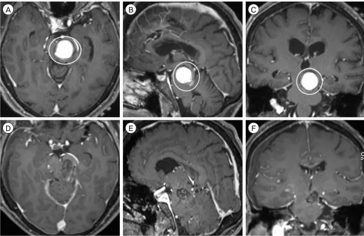
Fig. 2. Preoperative MRI, T1-weighted sequence with contrast, (A) axial, (B) sagittal, and (C) coronal views, shows a 1.7 × 1.5 × 1.7 cm CCM in left posterior medial thalamus and midbrain (circle). Postoperative MRI (follow-up interval 19 months) after resection via a supracerebellar infratentorial approach, T1-weighted sequence with contrast, (D) axial, (E) sagittal, and (F) coronal views, shows a 1.1
× 1.2 × 1.6 cm residual CCM (circle). MRI = magnetic resonance imaging; CCM = cerebral cavernous malformation.
supracollicular area was visualized. The supra- collicular area was opened at the site of hemosiderin staining in order to enter the CCM. The lesion was re- sected with standard microsurgical technique, in a piecemeal fashion. The CCM was initially dissected inferiorly and its capsule was retracted cephalad, away from the superior colliculus. Next, the lesion was retracted medially, so that the lateral border of the capsule could be dissected away from the pulvinar. At the conclusion of the resection, the third ventricle was visualized.
The patient had a complicated postoperative course, including a cerebellar hemorrhage treated with EVD placement and suboccipital craniectomy with expansion duraplasty, respiratory failure requiring tracheostomy, and persistent dysphagia requiring percutaneous en- doscopic gastrostomy (PEG) tube placement. The pa-
tient was discharged to an inpatient rehabilitation fa- cility (mRS score 3 at discharge) two weeks after CCM resection. Over the course of the next 18 months, the patient subsequently underwent CSF diversion with ventriculoperitoneal shunt placement for hydrocephalus, pseudomeningocele repair, multiple shunt revisions, and suboccipital titanium mesh cranioplasty with lysis of adhesions. In total, the patient underwent nine ad- ditional surgical procedures after the initial resection.
Follow-up MRI, performed 19 months after surgery, showed residual CCM. At 20 months clinical fol- low-up, the patient was able to walk independently, but continued to experience persistent headaches and diplopia, for a mRS score of 3.
Retrosigmoid approach for dorsolateral midbrain and pontine CCMs
Three patients with dorsolateral midbrain or pontine
A B C
D E F
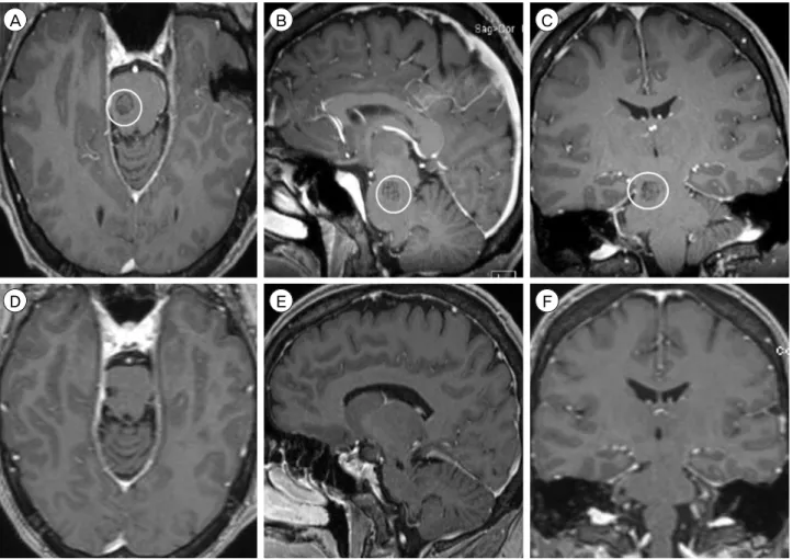
Fig. 3. Preoperative MRI, T1-weighted sequence with contrast, (A) axial, (B) sagittal, and (C) coronal views, shows a 2.1 × 1.9 × 1.9 cm CCM in the left midbrain, centered in the tegmentum (circle). Postoperative MRI (follow-up interval 1 day) after resection via a left-sided retrosigmoid approach, T1-weighted sequence with contrast, (D) axial, (E) sagittal, and (F) coronal views, shows post-surgical changes without evidence of residual CCM. MRI = magnetic resonance imaging; CCM = cerebral cavernous malformation.
CCMs underwent resection via a retrosigmoid ap- proach ipsilateral to the lesion (38%). For all patients, a lumbar drain was placed preoperatively and re- moved at the end of surgery. The patient was placed in a three-quarter prone position, with the head ro- tated to the side ipsilateral to the lesion and in capital flexion. A sigmoid incision was made from 3 cm pos- terior to the superior border of the pinna, across the asterion to the nuchal midline. A retrosigmoid cra- niectomy and partial posterior mastoidectomy were performed to expose the inferior border of the trans- verse sinus and posterior border of the sigmoid sinus, and 50 mL of CSF was released from the lumbar drain prior to dural opening to promote brain relaxation.
The dura was opened in a cruciate fashion, and the arachnoid of the cisterna magna was opened to re- lease additional CSF (i.e., Dandy's maneuver). The lat-
eral aspect of the cerebellar hemisphere was retracted medially to expose the facial and vestibulocochlear nerves. The junction of the tentorium with its attach- ment to the petrous bone was followed medially until the trigeminal nerve was encountered. The superior petrosal vein was coagulated and divided, and the arachnoid of the ambient and quadrigeminal cisterns was opened to allow further CSF egress. The details of each CCM resection varied among patients and are described below.
A 70 year-old female presented with six months of progressively worsening right-sided hemiparesis (mRS score 3 at presentation). MRI showed a 2.1 cm CCM in the left midbrain tegmentum (Fig. 3). From a left retrosigmoid approach, the CCM could not be readily visualized at the pial surface. The trochlear nerve was identified, and the tentorium was divided in order to
A B C
D E F
Fig. 4. Preoperative MRI, T1-weighted sequence with contrast, (A) axial, (B) sagittal, and (C) coronal views, shows a 1.0 × 1.0 × 1.1 cm CCM in the right pons, immediately inferior to the pontomesencephalic junction (circle). Postoperative MRI (follow-up interval 21 months) after resection via a right-sided retrosigmoid approach, T1-weighted sequence with contrast, (D) axial, (E) sagittal, and (F) coronal views, shows no evidence of residual CCM. MRI = magnetic resonance imaging; CCM = cerebral cavernous malformation.
explore cephalad and medially. The lateral pontome- sencephalic sulcus was opened longitudinally, and the CCM was immediately encountered below the pial surface. The lesion was resected with standard micro- surgical technique, in a piecemeal fashion. During the resection, the right-sided hemibody SSEPs were lost without changes in MEPs. No residual CCM was evi- dent on MRI performed on POD 1. Postoperatively, the patient developed contralateral, right frontal sub- dural and intracerebral hematomas, which were man- aged conservatively. She was discharged from the hospital to an inpatient rehabilitation facility (mRS score 3 at discharge) on POD 6. At 2 months clinical follow-up, the patient had improved to a mRS score of 2.
A 44 year-old female presented with one month of progressively worsening left-sided hemibody sensory loss, headache, diplopia, and ataxia (mRS score 3 at presentation). MRI showed a 1.1 cm CCM in the right pons, immediately caudal to the pontomesencephalic junction (Fig. 4). From a right retrosigmoid approach, the superior cerebellar artery (SCA) was noted to be coursing over the approximate location of the lesion and was therefore circumferentially dissected from its arachnoid attachments to allow for mobilization. Since the CCM was subpial, a longitudinal incision was made in the lateral pontomesencephalic sulcus to ac- cess the lesion. Dissecting above and below the SCA, the CCM was resected in a piecemeal fashion with standard microsurgical technique. Toward the end of
A B C
D E F
Fig. 5. Preoperative MRI, T1-weighted sequence with contrast, (A) axial, (B) sagittal, and (C) coronal views, shows a 1.4 × 1.2 × 1.3 cm CCM in the right midbrain and pons, centered at the pontomesencephalic junction (circle). Postoperative MRI (follow-up interval 8 months) after resection via a right-sided retrosigmoid approach, T1-weighted sequence with contrast, (D) axial, (E) sagittal, and (F) coronal views, shows no evidence of residual CCM. MRI = magnetic resonance imaging; CCM = cerebral cavernous malformation.
the resection, the left-sided hemibody SSEPs were lost, while the MEPs remained stable. The patient had an uncomplicated postoperative course and was dis- charged from the hospital to an inpatient rehabilitation facility (mRS score 3 at discharge) on POD 4. At 21 months radiologic and clinical follow-up, no residual CCM was evident on MRI, and the patient had im- proved to a mRS score of 1 and was able to return to work.
A 42 year-old female presented with three months of progressively worsening headaches, diplopia, and left-sided dysmetria (mRS score 3 at presentation).
MRI showed a 1.4 cm CCM in the right midbrain and pons, at the pontomesencephalic junction (Fig. 5).
From a right retrosigmoid approach, the CCM, which did not broach the pial surface, could not be readily visualized. After identifying the relevant local anat- omy, a myelotomy was made in the infracollicular
area, medial to the origin of the trochlear nerve. The CCM was encountered approximately 0.5 mm deep to the pial surface, and resected in a piecemeal fashion with standard microsurgical technique. During wound closure, the left-sided SSEPs and MEPs were lost. The patient had an uncomplicated postoperative course and was discharged from the hospital to an inpatient rehabilitation facility (mRS score 3 at discharge) on POD 5. At 8 and 12 months radiologic and clinical follow-up, respectively, no residual CCM was evident on MRI, and the patient had improved to a mRS score of 2.
Suboccipital trans-cerebellomedullary fissure ap- proach for dorsomedian pontine and medullary CCMs
Three patients with dorsomedian pontine or medul- lary CCMs underwent resection via a suboccipital
A B C
D E F
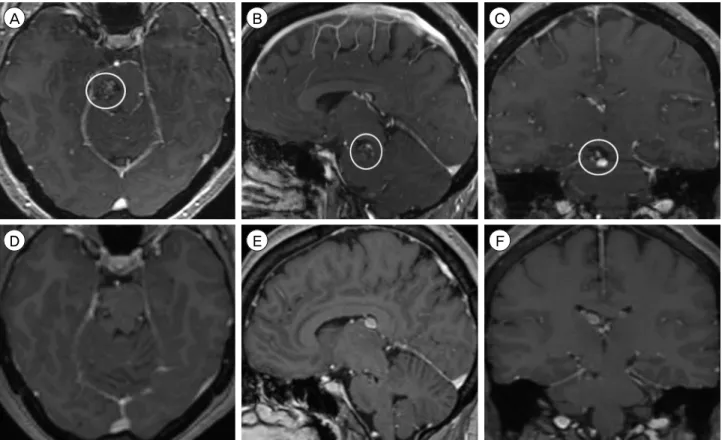
Fig. 6. Preoperative MRI, T1-weighted sequence with contrast, (A) axial, (B) sagittal, and (C) coronal views, shows a 1.5 × 1.4 × 1.3 cm CCM in the dorsomedian medulla (circle). Postoperative MRI (follow-up interval 23 months) after resection via a suboccipital trans-cerebellomedullary fissure approach, T1-weighted sequence with contrast, (D) axial, (E) sagittal, and (F) coronal views, shows no evidence of residual CCM. MRI = magnetic resonance imaging; CCM = cerebral cavernous malformation.
trans-cerebellomedullary fissure approach (38%). The patient was placed in a prone position with capital flexion. A longitudinal midline incision was made from the inion to the C4 spinous process, and a sub- occipital craniotomy or craniectomy and C1 lam- inectomy were performed. The dura was opened in a Y-shaped fashion, and the arachnoid of the cisterna magna was opened to release CSF and expose the cer- ebellar tonsils.
The cerebellomedullary fissures were opened bi- laterally to access the dorsal surface of the medulla and floor of the fourth ventricle. The cerebellar tonsils were retracted superolaterally, and the vermis was re- tracted superiorly to maintain the operative view.
Depending on the craniocaudal location of the CCM, the tela choroidea of the fourth ventricle was opened
bilaterally to increase the surgical corridor superiorly, up to the cerebral aqueduct. The details of each CCM resection varied among patients and are described below.
A 28 year-old male presented with one month of progressively worsening bilateral upper extremity sensory loss and ataxia (mRS score 3 at presentation). MRI showed a 1.5 cm CCM in the dorsomedian medulla (Fig. 6). From a suboccipital trans-cerebellomedullary fissure approach, the lesion could be visualized at the pial surface of the dorsal medulla. The posterior me- dian sulcus overlying the CCM was opened, and the lesion was resected in a piecemeal fashion with stand- ard microsurgical technique. During dural closure, the SSEPs of the bilateral upper extremities were lost, fol- lowed by the loss of bilateral lower extremity SSEPs.
A B C
D E F
Fig. 7. Preoperative MRI, T1-weighted sequence with contrast, (A) axial, (B) sagittal, and (C) coronal views, shows a 1.6 × 1.2 × 1.1 cm CCM in the right dorsomedian medulla (circle). Postoperative MRI (follow-up interval 1 day) after resection via a suboccipital trans-cerebellomedullary fissure approach, T1-weighted sequence with contrast, (D) axial, (E) sagittal, and (F) coronal views, shows post-surgical changes without evidence of residual CCM. MRI = magnetic resonance imaging; CCM = cerebral cavernous malformation.
The dura was re-opened in order to explore the surgi- cal cavity, but no hematoma or other anomaly was identified to explain the loss of electrophysiologic signals. The patient had an uncomplicated post- operative course and was discharged from the hospi- tal to an inpatient rehabilitation facility (mRS score 3 at discharge) on POD 6. At 23 and 21 months radio- logic and clinical follow-up, respectively, no residual CCM was evident on MRI, and the patient had im- proved to a mRS score of 2.
A 40 year-old male presented with six months of progressively worsening left-sided hemifacial and right-sided hemibody sensory alteration (mRS score 3 at presentation). MRI showed a 1.6 cm CCM in the right dorsomedian medulla (Fig. 7). From a sub- occipital trans-cerebellomedullary fissure approach, the lesion was readily visualized at the medulla's cau-
dal pial surface. A vertical myelotomy was performed along the posterior median sulcus, which was over- lying the lesion, and the CCM resection was initiated by internal debulking. A circumferential dissection was then performed around the collapsed CCM cap- sule with standard microsurgical technique, and the remainder of the lesion was removed in a piecemeal fashion. No residual CCM was evident on MRI per- formed on POD 1. Postoperatively, the patient suf- fered from persistent dysphagia, requiring PEG tube placement on POD 6. He was discharged to home two weeks after surgery, at which time he had im- proved to a mRS score of 2. Unfortunately, the patient was subsequently lost to follow-up.
A 28 year-old female presented with one week of progressively worsening right-sided headache and hear- ing loss, diplopia, bilateral upper extremity sensory
A B C
D E F
Fig. 8. Preoperative MRI, T1-weighted sequence with contrast, (A) axial, (B) sagittal, and (C) coronal views, shows a 2.8 × 2.7 × 2.3 cm CCM in the left pons. Postoperative MRI (follow-up interval 1 day) after resection via a suboccipital trans-cerebellomedullary fissure approach, T1-weighted sequence with contrast, (D) axial, (E) sagittal, and (F) coronal views, shows post-surgical changes without evi- dence of residual CCM. MRI = magnetic resonance imaging; CCM = cerebral cavernous malformation.
loss, dysarthria, and dysphagia (mRS score 4 at pre- sentation). MRI showed a 2.8 cm CCM in the left pons (Fig. 8). From a suboccipital trans-cerebellomedullary fissure approach, the anatomical landmarks at the floor of the fourth ventricle were identified and con- firmed with electrophysiologic stimulation and BSEPs.
A longitudinal myelotomy was performed in the in- frafacial triangle, bounded by the posterior median sulcus medially, facial colliculus superiorly and later- ally, and stria medullaris inferiorly. The opening was approximately 5 mm lateral to the posterior median sulcus and measured approximately 8 mm in length.
The CCM was noted beneath the pial surface, and subacute blood products extravasated under pressure after the capsule was opened. The remainder of the CCM was resected with standard microsurgical tech-
nique in a piecemeal fashion. At the end of the re- section, the BSEPs for the glossopharyngeal and va- gus nerves and the left-sided hemibody SSEPs were lost. No residual CCM was evident on MRI per- formed on POD 1. Postoperatively, the patient suf- fered from persistent dysphagia and pharyngeal ede- ma, requiring PEG tube placement and tracheostomy, respectively, on POD 4. She was transferred to a tran- sitional care hospital two weeks after surgery in neu- rological stable condition. At 2 months clinical fol- low-up, the patient is improved from her preoperative functional status and able to walk with assistance, al- though she remains with a mRS score of 4.
Summary of surgical outcomes for thalamic and brainstem CCMs
Table 1 summarizes the baseline characteristics and
Patient Age (years),
gender Clinical presentation, mRS at presentation
CCM location, maximum diameter,
volume*
Surgical approach, complete resection
Complications, number of additional
procedures
Clinical follow-up (months)
mRS Score at follow-up 1 69, Female Altered mental status,
ataxia, 3
Right anterior medial thalamus, 2.3 cm, 4.4 cm3
Anterior transcallosal
transchoroidal, No None, 0 11 3
2 50, Male Headache, diplopia, ataxia, 3
Left posterior medial thalamus
and midbrain, 1.7 cm, 2.2 cm3
Supracerebellar infratentorial, No
Cerebellar ICH, obstructive hydrocephalus, pseudomeningocele, dysphagia, respiratory
failure, 9
20 3
3 70, Female Hemiparesis, 3 Left midbrain,
2.1 cm, 3.8 cm3 Retrosigmoid, Yes Right frontal SDH
and ICH, 0 2 2
4 44, Female Hemibody sensory loss, headache, diplopia, ataxia, 3
Right pons,
1.1 cm, 0.6 cm3 Retrosigmoid, Yes None, 0 21 1
5 42, Female Headache, diplopia, dysmetria, 3
Right midbrain and pons, 1.4 cm,
1.1 cm3 Retrosigmoid, Yes None, 0 12 2
6 28, Male BUE sensory loss, ataxia, 3
Dorsomedian medulla, 1.5 cm,
1.4 cm3
Suboccipital trans-cerebellomed
ullary fissure, Yes None, 0 21 2
7 40, Male Hemifacial and hemibody sensory
alteration, 3
Right dorsomedian medulla, 1.6 cm,
1.1 cm3
Suboccipital trans-cerebellomed
ullary fissure, Yes Dysphagia, 1 0.5 2
8 26, Female
Headache, hearing loss, diplopia, BUE
sensory loss, dysarthria, dysphagia, 4
Left pons, 2.8 cm, 8.7 cm3
Suboccipital trans-cerebellomed
ullary fissure, Yes
Dysphagia,
pharyngeal edema, 2 2 4
BUE = bilateral upper extremity; CCM = cerebral cavernous malformation; ICH = intracerebral hemorrhage; mRS = modified Rankin Scale;
SDH = subdural hematoma.
*Volume was calculated by multiplying the axial, transverse, and craniocaudal dimensions of the CCM, and dividing the product by 2 Table 1. Summary surgical approaches and postoperative outcomes for patients with thalamic and brainstem CCMs
postoperative outcomes of the study cohort. Complete resection was achieved in six patients (75%), includ- ing all patients with brainstem CCMs (100%).
Perioperative complications were present in four pa- tients (50%), including three patients who required additional procedures (38%). At a median follow-up of 11.5 months (range 0.5 to 21 months), the median mRS score was 2 (range 1 to 4). Functional outcome was improved, at follow-up, in five patients (63%), stable in three (38%), and worse in none. Five patients were functionally independent, defined as mRS 2 or less (63%).
DISCUSSION
The management of thalamic and brainstem CCMs remains controversial. The elevated risk of both initial
and repeat hemorrhage for these deep-seated CCMs compared to cortical CCMs favors intervention, whereas the relatively high rate of neurological mor- bidity associated with surgical resection of these le- sions supports a more conservative approach.41)42) Although radiosurgery is a conceptually attractive al- ternative to surgical resection of eloquent CCMs, the radiosurgery-induced complication rate is significantly higher for CCMs than for AVMs, and CCMs cannot be radiologically monitored for obliteration after radiosurgery.3)5)6)8-10)12-23)25-37)43)44)49)50)52)53)56)59)63-68)
Instead, the proposed effect of radiosurgery on CCMs is a decreased risk of symptomatic hemorrhages, which is controversial due to temporal clustering of CCM hemorrhages.4) Thus, surgical resection remains the gold standard of CCM intervention and is war- ranted for appropriately selected patients harboring
thalamic and brainstem CCMs.
The following patient and lesion characteristics should be considered when deciding on the surgical approach for deep-seated CCMs. First, the patient's neurological and clinical status at presentation should dictate the decision for intervention versus conservative management. Patients with symptomatic CCM hemor- rhage, including progressive neurological deterio- ration from multiple hemorrhages, are preferred can- didates for intervention. In patients with focal neuro- logical deficits from CCM hemorrhage, an approach through a region related to the pre-existing deficit should be utilized in favor of one which may incur additional injury. We typically waited one to two weeks after a clinically symptomatic hemorrhage to perform surgical intervention, in order to allow lique- faction of the hematoma but with the goal of inter- vening before contraction and gliotic scarring of the hematoma. Additionally, medical comorbidities should be limited and clinical optimized to ensure the patient will be able to tolerate the physiologic stress of gen- eral anesthesia and major intracranial surgery. Next, the location of CCM presentation to the pial surface, or the closest pial border for subpial lesions, will de- termine the site of entry and, consequently, the surgi- cal approach. Finally, the size and morphology of the CCM should be carefully assessed at the time of inter- vention, as these lesions have been shown to be dy- namic, rather than static.7) Furthermore, a new hemor- rhage may yield a previously treacherous surgical route, and even small morphological alterations of brainstem CCMs can force revisions of the original surgical plan.
Numerous approaches have been proposed for to gain access to thalamic and brainstem CCMs.1)39)40)47)55)62)
When feasible, we advocate for the use of existing subarachnoid corridors to deep-seated lesions, such as the quadrigeminal cistern through a supracerebellar infratentorial approach (Patient 2) and the cer- ebellopontine cistern through a retrosigmoid approach (Patients 3-5).11) The retrosigmoid approach is the
workhorse of skull base surgery in the posterior fossa and, in combination with adequate brain relaxation, allows a fairly generous operative corridor for CCMs in the ventrolateral or dorsolateral pons.54) Although we generally avoid the sitting position due to the in- creased risk of venous air embolism and surgeon preference, it has certain advantages over prone and supine positions which may be considered by those who have gained sufficient experience and comfort with this position.46) Sharp dissection and opening of the tela choroidea, such as the choroidal fissure through an anterior transcallosal transchoroidal ap- proach (Patient 1) or the cerebellomedullary fissure through a suboccipital approach to the fourth ven- tricle (Patients 6-8), can minimize violation of normal brain parenchyma.45) The trans-cerebellomedullary fis- sure approach allows a broad exposure of the floor of the fourth ventricle without compromising the vermis, which can lead to cerebellar mutism and truncal ataxia.48) Some cerebrovascular surgeons caution against accessing brainstem CCMs through the floor of the fourth ventricle due to the high density of crit- ical structures, especially in the midline. However, we have found that the use of previously defined safe en- try zones on the posterior surface of the brainstem and floor of the fourth ventricle can be utilized for dorsally located brainstem CCMs with limited neuro- logical morbidity.40)
Regardless of the approach, routine use of intraoperative neuronavigation and electrophysiologic monitoring is crucial to supplementing intraoperative identification of landmarks for determining the safest site of entry, due to the distortion of normal anatomy by CCMs.
This is especially true for lesions which do not pres- ent to the pial surface or stain it with hemosiderin. By utilizing commonly employed surgical approaches, we were able to improve or maintain the functional sta- tus of all patients in our study cohort. Although 38%
of patients required additional procedures after CCM resection, five patients were functionally independent (63%) at clinical follow-up (median duration 11.5
months). The majority of perioperative complications for thalamic and brainstem CCMs have been shown to abate over time, but the combined rate of long-term surgical morbidity and mortality remains significant, exceeding 10% in most large series.41)42)58) Samii et al.
reported new cranial nerve, motor, and sensory defi- cits in 47%, 8%, and 33% of patients after surgical re- section of brainstem CCMs, respectively.61) Thus, de- spite every effort to optimize patient outcomes, safe and successful surgery of thalamic and brainstem CCMs remains one of the most formidable challenges facing modern neurosurgeons.
The limitations of our study should be noted. The study design is retrospective and describes the experi- ence of a single surgeon (K.C.L.) at a single center, which subjects our findings to the selection and re- ferral biases of the physician and institution. Since the surgeon was not present at our institution prior to 2010, the cohort size is relatively small, which restricts our ability to perform any meaningful statistical analysis. The number of cases was also too small to adequately assess the safety and efficacy of the surgi- cal techniques and approaches employed in this study. Additionally, certain surgical corridors, such as transpetrosal approaches which may be preferred for more cranially or ventrally located midbrain CCMs, which were not utilized for our patient cohort.60) However, we believe that highlighting the technical aspects of the common approaches employed in our study may provide valuable insights regarding the modern management of patients harboring CCMs of the thalamus and brainstem. Furthermore, prospective studies remain necessary to determine the long-term benefit of intervention for CCM patients and the pa- tient and lesion characteristics most likely to benefit from surgical resection.51)
CONCLUSION
The thalamus and brainstem are highly eloquent brain regions which exhibit poor tolerance to CCM
hemorrhage. Partial CCM resection may offer tempo- rary neurological benefit by decompressing the local mass effect of a hematoma, but since residual CCM remains prone to hemorrhage, the goal of surgery is complete extirpation. Selection of the optimal surgical approach is important for maximizing the operative field, which is typically quite small for deep-seated le- sions, thus limiting manipulation of the lesion and re- ducing the likelihood of total resection. Appreciation of the technical nuances of the surgical approaches for thalamic and brainstem CCM resection, identification of the anatomical landmarks of safe entry zones, and use of stereotactic image-guided neuronavigation may reduce operative morbidity and mortality and im- prove long-term patient outcomes.
Disclosure
The authors report no conflict of interest concerning the materials or methods used in this study or the findings specified in this paper.
REFERENCES
1. Abla AA, Turner JD, Mitha AP, Lekovic G, Spetzler RF.
Surgical approaches to brainstem cavernous malformations.
Neurosurg Focus. 2010 Sep;29(3):E8.
2. Aiba T, Tanaka R, Koike T, Kameyama S, Takeda N, Komata T. Natural history of intracranial cavernous malformations. J Neurosurg. 1995 Jul;83(1):56-9.
3. Awad AJ, Walcott BP, Stapleton CJ, Ding D, Lee CC, Loeffler JS. Repeat radiosurgery for cerebral arteriovenous malformations. J Clin Neurosci. 2015 Jun;22(6):945-50.
4. Barker FG 2nd, Amin-Hanjani S, Butler WE, Lyons S, Ojemann RG, Chapman PH, et al. Temporal clustering of hemorrhages from untreated cavernous malformations of the central nervous system. Neurosurgery. 2001 Jul;49(1):15-24; discussion 24-5.
5. Buell TJ, Ding D, Starke RM, Webster Crowley R, Liu KC.
Embolization-induced angiogenesis in cerebral arteriovenous malformations. J Clin Neurosci. 2014 Nov;21(11):1866-71.
6. Chen CJ, Chivukula S, Ding D, Starke RM, Lee CC, Yen CP, et al. Seizure outcomes following radiosurgery for cerebral arteriovenous malformations. Neurosurg Focus.
2014 Sep;37(3):E17.
7. Clatterbuck RE, Moriarity JL, Elmaci I, Lee RR, Breiter SN, Rigamonti D. Dynamic nature of cavernous malfor- mations: a prospective magnetic resonance imaging study with volumetric analysis. J Neurosurg. 2000 Dec;93(6):981-6.
8. Cohen-Inbar O, Ding D, Chen CJ, Sheehan JP.
Stereotactic radiosurgery for deep intracranial arterio-
venous malformations, part 1: brainstem arteriovenous malformations. J Clin Neurosci. 2016 Feb;24:30-6.
9. Cohen-Inbar O, Ding D, Sheehan JP. Stereotactic radio- surgery for deep intracranial arteriovenous malforma- tions, part 2: basal ganglia and thalamus arteriovenous malformations. J Clin Neurosci. 2016 Feb;24:37-42.
10. Conger JR, Ding D, Raper DM, Starke RM, Durst CR, Liu KC, et al. Preoperative embolization of cerebral arte- riovenous malformations with silk suture and particles:
technical considerations and outcomes. J Cerebrovasc Endovasc Neurosurg. 2016 Jun;18(2):90-9.
11. de Oliveira JG, Lekovic GP, Safavi-Abbasi S, Reis CV, Hanel RA, Porter RW, et al. Supracerebellar in- fratentorial approach to cavernous malformations of the brainstem: surgical variants and clinical experience with 45 patients. Neurosurgery. 2010 Feb;66(2):389-99.
12. Ding D. Mechanisms of cyst formation after radiosurgery for intracranial arteriovenous malformations. Clin Neurol Neurosurg. 2014 Sep;124:192-3.
13. Ding D. Pathogenesis of radiosurgery-induced cyst formation in patients with cerebral arteriovenous malformations.
Acta Neurochir (Wien). 2015 May;157(5):775-7.
14. Ding D, Liu KC. Predictive capability of the Spetzler-Martin versus supplementary grading scale for microsurgical outcomes of cerebellar arteriovenous malformations. J Cerebrovasc Endovasc Neurosurg. 2013 Dec;15(4):307-10.
15. Ding D, Quigg M, Starke RM, Xu Z, Yen CP, Przybylowski CJ, et al. Radiosurgery for temporal lobe arteriovenous malformations: effect of temporal location on seizure outcomes. J Neurosurg. 2015 Oct;123(4):924-34.
16. Ding D, Quigg M, Starke RM, Yen CP, Przybylowski CJ, Dodson BK, et al. Cerebral arteriovenous malforma- tions and epilepsy, part 2: predictors of seizure out- comes following radiosurgery. World Neurosurg. 2015 Sep;84(3):653-62.
17. Ding D, Sheehan JP, Starke RM, Durst CR, Raper DM, Conger JR, et al. Embolization of cerebral arteriovenous malformations with silk suture particles prior to stereo- tactic radiosurgery. J Clin Neurosci. 2015 Oct;22(10):1643-9.
18. Ding D, Starke RM, Kano H, Lee JY, Mathieu D, Pierce J, et al. Stereotactic radiosurgery for Spetzler-Martin Grade III arteriovenous malformations: an international multicenter study. J Neurosurg. 2017 Mar;126(3):859-71.
19. Ding D, Starke RM, Kano H, Mathieu D, Huang P, Kondziolka D, et al. Radiosurgery for cerebral arterio- venous malformations in a randomized trial of unruptured brain arteriovenous malformations (ARUBA)-eligible pa- tients: a multicenter study. Stroke. 2016 Feb;47(2):342-9.
20. Ding D, Starke RM, Kano H, Mathieu D, Huang PP, Feliciano C, et al. International multicenter cohort study of pediatric brain arteriovenous malformations. Part 1:
predictors of hemorrhagic presentation. J Neurosurg Pediatr. 2017 Feb;19(2):127-35.
21. Ding D, Starke RM, Kano H, Mathieu D, Huang PP, Kondziolka D, et al. Stereotactic radiosurgery for a randomized trial of unruptured brain arteriovenous mal- formations (ARUBA)-Eligible Spetzler-Martin Grade I and II arteriovenous malformations: a multicenter study. World Neurosurg. 2017 Mar 23. [Epub ahead of print]
22. Ding D, Starke RM, Liu KC, Crowley RW. Cortical plas-
ticity in patients with cerebral arteriovenous malformations.
J Clin Neurosci. 2015 Dec;22(12):1857-61.
23. Ding D, Starke RM, Quigg M, Yen CP, Przybylowski CJ, Dodson BK, et al. Cerebral arteriovenous malforma- tions and epilepsy, part 1: predictors of seizure presentation.
World Neurosurg. 2015 Sep;84(3):645-52.
24. Ding D, Starke RM, Crowley RW, Liu KC. Endoport-as- sisted microsurgical resection of cerebral cavernous malformations. J Clin Neurosci. 2015 Jun;22(6):1025-9.
25. Ding D, Starke RM, Yen CP, Sheehan JP. Radiosurgery for cerebellar arteriovenous malformations: does in- fratentorial location affect outcome? World Neurosurg.
2014 Jul-Aug;82(1-2):e209-17.
26. Ding D, Xu Z, Shih HH, Starke RM, Yen CP, Cohen-Inbar O, et al. Worse outcomes after repeat vs initial stereotactic radiosurgery for cerebral arteriovenous malformations: a retrospective matched-cohort study.
Neurosurgery. 2016 Nov;79(5):690-700.
27. Ding D, Xu Z, Shih HH, Starke RM, Yen CP, Sheehan JP. Stereotactic radiosurgery for partially resected cere- bral arteriovenous malformations. World Neurosurg. 2016 Jan;85:263-72.
28. Ding D, Xu Z, Starke RM, Yen CP, Shih HH, Buell TJ, et al. Radiosurgery for cerebral arteriovenous malforma- tions with associated arterial aneurysms. World Neurosurg.
2016 Mar;87:77-90.
29. Ding D, Xu Z, Yen CP, Starke RM, Sheehan JP.
Radiosurgery for cerebral arteriovenous malformations in elderly patients: effect of advanced age on outcomes after intervention. World Neurosurg. 2015 Sep;84(3):795-804.
30. Ding D, Xu Z, Yen CP, Starke RM, Sheehan JP.
Radiosurgery for unruptured cerebral arteriovenous mal- formations in pediatric patients. Acta Neurochir (Wien).
2015 Feb;157(2):281-91.
31. Ding D, Yen CP, Starke RM, Xu Z, Sheehan JP. Effect of prior hemorrhage on intracranial arteriovenous mal- formation radiosurgery outcomes. Cerebrovasc Dis.
2015;39(1):53-62.
32. Ding D, Yen CP, Starke RM, Xu Z, Sheehan JP.
Radiosurgery for ruptured intracranial arteriovenous malformations. J Neurosurg. 2014 Aug;121(2):470-81.
33. Ding D, Yen CP, Starke RM, Xu Z, Sun X, Sheehan JP.
Outcomes following single-session radiosurgery for high-grade intracranial arteriovenous malformations. Br J Neurosurg. 2014 Oct;28(5):666-74.
34. Ding D, Yen CP, Starke RM, Xu Z, Sun X, Sheehan JP.
Radiosurgery for Spetzler-Martin Grade III arteriovenous malformations. J Neurosurg. 2014 Apr;120(4):959-69.
35. Ding D, Yen CP, Xu Z, Starke RM, Sheehan JP.
Radiosurgery for low-grade intracranial arteriovenous malformations. J Neurosurg. 2014 Aug;121(2):457-67.
36. Ding D, Yen CP, Xu Z, Starke RM, Sheehan JP.
Radiosurgery for patients with unruptured intracranial arteriovenous malformations. J Neurosurg. 2013 May;118(5):
958-66.
37. Ding D, Yen CP, Xu Z, Starke RM, Sheehan JP.
Radiosurgery for primary motor and sensory cortex arte- riovenous malformations: outcomes and the effect of elo- quent location. Neurosurgery. 2013 Nov;73(5):816-24.
38. Garcia RM, Ivan ME, Lawton MT. Brainstem cavernous malformations: surgical results in 104 patients and a proposed grading system to predict neurological outcomes.
Neurosurgery. 2015 Mar;76(3):265-77; discussion 277-8.
39. Garrett M, Spetzler RF. Surgical treatment of brainstem cavernous malformations. Surg Neurol. 2009 Dec;72 Suppl 2:S3-9; discussion S9-10.
40. Giliberto G, Lanzino DJ, Diehn FE, Factor D, Flemming KD, Lanzino G. Brainstem cavernous malformations: ana- tomical, clinical, and surgical considerations. Neurosurg Focus. 2010 Sep;29(3):E9.
41. Gross BA, Batjer HH, Awad IA, Bendok BR. Brainstem cav- ernous malformations. Neurosurgery. 2009 May;64(5):E805-18;
discussion E818.
42. Gross BA, Batjer HH, Awad IA, Bendok BR. Cavernous malformations of the basal ganglia and thalamus.
Neurosurgery. 2009 Jul;65(1):7-18; discussion 18-9.
43. Hong CS, Peterson EC, Ding D, Sur S, Hasan D, Dumont AS, et al. Intervention for a randomized trial of unruptured brain arteriovenous malformations (ARUBA) - Eligible patients: an evidence-based review. Clin Neurol Neurosurg. 2016 Nov;150:133-8.
44. Ilyas A, Chen CJ, Ding D, Taylor DG, Moosa S, Lee CC, et al. Volume-staged versus dose-staged stereotactic radiosurgery outcomes for large brain arteriovenous malformations: a systematic review. J Neurosurg. 2017 Jan:1-11. [Epub ahead of print]
45. Kawashima M, Matsushima T, Nakahara Y, Takase Y, Masuoka J, Ohata K. Trans-cerebellomedullary fissure approach with special reference to lateral route. Neurosurg Rev. 2009 Oct;32(4):457-64.
46. Leslie K, Hui R, Kaye AH. Venous air embolism and the sitting position: a case series. J Clin Neurosci. 2006 May;13(4):419-22.
47. Li D, Zhang J, Hao S, Tang J, Xiao X, Wu Z, et al.
Surgical treatment and long-term outcomes of thalamic cavernous malformations. World Neurosurg. 2013 May-Jun;79(5-6):704-13.
48. Matsushima T, Abe H, Kawashima M, Inoue T.
Exposure of the wide interior of the fourth ventricle without splitting the vermis: importance of cutting pro- cedures for the tela choroidea. Neurosurg Rev. 2012 Oct;35(4):563-71; discussion 571-2.
49. Moosa S, Chen CJ, Ding D, Lee CC, Chivukula S, Starke RM, et al. Volume-staged versus dose-staged ra- diosurgery outcomes for large intracranial arteriovenous malformations. Neurosurg Focus. 2014 Sep;37(3):E18.
50. Mouchtouris N, Jabbour PM, Starke RM, Hasan DM, Zanaty M, Theofanis T, et al. Biology of cerebral arte- riovenous malformations with a focus on inflammation.
J Cereb Blood Flow Metab. 2015 Feb;35(2):167-75.
51. Moultrie F, Horne MA, Josephson CB, Hall JM, Counsell CE, Bhattacharya JJ, et al. Outcome after surgical or conservative management of cerebral cavernous malformations. Neurology. 2014 Aug;83(7):582-9.
52. Oermann EK, Ding D, Yen CP, Starke RM, Bederson JB, Kondziolka D, et al. Effect of prior embolization on cer- ebral arteriovenous malformation radiosurgery outcomes:
a case-control study. Neurosurgery. 2015 Sep;77(3):406-17;
discussion 417.
53. Oermann EK, Rubinsteyn A, Ding D, Mascitelli J, Starke RM, Bederson JB, et al. Using a machine learning ap- proach to predict outcomes after radiosurgery for cere- bral arteriovenous malformations. Sci Rep. 2016 Feb 09;6:21161.
54. Ohue S, Fukushima T, Friedman AH, Kumon Y, Ohnishi T. Retrosigmoid suprafloccular transhorizontal fissure ap- proach for resection of brainstem cavernous malformation.
Neurosurgery. 2010 Jun;66(6 Suppl Operative):306-12; dis- cussion 312-3.
55. Pandey P, Westbroek EM, Gooderham PA, Steinberg GK.
Cavernous malformation of brainstem, thalamus, and bas- al ganglia: a series of 176 patients. Neurosurgery. 2013 Apr;72(4):573-89; discussion 588-9.
56. Patibandla MR, Ding D, Xu Z, Sheehan JP. Stereotactic radiosurgery for pediatric high-grade brain arteriovenous malformations: our experience and review of literature.
World Neurosurg. 2017 Mar 23. [Epub ahead of print]
57. Porter PJ, Willinsky RA, Harper W, Wallace MC. Cerebral cavernous malformations: natural history and prognosis after clinical deterioration with or without hemorrhage. J Neurosurg. 1997 Aug;87(2):190-7.
58. Porter RW, Detwiler PW, Spetzler RF, Lawton MT, Baskin JJ, Derksen PT, et al. Cavernous malformations of the brainstem: experience with 100 patients. J Neurosurg.
1999 Jan;90(1):50-8.
59. Przybylowski CJ, Ding D, Starke RM, Yen CP, Quigg M, Dodson B, et al. Seizure and anticonvulsant outcomes following stereotactic radiosurgery for intracranial arterio- venous malformations. J Neurosurg. 2015 Jun;122(6):1299-305.
60. Recalde RJ, Figueiredo EG, de Oliveira E. Microsurgical anatomy of the safe entry zones on the anterolateral brainstem related to surgical approaches to cavernous malformations. Neurosurgery. 2008 Mar;62(3 Suppl 1):9-15;
discussion 15-7.
61. Samii M, Eghbal R, Carvalho GA, Matthies C. Surgical management of brainstem cavernomas. J Neurosurg. 2001 Nov;95(5):825-32.
62. Sinson G, Zager EL, Grossman RI, Gennarelli TA, Flamm ES. Cavernous malformations of the third ventricle.
Neurosurgery. 1995 Jul;37(1):37-42.
63. Starke RM, Ding D, Kano H, Mathieu D, Huang PP, Feliciano C, et al. International multicenter cohort study of pediatric brain arteriovenous malformations. Part 2:
Outcomes after stereotactic radiosurgery. J Neurosurg Pediatr. 2017 Feb;19(2):136-48.
64. Starke RM, Kano H, Ding D, Lee JY, Mathieu D, Whitesell J, et al. Stereotactic radiosurgery for cerebral arteriovenous malformations: evaluation of long-term outcomes in a multicenter cohort. J Neurosurg. 2017 Jan;126(1):36-44.
65. Starke RM, Sheehan JP, Ding D, Liu KC, Kondziolka D, Crowley RW, et al. Conservative management or inter- vention for unruptured brain arteriovenous malformations.
World Neurosurg. 2014 Nov;82(5):e668-9.
66. Starke RM, Yen CP, Chen CJ, Ding D, Mohila CA, Jensen ME, et al. An updated assessment of the risk of radiation-induced neoplasia after radiosurgery of arterio- venous malformations. World Neurosurg. 2014 Sep-Oct;82
(3-4):395-401.
67. Starke RM, Yen CP, Ding D, Sheehan JP. A practical grading scale for predicting outcome after radiosurgery for arteriovenous malformations: analysis of 1012 treated patients. J Neurosurg. 2013 Oct;119(4):981-7.
68. Yen CP, Ding D, Cheng CH, Starke RM, Shaffrey M, Sheehan J. Gamma Knife surgery for incidental cerebral arteriovenous malformations. J Neurosurg. 2014 Nov;121(5):
1015-21.