Immunoglobulin G Subclass Deficiencies in Adult Patients with Chronic Airway Diseases
Immunoglobulin G subclass deficiency (IgGSCD) is a relatively common primary
immunodeficiency disease (PI) in adults. The biological significance of IgGSCD in patients with chronic airway diseases is controversial. We conducted a retrospective study to characterize the clinical features of IgGSCD in this population. This study examined the medical charts from 59 adult patients with IgGSCD who had bronchial asthma or chronic obstructive pulmonary disease (COPD) from January 2007 to December 2012. Subjects were classified according to the 10 warning signs developed by the Jeffrey Modell Foundation (JMF) and divided into two patient groups: group I (n = 17) met ≥ two JMF criteria, whereas group II (n = 42) met none. IgG3 deficiency was the most common subclass deficiency (88.1%), followed by IgG4 (15.3%). The most common infectious complication was pneumonia, followed by recurrent bronchitis, and rhinosinusitis. The numbers of infections, hospitalizations, and exacerbations of asthma or COPD per year were significantly higher in group I than in group II (P < 0.001, P = 0.012, and P < 0.001, respectively). The follow-up mean forced expiratory volume (FEV1) level in group I was significantly lower than it was at baseline despite treatment of asthma or COPD
(P = 0.036). In conclusion, IgGSCD is an important PI in the subset of patients with chronic airway diseases who had recurrent upper and lower respiratory infections as they presented with exacerbation-prone phenotypes, decline in lung function, and subsequently poor prognosis.
Keywords: Asthma; Chronic Obstructive Lung Diseases; IgG Subclass Deficiency;
Respiratory Tract Infection Joo-Hee Kim,1 Sunghoon Park,1
Yong Il Hwang,1 Seung Hun Jang,1 Ki-Suck Jung,1 Yun Su Sim,1 Cheol-Hong Kim,1 Changhwan Kim,2 and Dong-Gyu Kim1
1Division of Pulmonary, Allergy, and Critical Care Medicine, Department of Medicine, Hallym University Medical Center, Anyang, Korea; 2Division of Pulmonary and Allergy, Department of Medicine, Jeju National University Hospital, Jeju, Korea Received: 12 February 2016
Accepted: 12 June 2016 Address for Correspondence:
Dong-Gyu Kim, MD
Division of Pulmonary, Allergy, and Critical Care Medicine, Department of Medicine, Hallym University Medical Center, 22 Gwanpyeong-ro 170-beon-gil, Dongan-gu, Anyang 14068, Korea
E-mail: [email protected]
Funding: This study was supported by Hallym University Research Fund 2014 (HURF-2014-58).
http://dx.doi.org/10.3346/jkms.2016.31.10.1560 • J Korean Med Sci 2016; 31: 1560-1565
INTRODUCTION
Asthma and chronic obstructive pulmonary disease (COPD) are the most common chronic airway diseases worldwide. They differ from each other in their patterns of inflammation, immu- nological mechanisms, and the extent of reversible airflow limi- tations (1). However, exacerbations of both asthma and COPD result in significant morbidity and healthcare costs and, in some cases, death. Upper respiratory infections are known as factors that trigger acute exacerbations, with viruses as the primary causative agents (2). In addition, bacterial infections are impor- tant factors in the exacerbation of COPD; such infections can act as the primary cause of lower respiratory infections or result from secondary complications of viral infections (3).
Recurrent exacerbations and poorly controlled conditions cause declining lung function and subsequent airway remodel- ing (4). Previous asthma cohort studies have described the pres- ence of several asthma phenotypes in asthmatics, with one sub- set showing an intrinsic phenotype of exacerbation-prone asth- ma (2). This subset had a history of multiple exacerbations re- quiring three or more bursts of oral corticosteroids per year and
characteristic features including chronic rhinosinusitis, irre- versible airflow limitation, and bronchiectatic changes on ra- diologic studies. Previous studies using COPD cohorts suggest- ed that patients suffering from frequent exacerbations belonged to a distinct phenotype associated with an accelerated decline in lung function, reduced physical activity, poorer quality of life (QOL), and increased risk of mortality (5). In patients with mod- erate-to-severe COPD, the prevalence of bronchiectasis was higher and it was associated with COPD exacerbation and hos- pitalization (6). These exacerbations were likely caused by the complex interactions between the host, respiratory viruses, air- way bacteria, environmental pollution, increased susceptibility to viruses and bacteria, and both innate and adaptive immune dysfunction.
Adult-onset primary immunodeficiency diseases (PIs) are mostly humoral immune deficiencies, such as common vari- able immunodeficiencies, hypogammaglobulinemia, immu- noglobulin G subclass deficiency (IgGSCD), and selective IgA deficiency (7,8). Previous studies have shown that IgGSCD may be associated with increased susceptibility to sinopulmonary infections and airway obstruction in patients with chronic bron- Immunology, Allergic Disorders & Rheumatology
chitis or COPD (9-11). In severe asthmatics, immunoglobulin replacement provided corticosteroid sparing effect and reduc- ed hospital admissions (12). However, asymptomatic individu- als who have decreased levels of one or more IgG subclasses can have normal responses to polysaccharide antigens and pres- ent no infectious complications. Presentation due to recurrent infections has long been the hallmark of immunodeficiency, yet, more recently it is becoming recognized that some patients may present with other alterations in immunity, such as auto- immunity (13). Diagnosis of PI is challenging and relies on clin- ical suspicion and laboratory confirmation. The 10 Warning Signs of PI, developed for the purpose of early diagnosis and awareness of PI, are based on expert consensus developed at the Jeffrey Modell Foundation (JMF). This system is currently used as a screening tool for the diagnosis of PI (14).
We conducted this study to assess IgGSCD and its correla- tion with infectious complications in patients with chronic air- way diseases such as asthma or COPD using the 10 warning signs of PI. Furthermore, we aimed to evaluate the clinical value of serum IgG subclass levels in this population.
MATERIALS AND METHODS Study subjects
We enrolled 59 patients with IgGSCD who were diagnosed with bronchial asthma, asthma COPD overlap syndrome (ACOS) or COPD between January 2007 and December 2012. Asthma was defined as episodic respiratory symptoms and reversible air- flow obstruction with bronchodilator response (BDR) or posi- tivity to the methacholine bronchoprovocation test. Patients with COPD had incompletely reversible airflow obstruction with a post-bronchodilator ratio of forced expiratory volume in 1 second (FEV1) to forced vital capacity (FVC) < 70%, a post- bronchodilator FEV1 < 80% of predicted, and no airway hyper- responsiveness (AHR) or BDR. Patients with ACOS had respira- tory symptoms and positive bronchodilator responses as well as incompletely reversible airflow obstruction (post-broncho- dilator FEV1/FVC < 70%). Each patient was followed in our clin- ic for the treatment of asthma or COPD for at least 2 consecu- tive years, based on the Global Initiative for Asthma (GINA) (15) or the Global Initiative for Chronic Obstructive Lung Disease (GOLD) guidelines (16). The diagnosis of IgGSCD was made according to the published guidelines (17). Patients had an IgG subclass level that was > 2 standard deviations below the mean on at least two separate occasions. Patients with secondary im- munodeficiency diseases, such as human immunodeficiency virus (HIV) infection, and chronic systemic corticosteroid or other immunosuppressant users were excluded. Quantification of IgG subclasses was done by using turbidimetric assay and normal ranges for IgG subclasses were presented in Table 1.
Baseline data and exacerbation variables
The medical records of each patient included age, sex, smoking history, previous medications, comorbidity, type of infection, exacerbation, sputum eosinophil count, total IgE, and atopic status. Asthma or COPD exacerbations were defined as wors- ening of respiratory symptoms that required treatment with an- tibiotics or systemic glucocorticoids alone or in combination for > 3 days in an outpatient clinic or hospitalization. Sputum specimens for bacteria culture were obtained during the exac- erbations. Pneumonia was defined as a new chest radiographic infiltration plus respiratory symptoms such as cough, dyspnea, fever, discolored sputum or pleuritic chest pain. Patients with- out the evidence of pneumonic infiltration on chest X-ray were diagnosed as having bronchitis and recurrent bronchitis was defined as three or more episodes of acute bronchitis per year.
Atopy was defined as 1) at least one positive specific IgE, or 2) one positive skin-prick test to aeroallergens (cats, dogs, house dust mites, trees, grasses, weeds and fungi). The skin-prick test was considered positive with a wheal diameter ≥ 3 mm and er- ythema ≥ 10 mm compared with the negative control.
Pulmonary function tests
Spirometry was conducted using handheld spirometers (Vmax 2130; Sensor Medics, Yorba Linda, CA, USA) and was perform- ed and interpreted according to the American Thoracic Society recommendations (18). The instruments were calibrated ac- cording to the manufacturers’ instructions before each testing day. All spirometry tests were performed by three trained tech- nicians. The spirometry results were reevaluated and finally in- terpreted by respiratory specialists. Forced expiratory maneu- vers were repeated until three reproducible acceptable readings were obtained, and the best FEV1, FVC, and FEV1/FVC ratios were analyzed. Reversibility testing to exclude individuals with asthma was performed 15 minutes after inhalation of 400 μg Ventolin® via metered-dose inhaler (salbutamol; GlaxoSmith- Kline plc, London, UK) in all patients. Airway hyperresponsive- ness to methacholine was determined by a method described previously (19). An aerosol of 0.9% NaCl, followed by serial dou- bling concentrations of methacholine (0.625-25.0 mg/mL), was inhaled. FEV1 was measured 5 minutes after each inhalation until the FEV1 had fallen by 20% from the post saline value. Air- way hyperresponsiveness was considered present if a patient Table 1. Immunoglobulin G subclass deficiencies in the study subjects
IgG subclasses
No. (%) of patients
P value All
(n = 59) Group I
(n = 17) Group II (n = 42)
IgG1 (382.4-928.6 mg/dL) 1 (1.7) 0 (0.0) 1 (2.4) 1.000 IgG2 (241.8-700.3 mg/dL) 1 (1.7) 0 (0.0) 1 (2.4) 1.000 IgG3 (21.8-176.1 mg/dL) 52 (88.1) 14 (82.4) 38 (90.5) 0.399 IgG4 (3.9-86.4 mg/dL) 9 (15.3) 6 (35.2) 3 (7.1) 0.013
demonstrated more than a 20% decrease in FEV1 after inhala- tion of any concentration (0.625-25 mg/mL).
Statistical analysis
Continuous variables are presented as means ± standard devi- ations of the mean and analyzed using the Mann-Whitney U- test for two groups (group I and II) and Kruskal-Wallis test for three subgroups (asthma, ACOS, COPD). Categorical variables are presented as frequencies and percentages and analyzed us- ing Pearson’s χ2 tests or Fisher’s exact tests. Wilcoxon signed rank test was used to evaluate whether the decline of FEV1 dur- ing the study period. All tests were two-sided, and P < 0.05 was considered statistically significant. All analyses were performed using the SPSS computer package, version 18.0 (SPSS Inc., Chi- cago, IL, USA).
Ethics statement
This study was approved by the institutional review board of the Hallym Sacred Heart Hospital for the evaluation and publica- tion of information from patient records (IRB No. 2014-I004).
The requirement for informed consent was waived because of the retrospective nature of the study.
RESULTS
Patient characteristics
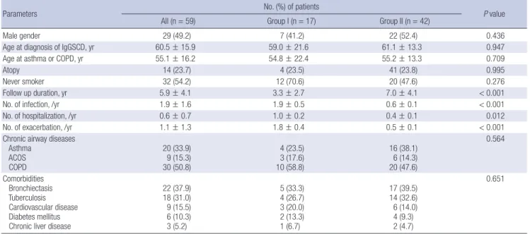
This study enrolled a total of 59 patients (30 females and 29 males) with a mean age of 60.5 ± 15.9 years. Most patients were diagnosed as having IgGSCD 5.4 years after initial treatment of asthma or COPD. Thirty-three percent of patients were diag- nosed as having bronchial asthma and half of the subjects had
never smoked. The major underlying diseases were bronchiec- tasis (37.9%), history of pulmonary tuberculosis (31.0%), car- diovascular disease (15.5%), and diabetes (10.3%). According to the 10 JMF warning signs, patients were classified into two groups:
group I (n = 17) met ≥ 2 criteria, and group II (n = 42) met none of the criteria as they had no recurrent infections even though they had IgGSCD (Supplementary Table 1). The numbers of in- fections, hospitalizations, and exacerbations of asthma or COPD in a year were significantly higher in group I than in group II (P < 0.001, P = 0.012, and P < 0.001, respectively). Other base- line characteristics such as atopy, smoking status, underlying airway diseases, and comorbidities did not differ between the two groups (Table 2). When we compared the number of infec- tion, hospitalization and exacerbation in patients with asthma, ACOS, and COPD of each group, there were no significant dif- ferences (Supplementary Table 2).
Immunoglobulin levels and IgG subclass deficiencies Among the types of IgGSCD, IgG3 subclass deficiency was the most common (n = 52, 88.1%), followed by IgG4 subclass defi- ciency (n = 9, 15.3%). Four patients showed at least two or more IgG subclass deficiencies. Three patients had both IgG3 and IgG4 deficiency and one patient showed both IgG1 and IgG3 deficiency. All patients with both IgG3 and IgG4 deficiency be- longed to group I. When we compared the types of IgG subclass deficiencies between the two groups, the prevalence of IgG4 was significantly higher in group I than group II (P = 0.013, Ta- ble 1). There was no significant difference in the IgG subclass deficiency among the patients with asthma, ACOS, and COPD (Supplementary Table 3).
Table 2. Baseline characteristics of the study subjects
Parameters No. (%) of patients
P value
All (n = 59) Group I (n = 17) Group II (n = 42)
Male gender 29 (49.2) 7 (41.2) 22 (52.4) 0.436
Age at diagnosis of IgGSCD, yr 60.5 ± 15.9 59.0 ± 21.6 61.1 ± 13.3 0.947
Age at asthma or COPD, yr 55.1 ± 16.2 54.8 ± 22.4 55.2 ± 13.3 0.709
Atopy 14 (23.7) 4 (23.5) 41 (23.8) 0.995
Never smoker 32 (54.2) 12 (70.6) 20 (47.6) 0.276
Follow up duration, yr 5.9 ± 4.1 3.3 ± 2.7 7.0 ± 4.1 < 0.001
No. of infection, /yr 1.9 ± 1.6 1.9 ± 0.5 0.6 ± 0.1 < 0.001
No. of hospitalization, /yr 0.6 ± 0.7 1.0 ± 0.2 0.4 ± 0.1 0.012
No. of exacerbation, /yr 1.1 ± 1.3 1.8 ± 0.4 0.5 ± 0.1 < 0.001
Chronic airway diseases Asthma
ACOS COPD
20 (33.9) 9 (15.3) 30 (50.8)
4 (23.5) 3 (17.6) 10 (58.8)
16 (38.1) 6 (14.3) 20 (47.6)
0.564
Comorbidities Bronchiectasis Tuberculosis Cardiovascular disease Diabetes mellitus Chronic liver disease
22 (37.9) 18 (31.0) 9 (15.5) 6 (10.3) 3 (5.2)
5 (33.3) 4 (26.7) 3 (20.0) 2 (13.3) 1 (6.7)
17 (39.5) 14 (32.6) 6 (14.0) 4 (9.3) 2 (4.7)
0.651
IgGSCD, immunoglobulin G subclass deficiency; COPD, chronic obstructive pulmonary disease; ACOS, asthma COPD overlap syndrome.
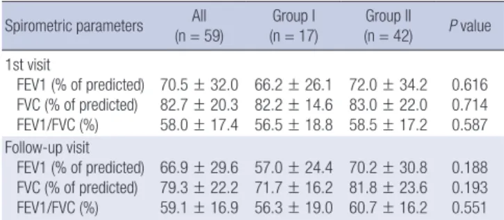
Pulmonary function tests at base line and follow-up The mean post- albuterol FEV1 value was 70.5% ± 32.0% and 66.9% ± 29.6% predicted at baseline and follow-up visits in all subjects. There were no significant differences between the two groups regarding FEV1, FVC, the ratio of FEV1/FVC, and BDR at baseline and follow-up visits (Table 3). However, the mean values of FEV1, FVC, and the ratio tended to be lower in group I than in group II at both baseline and follow-up visits. The follow up mean FEV1 was significantly decreased in patients in group I compared with their baseline values despite regular treatment of asthma or COPD (P = 0.036). No significant changes were noted between the baseline and follow-up FEV1 values in group II (P = 0.321) (Fig. 1). When we compared the FEV1 values be- tween the initial and follow up visits among the three subgroups (asthma, ACOS, and COPD) of group I and group II, there were significant differences, with ACOS poorer than asthma, and COPD worst of all groups. Longitudinal changes of the FEV1 values in the three subgroups of each group did not differ be- tween baseline and follow up visits (Supplementary Fig. 1).
Frequencies of infectious complications and identified organisms
The most common infectious complication was pneumonia, followed by recurrent bronchitis, rhinosinusitis, and extrapul-
monary site infections including herpes zoster, urinary tract in- fection, and gasteroenteritis (Table 4). The types of infectious complications were similar between the groups. In patients with pneumonia, 17 organisms were isolated in cultured spu- tum specimens during exacerbations. Pseudomonas aerugino- sa was the most commonly detected organism (n = 11), follow- ed by Klebsiella pneumonia (n = 4), Streptococcal pneumonia (n = 1), and Moraxella catarrhalis (n = 1). The number and type of isolated organisms were similar between group I and group II. The prevalence of P. aeruginosa tended to be higher in pati- ents with COPD compared to those with asthma or ACOS with- out a statistical significance.
DISCUSSION
In this study, we found that one-third of patients with chronic airway diseases who had been diagnosed with IgGSCD showed recurrent respiratory infections. In addition, these patients show- ed an increased number of exacerbations, hospitalizations and a subsequent decline in lung function despite regular anti asth- matic or COPD treatment.
IgGSCD is defined as an abnormally low level of one or more IgG subclasses in patients with normal levels of total IgG level.
The IgG subclasses are composed of four different isotypes, and each subclass has different structural and biological properties (20). IgG3 comprises only 4%-8% of total serum IgG and has a shorter half-life in comparison with other IgG subclasses. IgG3 plays an important role in complement system activation. More- over, it binds with high affinity to Fc receptors on macrophages and may be important in antibody-mediated phagocytosis (21).
Selective IgG3 deficiencies are a commonly detected type of IgGSCD in adults and recurrent respiratory tract infections and allergy are major clinical associations with IgGSCD (21,22). While the role of selective IgG4 deficiency in infection susceptibility is still unknown, IgG4 deficiency usually occurs in association with other isotype deficiencies and increased risk of sinopul- monary infection (8,11). In this study, we found isolated IgG3 was the most common type of IgGSD, followed by IgG4, and IgG3 combined with other types.
Exacerbations, caused by respiratory viruses, airway bacteria, Fig. 1. FEV1% predicted at baseline and follow-up visit in patients of group I and II.
Baseline Follow-up
FEV1(%)
0 20 40 60 80 100
Group I Group II P = 0.036
P = 0.321
Fig. 1. FEV1% predicted at baseline and follow-up visit in patients of group I and II
FEV1(%)
Baseline Follow-up
100
80
60
40 20
0
P = 0.036 P = 0.321
Baseline Follow-up
FEV1(%)
0 20 40 60 80 100
Group I Group II P = 0.036
P = 0.321
Fig. 1. FEV1% predicted at baseline and follow-up visit in patients of group I and II
Group I Group II Table 3. Spirometric results in the study subjects
Spirometric parameters All
(n = 59) Group I
(n = 17) Group II
(n = 42) P value 1st visit
FEV1 (% of predicted) FVC (% of predicted) FEV1/FVC (%)
70.5 ± 32.0 82.7 ± 20.3 58.0 ± 17.4
66.2 ± 26.1 82.2 ± 14.6 56.5 ± 18.8
72.0 ± 34.2 83.0 ± 22.0 58.5 ± 17.2
0.616 0.714 0.587 Follow-up visit
FEV1 (% of predicted) FVC (% of predicted) FEV1/FVC (%)
66.9 ± 29.6 79.3 ± 22.2 59.1 ± 16.9
57.0 ± 24.4 71.7 ± 16.2 56.3 ± 19.0
70.2 ± 30.8 81.8 ± 23.6 60.7 ± 16.2
0.188 0.193 0.551 FEV1, Forced expiratory volume in one second; FVC, Forced vital capacity.
Table 4. The frequencies of infectious complications
Infections No. (%) of patients
All (n = 88) Group I (n = 28) Group II (n = 60)
Pneumonia 53 (60.2) 14 (50.0) 39 (65.0)
Recurrent bronchitis 14 (15.9) 5 (17.9) 9 (15.0)
Rhinosinusitis 11 (12.5) 4 (14.3) 7 (11.7)
Others 4 (4.5) 1 (3.6) 3 (5.0)
Herpes zoster 3 (3.4) 1 (3.6) 2 (3.3)
Urinary tract infection 2 (2.3) 2 (7.1) 0 (0.0)
Gastroenteritis 1 (1.1) 1 (3.6) 0 (0.0)
allergens, and environmental pollution, can lead to increased airway inflammation. Such inflammatory processes may be amplified by host factors such as immune deficiencies and im- paired lung functions. In our study, one-third of patients with reduced IgG3 or G4 levels experienced frequent exacerbations due to upper or lower respiratory infections and subsequent declining lung functions. A recent study demonstrated that a subset of patients with selective IgG3 deficiency had combined T and B cell defects, suggesting the propensity for infections may not be solely attributable to IgG3 deficiency but may in- volve more complicated immune dysfunctions (21). Neverthe- less, a clinical diagnosis of IgGSCD is controversial and some patients with IgGSCD also showed specific antibody deficiency (SAD) as well (17). In this study, we confirmed IgGSCD based on laboratory findings; however, we did not measure antibody responses after vaccination to exclude SAD, which is a major limitation of our study.
Exacerbations appear to accelerate the decline in lung func- tion, resulting in reduced physical activity, poor QOL, and high healthcare costs. Thus, the prevention of exacerbations is a key component of the management strategies for both asthma and COPD. In a comparison of lung functions between the two groups, we found no significant change between baseline and follow- up visits. However, a longitudinal analysis of lung function over 3 years showed a significant difference between groups I and II.
The mean FEV1 level in group I, which had IgGSCD and a his- tory of recurrent infections, was significantly reduced from base- line despite treatment of asthma or COPD; in contrast, we found no significant change in the follow-up mean FEV1 level in group II. These results were consistent with prior studies showing that repeated infectious exacerbations had negative effects on lung functions (2).
One important phenotype is the frequent exacerbator in CO- PD or exacerbation-prone asthma, which is now recognized as a distinct clinical subgroup in COPD or asthma cohort studies (23-25). In this study, group I had more than three times as many exacerbations per person per year as group II (1.8 vs. 0.5 exac- erbation/person/year). Such symptoms are compatible with the typical characteristics of exacerbation-prone phenotypes, implying that these patients require basic immunological eval- uations, in particular, if they have histories of recurrent exacer- bations triggered by virus or bacterial infections.
The principle of IgGSCD management includes infection con- trol and use of gammaglobulin in selected patients (26). Althou- gh few reports have suggested the efficacy of immunoglobulin replacement therapy in patients with IgGSCD (21,27), these re- ports demonstrate that immunoglobulin replacement therapy significantly improved the QOL, reduced the number of infec- tions, and decreased the need for antibiotics and hospitaliza- tion (22,27). In addition, a recent retrospective study showed patients with selective IgG3 deficiency responded clinically to
immunoglobulin replacement (21). Several open trials suggest- ed that immunoglobulin replacement had corticosteroid-spar- ing effects in severe asthmatics because of the potent anti-in- flammatory properties of immunoglobulin (28,29). However, randomized controlled studies failed to demonstrate the effect of immunoglobulin in asthmatics, although significant cortico- steroid-sparing effect was noted in a subgroup that required high daily doses of oral corticosteroids (12,29). To evaluate the effect of immunoglobulin replacement on the exacerbations and the lung functions of patients with chronic airway diseases, future research should include a prospective study among care- fully defined patients.
In this study, we investigated the clinical features of patients with IgGSCD and chronic airway diseases. IgGSCD exists as the subset of exacerbation prone phenotype of asthma or COPD patients. Detailed historical data regarding infectious complica- tions in both upper and lower airway is the only predictable fac- tor and the measurements of immunoglobulin levels are sup- portive to detect symptomatic patients with IgGSCD in chronic airway disease. Immunoglobulin replacement has beneficial effects when patients with IgGSCD demonstrate recurrent in- fections. However, further studies are required to determine whe- ther immunoglobulin replacement therapy could be a thera- peutic option in this population. Furthermore, a detailed study evaluating B cells, T cells, and other components of the innate immune system of a large cohort of these patients is needed.
DISCLOSURE
The authors have no potential conflicts of interest to disclose.
AUTHOR CONTRIBUTION
Study concept and design: Kim JH, Park S, Hwang YI, Jang SH, Jung KS, Kim DG. Study implementation, analysis and interpre- tation of data: Kim JH, Sim YS, Kim CH, Kim DG. Drafting of ar- ticle or critical revision: Kim JH, Jung KS, Kim CH, Kim DG. Fi- nal approval of the version to be published: all authors.
ORCID
Joo-Hee Kim http://orcid.org/0000-0002-1572-5149 Sunghoon Park http://orcid.org/0000-0001-7004-6985 Yong Il Hwang http://orcid.org/0000-0002-3502-5211 Seung Hun Jang http://orcid.org/0000-0001-5457-5780 Ki-Suck Jung http://orcid.org/0000-0002-6878-6543 Yun Su Sim http://orcid.org/0000-0002-3746-4947 Cheol-Hong Kim http://orcid.org/0000-0002-8815-800X Changhwan Kim http://orcid.org/0000-0003-3318-2528 Dong-Gyu Kim http://orcid.org/0000-0002-9588-8748
REFERENCES
1. Barnes PJ. The cytokine network in asthma and chronic obstructive pul- monary disease. J Clin Invest 2008; 118: 3546-56.
2. Saturni S, Contoli M, Spanevello A, Papi A. Models of respiratory infec- tions: virus-induced asthma exacerbations and beyond. Allergy Asthma Immunol Res 2015; 7: 525-33.
3. Wedzicha JA, Seemungal TA. COPD exacerbations: defining their cause and prevention. Lancet 2007; 370: 786-96.
4. Holgate ST. Mechanisms of asthma and implications for its prevention and treatment: a personal journey. Allergy Asthma Immunol Res 2013; 5:
343-7.
5. Hurst JR, Vestbo J, Anzueto A, Locantore N, Müllerova H, Tal-Singer R, Miller B, Lomas DA, Agusti A, Macnee W, et al. Susceptibility to exacer- bation in chronic obstructive pulmonary disease. N Engl J Med 2010; 363:
1128-38.
6. Martínez-García MA, de la Rosa Carrillo D, Soler-Cataluña JJ, Donat-Sanz Y, Serra PC, Lerma MA, Ballestín J, Sánchez IV, Selma Ferrer MJ, Dalfo AR, et al. Prognostic value of bronchiectasis in patients with moderate-to-se- vere chronic obstructive pulmonary disease. Am J Respir Crit Care Med 2013; 187: 823-31.
7. Bonilla FA, Bernstein IL, Khan DA, Ballas ZK, Chinen J, Frank MM, Ko- brynski LJ, Levinson AI, Mazer B, Nelson RP Jr, et al. Practice parameter for the diagnosis and management of primary immunodeficiency. Ann Allergy Asthma Immunol 2005; 94: S1-63.
8. Bonilla FA, Khan DA, Ballas ZK, Chinen J, Frank MM, Hsu JT, Keller M, Kobrynski LJ, Komarow HD, Mazer B, et al. Practice parameter for the di- agnosis and management of primary immunodeficiency. J Allergy Clin Immunol 2015; 136: 1186-1205.e1-78.
9. Popa V. Airway obstruction in adults with recurrent respiratory infections and IgG deficiency. Chest 1994; 105: 1066-72.
10. O’Keeffe S, Gzel A, Drury R, Cullina M, Greally J, Finnegan P. Immuno- globulin G subclasses and spirometry in patients with chronic obstruc- tive pulmonary disease. Eur Respir J 1991; 4: 932-6.
11. Schwitzguébel AJ, Jandus P, Lacroix JS, Seebach JD, Harr T. Immunoglob- ulin deficiency in patients with chronic rhinosinusitis: Systematic review of the literature and meta-analysis. J Allergy Clin Immunol 2015; 136: 1523- 31.
12. Salmun LM, Barlan I, Wolf HM, Eibl M, Twarog FJ, Geha RS, Schneider LC. Effect of intravenous immunoglobulin on steroid consumption in pa- tients with severe asthma: a double-blind, placebo-controlled, random- ized trial. J Allergy Clin Immunol 1999; 103: 810-5.
13. Lehman H, Hernandez-Trujillo V, Ballow M. Diagnosing primary immu- nodeficiency: a practical approach for the non-immunologist. Curr Med Res Opin 2015; 31: 697-706.
14. Costa-Carvalho BT, Grumach AS, Franco JL, Espinosa-Rosales FJ, Leiva LE, King A, Porras O, Bezrodnik L, Oleastro M, Sorensen RU, et al. Attend- ing to warning signs of primary immunodeficiency diseases across the range of clinical practice. J Clin Immunol 2014; 34: 10-22.
15. Bateman ED, Hurd SS, Barnes PJ, Bousquet J, Drazen JM, FitzGerald M, Gibson P, Ohta K, O’Byrne P, Pedersen SE, et al. Global strategy for asth- ma management and prevention: GINA executive summary. Eur Respir J 2008; 31: 143-78.
16. Vestbo J, Hurd SS, Agustí AG, Jones PW, Vogelmeier C, Anzueto A, Barnes PJ, Fabbri LM, Martinez FJ, Nishimura M, et al. Global strategy for the di- agnosis, management, and prevention of chronic obstructive pulmonary disease: GOLD executive summary. Am J Respir Crit Care Med 2013; 187:
347-65.
17. Al-Herz W, Bousfiha A, Casanova JL, Chatila T, Conley ME, Cunningham- Rundles C, Etzioni A, Franco JL, Gaspar HB, Holland SM, et al. Primary immunodeficiency diseases: an update on the classification from the in- ternational union of immunological societies expert committee for pri- mary immunodeficiency. Front Immunol 2014; 5: 162.
18. Pellegrino R, Viegi G, Brusasco V, Crapo RO, Burgos F, Casaburi R, Coates A, van der Grinten CP, Gustafsson P, Hankinson J, et al. Interpretative strat- egies for lung function tests. Eur Respir J 2005; 26: 948-68.
19. Popa V. ATS guidelines for methacholine and exercise challenge testing.
Am J Respir Crit Care Med 2001; 163: 292-3.
20. Buckley RH. Immunoglobulin G subclass deficiency: fact or fancy? Curr Allergy Asthma Rep 2002; 2: 356-60.
21. Abrahamian F, Agrawal S, Gupta S. Immunological and clinical profile of adult patients with selective immunoglobulin subclass deficiency: response to intravenous immunoglobulin therapy. Clin Exp Immunol 2010; 159:
344-50.
22. Olinder-Nielsen AM, Granert C, Forsberg P, Friman V, Vietorisz A, Björ- kander J. Immunoglobulin prophylaxis in 350 adults with IgG subclass deficiency and recurrent respiratory tract infections: a long-term follow- up. Scand J Infect Dis 2007; 39: 44-50.
23. Moore WC, Meyers DA, Wenzel SE, Teague WG, Li H, Li X, D’Agostino R Jr, Castro M, Curran-Everett D, Fitzpatrick AM, et al. Identification of asth- ma phenotypes using cluster analysis in the Severe Asthma Research Pro- gram. Am J Respir Crit Care Med 2010; 181: 315-23.
24. Han MK, Agusti A, Calverley PM, Celli BR, Criner G, Curtis JL, Fabbri LM, Goldin JG, Jones PW, Macnee W, et al. Chronic obstructive pulmonary disease phenotypes: the future of COPD. Am J Respir Crit Care Med 2010;
182: 598-604.
25. Weatherall M, Travers J, Shirtcliffe PM, Marsh SE, Williams MV, Nowitz MR, Aldington S, Beasley R. Distinct clinical phenotypes of airways dis- ease defined by cluster analysis. Eur Respir J 2009; 34: 812-8.
26. Orange JS, Hossny EM, Weiler CR, Ballow M, Berger M, Bonilla FA, Buck- ley R, Chinen J, El-Gamal Y, Mazer BD, et al. Use of intravenous immuno- globulin in human disease: a review of evidence by members of the Pri- mary Immunodeficiency Committee of the American Academy of Aller- gy, Asthma and Immunology. J Allergy Clin Immunol 2006; 117: S525- 53.
27. Abdou NI, Greenwell CA, Mehta R, Narra M, Hester JD, Halsey JF. Effica- cy of intravenous gammaglobulin for immunoglobulin G subclass and/
or antibody deficiency in adults. Int Arch Allergy Immunol 2009; 149:
267-74.
28. Landwehr LP, Jeppson JD, Katlan MG, Esterl B, McCormick D, Hamilos DL, Gelfand EW. Benefits of high-dose i.v. immunoglobulin in patients with severe steroid-dependent asthma. Chest 1998; 114: 1349-56.
29. Haque S, Boyce N, Thien FC, O’Hehir RE, Douglass J. Role of intravenous immunoglobulin in severe steroid-dependent asthma. Intern Med J 2003;
33: 341-4.
Supplementary Table 1. Proportion of patients who met the 10 warning signs for adults by Jeffrey Modell Foundation
No. Warning signs Group I (n = 17) Group II (n = 42)
1 Two or more new ear infections within 1 year 1 (5.9) 0 (0.0)
2 Two or more new sinus infections within 1 year, in the absence of allergy 4 (23.5) 7 (16.7)
3 One pneumonia per year for more than 1 year 11 (64.7) 30 (71.4)
4 Chronic diarrhea with weight loss 0 (0.0) 0 (0.0)
5 Recurrent viral infections (colds, herpes, warts, condyloma) 6 (35.3) 11 (26.2)
6 Recurrent need for intravenous antibiotics to clear infections 3 (17.6) 0 (0.0)
7 Recurrent, deep abscesses of the skin or internal organs 0 (0.0) 0 (0.0)
8 Persistent thrush or fungal infection on skin or elsewhere 0 (0.0) 0 (0.0)
9 Infection with normally harmless tuberculosis-like bacteria 0 (0.0) 0 (0.0)
10 A family history of PI 0 (0.0) 0 (0.0)
Supplementary Table 2. Frequency of infection, hospitalization and exacerbation according to the obstructive airway diseases (asthma, ACOS, and COPD)
Group and variables Asthma ACOS COPD P value
Group I
No. of infection/yr No. of hospitalization/yr No. of exacerbation/yr
n = 4 4.5 ± 1.6 1.4 ± 1.6 1.5 ± 1.1
n = 3 2.8 ± 0.4 1.0 ± 0.5 1.8 ± 0.3
n = 10 3.8 ± 2.2 1.0 ± 0.9 2.9 ± 2.1
0.315 0.885 0.415 Group II
No. of infection/yr No. of hospitalization/yr No. of exacerbation/yr
n = 16 1.0 ± 0.6 0.3 ± 0.4 0.5 ± 0.5
n = 6 1.0 ± 0.7 0.3 ± 0.2 0.7 ± 0.6
n = 20 1.3 ± 0.6 0.5 ± 0.5 0.7 ± 0.5
0.116 0.223 0.694 ACOS, asthma COPD overlap syndrome; COPD, chronic obstructive pulmonary disease.
Supplementary Table 3. Immunoglobulin G subclass deficiency of the patients with asthma, ACOS, and COPD
IgG subclasses (normal range) No. (%) of deficient patients
P value
Asthma (n = 20) ACOS (n = 9) COPD (n = 30)
IgG1 (382.4-928.6 mg/dL) 1 (4.5) 0 (0.0) 0 (0.0) 0.398
IgG2 (241.8-700.3 mg/dL) 0 (0.0) 0 (0.0) 1 (3.1) 0.612
IgG3 (21.8-176.1 mg/dL) 19 (86.4) 8 (88.9) 25 (78.1) 0.397
IgG4 (3.9-86.4 mg/dL) 2 (9.1) 1 (11.1) 6 (18.8) 0.576
Kim J-H, et al. • IgG Subclass Deficiencies in Chronic Airway Diseases
Supplementary Fig. 1. FEV1% predicted in patients with asthma, ACOS, and COPD at baseline and follow-up visit in group I (A) and group II (B).
Baseline Follow-up
FVC (%pred)
0 20 40 60 80 100
120 P = 0.180
P = 0.180 P = 0.176
Asthma ACOSCOPD
Baseline Follow-up
FEV1(%pred)
0 20 40 60 80 100
120 P = 0.091
P = 0.345 P = 0.670
(A) (B)
FVC (%pred)
Baseline Follow-up
120 100 80 60 40 20 0
P = 0.180 P = 0.180
P = 0.176
Baseline Follow-up
FVC (%pred)
0 20 40 60 80 100
120 P = 0.180
P = 0.180 P = 0.176
Asthma ACOSCOPD
Baseline Follow-up
FEV1(%pred)
0 20 40 60 80 100
120 P = 0.091
P = 0.345 P = 0.670
(A) (B)
Baseline Follow-up
FVC (%pred)
0 20 40 60 80 100
120 P = 0.180
P = 0.180 P = 0.176
Asthma ACOSCOPD
Baseline Follow-up
FEV1(%pred)
0 20 40 60 80 100
120 P = 0.091
P = 0.345 P = 0.670
(A) (B)
Asthma ACOSCOPD
Baseline Follow-up
FVC (%pred)
0 20 40 60 80 100
120 P = 0.180
P = 0.180 P = 0.176
Asthma ACOSCOPD
Baseline Follow-up
FEV1(%pred)
0 20 40 60 80 100
120 P = 0.091
P = 0.345 P = 0.670
(A) (B)
Asthma ACOSCOPD
FEV1 (%pred)
Baseline Follow-up
120 100 80 60 40 20 0
P = 0.345 P = 0.091
P = 0.670
A B