Tandem High-Dose Chemotherapy and Autologous Stem Cell Transplantation for High-Grade Gliomas in Children and
Adolescents
With the aim to investigate the outcome of tandem high-dose chemotherapy and autologous stem cell transplantation (HDCT/auto-SCT) for high-grade gliomas (HGGs), we retrospectively reviewed the medical records of 30 patients with HGGs (16 glioblastomas, 7 anaplastic astrocytomas, and 7 other HGGs) between 2006 and 2015. Gross or near total resection was possible in 11 patients. Front-line treatment after surgery was radiotherapy (RT) in 14 patients and chemotherapy in the remaining 16 patients including 3 patients less than 3 years of age. Eight of 12 patients who remained progression free and 5 of the remaining 18 patients who experienced progression during induction treatment underwent the first HDCT/auto-SCT with carboplatin + thiotepa + etoposide (CTE) regimen and 11 of them proceeded to the second HDCT/auto-SCT with cyclophosphamide + melphalan (CyM) regimen. One patient died from hepatic veno-occlusive disease (VOD) during the second HDCT/auto-SCT; otherwise, toxicities were manageable. Four patients in complete response (CR) and 3 of 7 patients in partial response (PR) or second PR at the first HDCT/auto-SCT remained event free: however, 2 patients with progressive tumor experienced progression again. The probabilities of 3-year overall survival (OS) after the first HDCT/auto-SCT in 11 patients in CR, PR, or second PR was 58.2% ± 16.9%. Tumor status at the first HDCT/
auto-SCT was the only significant factor for outcome after HDCT/auto-SCT. There was no difference in survival between glioblastoma and other HGGs. This study suggests that the outcome of HGGs in children and adolescents after HDCT/auto-SCT is encouraging if the patient could achieve CR or PR before HDCT/auto-SCT.
Keywords: High-grade Glioma; Brain Tumor; High-dose Chemotherapy; Autologous Stem Cell Transplantation; Children
Ji Won Lee,1* Do Hoon Lim,2* Ki Woong Sung,1 Hyeong Jin Lee,1 Eun Sang Yi,1 Keon Hee Yoo,1 Hong Hoe Koo,1 Yeon-Lim Suh,3 and Hyung Jin Shin4
1Department of Pediatrics, Samsung Medical Center, Sungkyunkwan University School of Medicine, Seoul, Korea; 2Department of Radiation Oncology, Samsung Medical Center, Sungkyunkwan University School of Medicine, Seoul, Korea; 3Department of Pathology, Samsung Medical Center, Sungkyunkwan University School of Medicine, Seoul, Korea;
4Department of Neurosurgery, Samsung Medical Center, Sungkyunkwan University School of Medicine, Seoul, Korea
* Ji Won Lee and Do Hoon Lim contributed equally to this work.
Received: 3 June 2016 Accepted: 20 October 2016 Address for Correspondence:
Hyung Jin Shin, MD
Department of Neurosurgery, Samsung Medical Center, Sungkyunkwan University School of Medicine, 81 Irwon-ro, Gangnam-gu, Seoul 06351, Korea
E-mail: [email protected]
Funding: This study was supported by a Samsung Medical Center Grant (#PHO3110265).
https://doi.org/10.3346/jkms.2017.32.2.195 • J Korean Med Sci 2017; 32: 195-203
INTRODUCTION
While high-grade gliomas (HGGs) represent one of the most common central nervous system (CNS) tumors in adults, HGGs are less common in children and adolescents (1). HGGs include a variety of heterogeneous lesions with differing histologies, but the most common histologies are anaplastic astrocytoma (World Health Organization [WHO] grade III) and glioblastoma (WHO grade IV) (2). The prognosis of HGGs has been very poor and there is no universally accepted standard care for HGGs in chil- dren. Despite numerous treatment approaches, outcomes have remained dismal and the far majority of children are succumb- ing to their disease (3-5). Previous children’s cancer group study showed the effectiveness of adjuvant chemotherapy but current conventional therapies are not yet sufficient for survival (4,6,7).
In adults, concomitant temozolomide (TMZ) and radiotherapy (RT) prolonged survival duration and is now considered the
standard of treatment (8); however, multiple studies failed to demonstrate the benefits of TMZ on long-term survival in chil- dren (4,9,10). In addition, prolongation of survival, not cure, is less meaningful in children than in adults.
A treatment strategy using high-dose chemotherapy and au- tologous stem cell transplantation (HDCT/auto-SCT) has shown clinical benefit in children with high-risk or recurrent solid tu- mors (11,12). HDCT/auto-SCT has also been used successfully in children with high-risk or recurrent brain tumors in children (13-15). Recently, several studies have suggested that further dose-escalation using tandem HDCT/auto-SCT might improve outcomes for patients with recurrent or high-risk solid tumors including brain tumors (16-18). In the present study, we reviewed retrospectively our experience with HDCT/auto-SCT for HGGs in children and adolescents to investigate the outcome and risk factors of the results after HDCT/auto-SCT.
MATERIALS AND METHODS Patients
We reviewed the medical records of all patients younger than 18 years, who were diagnosed with HGGs at Samsung Medical Center between February 2006 and October 2015. Inclusion criteria were WHO grade 3 or 4 astrocytic tumors (glioblastoma, anaplastic astrocytoma, and gliomatosis cerebri) and other rare HGGs. Pontine glioma was not included in the analysis. During the study period, most patients were recommended to undergo HDCT/auto-SCT if they remained progression free during in- duction treatment. The subjects were retrospectively identified through survey of our institutional database. A detailed review of the clinical data was performed to ascertain the presenting features, degree of surgical resection, pathology, chemotherapy regimen, RT, and the response to treatment before and after HDCT/auto-SCT.
Response and toxicity criteria
We evaluated disease response using brain magnetic resonance imaging (MRI) with or without spine MRI. We estimated tumor size by MRI as the product of the greatest diameter and the lon- gest perpendicular diameter. We categorized disease response as follows: 1) progressive disease (PD): greater than 25% increase in tumor size or the appearance of a new tumor; 2) stable disease (SD): less than 50% reduction in tumor size or less than 25% in- crease in tumor size; 3) partial response (PR): greater than 50%
decrease in tumor size; and 4) complete response (CR): com- plete disappearance of all previously measurable tumors. Tox- icities during tandem HDCT/auto-SCT were graded using the National Cancer Institute’s Common Terminology Criteria (ver- sion 4.0).
Statistics
Event-free survival (EFS) was calculated from the date of diag- nosis until the date of relapse, progression, or death, whichever occurred first. Overall survival (OS) was calculated from the date of diagnosis until death from any cause. Survival rates and standard errors were estimated using the Kaplan-Meier meth- od. Differences in survival rates between groups were compared using the log-rank test. We performed multivariate analysis us- ing Cox-regression analysis to find independent prognostic fac- tors for survival. We analyzed differences in the frequency of toxicity between the first and second HDCT/auto-SCT using the χ2 test or Fisher’s exact test. Differences in continuous vari- ables between the first and second HDCT/auto-SCT were ana- lyzed using the Mann-Whitney U test. P values less than 0.05 were considered significant.
Ethics statement
The study protocol was approved by the Institutional Review
Board at Samsung Medical Center, Seoul, Korea (IRB No. 2016- 05-009). The need for informed consent was waived by the board.
RESULTS
Patient characteristics
A total of 30 patients (21 boys and 9 girls) were diagnosed with HGGs during the study period. Patient characteristics are sum- marized in Table 1. The median age at diagnosis was 12.5 years (range 0.3–18.0), and 3 patients were younger than 3 years at di- agnosis. Glioblastoma (n = 16) was the most frequent pathology, followed by anaplastic astrocytoma (n = 7), high-grade astroblas- Table 1. Patient characteristics
Characteristics No. (%)
Sex Male Female
21 (66.7) 9 (33.3) Age at diagnosis, yr
< 3 > 3
3 (10.0) 27 (90.0) Histology
Glioblastoma Anaplastic astrocytoma Gliomatosis cerebri High-grade astroblastoma Anaplastic pleomorphic astrocytoma Anaplastic glioneuronal tumor
16 (53.3) 7 (23.3) 2 (6.7) 3 (10.0) 1 (3.3) 1 (3.3) Location
Cerebral hemisphere Midline structures Basal ganglia Midbrain Cerebellum
19 (63.3) 9 (30.0)
8 1 2 (6.7) Leptomeningeal seeding
No Yes
26 (86.7) 4 (13.3) Results of surgery
GTR/NTR STR Biopsy
11 (36.7) 6 (20.0) 13 (43.3) Front-line treatment after surgery
RT
Chemotherapy 14 (46.7)
16 (53.3) Results of induction treatment
CR PR SD
Transfer to other hospital in SD TRM (ICH) in SD
PD PR2 after PD
5 (16.7) 4 (13.3) 1 (3.3) 1 (3.3) 1 (3.3) 15 (50.0)
3 (10.0) Tumor status at first HDCT/auto-SCT (n = 13)
CR PR PR2 after PD PD
4 (30.8) 4 (30.8) 3 (23.1) 2 (15.4) GTR = gross total resection, NTR = near total resection, STR = subtotal resection, RT = radiotherapy, CR = complete response, PR = partial response, SD = stable dis- ease, PR2 = second PR, PD = progressive disease, TRM = treatment-related mortal- ity, ICH = intracranial hemorrhage, HDCT = high dose chemotherapy, SCT = stem cell transplantation.
toma (n = 3), gliomatosis cerebri (n = 2) and other HGGs (n = 2).
Primary tumor originated from cerebral hemisphere in 19 pa- tients, midline structures including basal ganglia in 9, and cere- bellum in 2. Gross total resection or near total resection (> 90%
resection) was possible in 11 patients and subtotal resection (50%–
90% resection) or biopsy (< 50% resection) was performed in
the remaining 19 patients. Four patients had leptomeningeal seeding at diagnosis.
Induction treatment
Chemotherapy was the front-line treatment after surgery in 16 patients including 3 patients younger than 3 years. Among them,
Table 2. Chemotherapy regimens
Regimens Drugs Doses, mg/m2/day Schedules Total doses, mg/m2
Induction regimens
CECV* Cisplatin 90 Day 0 90
Etoposide 75 Days 0–2 225
Cyclophosphamide 1,500 Days 1 and 2 3,000
Vincristine 1.5 Days 0 and 7 3.0
VICE* Carboplatin 300 Days 0 and 1 600
Etoposide 75 Days 0–4 375
Ifosfamide 1,500 Days 0–4 7,500
Vincristine 1.5 Days 0 and 7 3.0
First HDCT regimen
CTE Carboplatin 500 Days –8, –7, –6 1,500
Thiotepa 300 Days –5, –4, –3 900
Etoposide 250 Days –5, –4, –3 750
Second HDCT regimen
CyM Cyclophosphamide 1,500 Days –8, –7, –6, –5 6,000
Melphalan 60 Days –4, –3, –2 180
CECV = cisplatin + etoposide + cyclophosphamide + vincristine, VICE = vincristine + ifosfamide + carboplatin + etoposide, CTE = carboplatin + thiotepa + etoposide, CyM = cyclophosphamide + melphalan, HDCT = high-dose chemotherapy.
*Dose was determined based on body weight in children under 3 years of age.
Fig. 1. Flow of patients. Treatment flow and outcome of all patients are illustrated.
HGGs = high-grade gliomas, PD = progressive disease, HDCT = high-dose chemotherapy, ICH = intracranial hemorrhage, HDCT1 = first high-dose chemotherapy, Tx = Treat- ment, HDCT2 = second high-dose chemotherapy, Ds = disease, DOD = died of disease, TRM = treatment-related mortality, VOD = veno-occlusive disease.
1 Refuse HDCT
During induction treatment
1 PD free
1 Censored without PD
1 Dead
1 PD free
1 PD free d/t insufficient stem cells
4 PD free 1 Relapse (alive) 1 DOD 1 TRM (VOD)
1 DOD
10 DODs 10 PDs
3 DODs d/t PD 3 PR2s
2 PDs 1 PD free
2 DODs 1 Alive with Ds 1 Off-Tx
7 HDCT2
4 HDCT2
1 Off-Tx
10 Off-Tx 1 Transfer
1 ICH
1 Waiting HDCT
8 HDCT1
5 HDCT1
13 Salvage Tx
3 Off-Tx 12 PD free
18 PDs 30 HGGs
concomitant (n = 8) or subsequent (n = 4) RT was given later except 4 patients who refused further treatment after progres- sion (n = 1) or who were younger than 3 years (n = 3). RT was the front-line treatment after surgery in the remaining 14 pati- ents and subsequent chemotherapy was given except 2 patients who was transferred to other hospital or refused further treat- ment after RT. Cisplatin + etoposide + cyclophosphamide + vin- cristine (CECV) and vincristine + ifosfamide + carboplatin + et- oposide (VICE) regimens were used as front-line chemothera- py in 24 patients (Table 2). Both regimens were used in alterna- tion (17,18). Other front-line regimens were prednisolone + CC- NU + vincristine (PCV) in 2, carboplatin + etoposide (CE) in one, and TMZ in one. Local RT dose to the primary site was in the range of 54.0–60.0 Gy and craniospinal RT (23.4–44.0 Gy) was given in 4 patients with leptomeningeal seeding.
Response to induction treatment
Fig. 1 summarizes the flow of patients. A total of 12 patients re- mained progression free during induction treatment. Among them, 8 patients (4 CRs and 4 PRs) proceeded to the first HDCT/
auto-SCT and one is waiting HDCT/auto-SCT. However, the re- maining 3 patients could not proceed to the HDCT/auto-SCT
due to death from intracranial hemorrhage during induction treatment in one, transfer to other hospital in one, and refusal of further treatment in one. The remaining 18 patients experi- enced progression during induction treatment. Among them, 13 received conventional salvage treatment including second- look surgery, RT, or chemotherapy, and 3 of them achieved sec- ond PR (PR2) and proceeded to the first HDCT/auto-SCT. An- other 2 patients proceeded to the first HDCT/auto-SCT as sal- vage treatment without preceding conventional salvage treat- ment. The other 3 patients gave up further treatment after initial progression.
Tumor progression during induction treatment was more frequent in subtotal or less resection group than in gross or near total resection group (78.9% vs. 27.3%, P = 0.009). Glioblastoma group as compared to other HGG group and chemotherapy as compared to RT as front-line treatment after surgery were not associated with a higher progression rate during induction treat- ment. Peripheral blood stem cells (PBSCs) were collected dur- ing the recovery phase of chemotherapy cycle and the aim was to collect a minimum of 2 × 10⁶ CD34+ cells/kg, with an optimal collection of greater than 5 × 10⁶/kg to be used for bone mar- row rescue during tandem HDCT/auto-SCT. The median num-
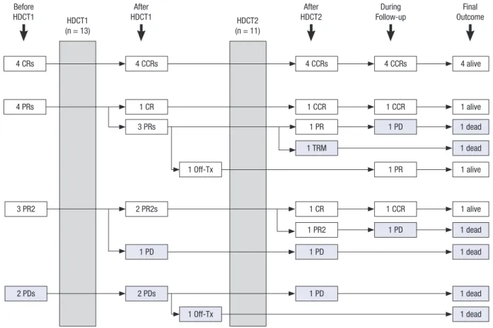
Fig. 2. Response to tandem HDCT/auto-SCT. Responses before and after HDCT/auto-SCT are illustrated. A total of 13 patients (CR in 4, PR in 4, PR2 in 3, and PD in 2) under- went the first HDCT/auto-SCT and 11 of them proceeded to the second HDCT/auto-SCT. Overall, 7 patients are alive after HDCT/auto-SCT.
HDCT/auto-SCT = high-dose chemotherapy and autologous stem cell transplantation, CR = complete response, PR = partial response, PR2 = second PR, PD = progressive disease, CCR = continuous, HDCT1 = first high-dose chemotherapy, HDCT2 = second high-dose chemotherapy, TRM = treatment-related mortality.
HDCT1
(n = 13) HDCT2
(n = 11)
1 PD
2 PDs 2 PDs
1 Off-Tx
1 PD
1 PD 1 dead
1 dead
1 dead 1 dead
1 dead 1 dead 1 TRM
1 CCR 1 PR 1 CCR 1 CCR
4 CCRs 4 alive
1 alive
1 alive
1 alive
4 CRs 4 CCRs
4 PRs 1 CR
3 PRs
3 PR2 2 PR2s
Before HDCT1
After HDCT1
After HDCT2
During Follow-up
Final Outcome
1 PR
1 Off-Tx
4 CCRs
1 PR2 1 CR
1 PD
1 PD
ber of CD34+ cells collected was 40.0 × 10⁶ cells/kg (range 2.4–
127.1).
Tandem HDCT/auto-SCT
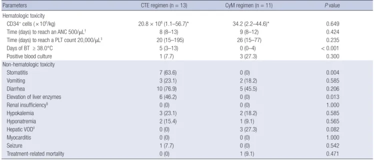
Fig. 2 summarizes the flow of patients during and after tandem HDCT/auto-SCT. A total of 13 patients (CR in 4, PR in 4, PR2 in 3, and PD in 2) underwent the first HDCT/auto-SCT and 11 of them proceeded to the second HDCT/auto-SCT. The remaining 2 patients could not proceed to the second HDCT/auto-SCT due to insufficient stem cells in one and tumor progression af- ter the first HDCT/auto-SCT in 1. Tumor status at the second HDCT/auto-SCT was CR in 5, PR in 2, PR2 in 2, and PD in 2. Car- boplatin + thiotepa + etoposide (CTE) was used for the first HD- CT/auto-SCT and cyclophosphamide + melphalan (CyM) regi- men was used for the second HDCT/auto-SCT (Table 2). The median interval from the first PBSC infusion to initiation of the second HDCT/auto-SCT was 84 days (range 79–102). Table 3 compares the frequency of grade 3 and 4 toxicities that devel- oped during tandem HDCT/auto-SCT. A median of 20.8 × 10⁶ CD34+ cells/kg (range 1.1–56.7), and 15.2 × 10⁶ CD34+ cells/kg (range 2.2–44.6) were infused for the first and second HDCT/
auto-SCT, respectively. Neutrophil and platelet counts recov- ered rapidly during the first and second HDCT/auto-SCT. The number of days with fever was higher in the first HDCT/auto- SCT than in the second (P < 0.001); however, we found no dif- ference in the number of positive blood cultures. Grade 3 and 4 stomatitis and elevation of liver enzymes were more frequent in the first HDCT/auto-SCT than in the second. In the second HD-
CT/auto-SCT, the frequency and severity of mucositis-related toxicity was lower; however, the frequency of hepatic veno-oc- clusive disease (VOD) was higher with borderline significance (P = 0.082) and one patient died from hepatic VOD during the second HDCT/auto-SCT.
Survival
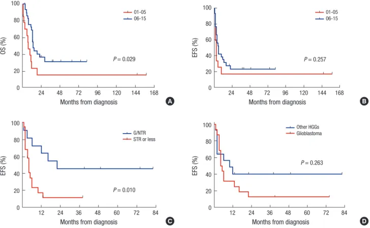
Overall, 9 patients remain progression free, 19 patients experi- enced progression at least once, and 2 patients died from toxici- ties (intracranial hemorrhage and hepatic VOD). Two of 19 pa- tients who experienced progression remain alive after salvage treatment (one in CR and one in PD). Therefore, a total of 11 pa- tients remain alive for a median of 38 months (range 3–82) after diagnosis. The probabilities of 3-year OS and EFS after diagno- sis were 31.5% ± 9.2% and 23.7% ± 8.1%, respectively. The prob- ability of 3-year OS is higher than that in 14 patients during 5 years (2001–2005) prior to the present study period, most of whom were treated with conventional modalities alone (Fig. 3A), al- though the EFS rate was not different (Fig. 3B). Subtotal or less resection of the primary tumor was associated with inferior EFS (Fig. 3C). However, glioblastoma (Fig. 3D), age under 3 years, chemotherapy compared to RT as front-line treatment after sur- gery were not associated with inferior OS or EFS. In the multi- variate analysis for EFS, subtotal or less resection was the only significant unfavorable factor (hazard ratio [HR] 3.95, 95% con- fidence interval [CI] 1.19–13.18, P = 0.025, Table 4). In analysis confined only to 13 patients who underwent the first HDCT/
auto-SCT, 8 patients remain alive for a median of 27 months
Table 3. Characteristics of tandem HDCT/auto-SCT
Parameters CTE regimen (n = 13) CyM regimen (n = 11) P value
Hematologic toxicity
CD34+ cells ( × 106/kg) 20.8 × 10⁶ (1.1–56.7)* 34.2 (2.2–44.6)* 0.649
Time (days) to reach an ANC 500/μL† 8 (8–13) 9 (8–12) 0.424
Time (days) to reach a PLT count 20,000/μL‡ 20 (15–195) 26 (15–77) 0.235
Days of BT ≥ 38.0°C 5 (3–13) 0 (0–4) < 0.001
Positive blood culture 1 (7.7) 3 (27.3) 0.300
Non-hematologic toxicity
Stomatitis 7 (63.6) 0 (0) 0.004
Vomiting 3 (23.1) 2 (18.2) 0.585
Diarrhea 10 (76.9) 5 (45.5) 0.206
Elevation of liver enzymes 6 (46.2) 0 (0) 0.013
Renal insufficiency§ 0 (0) 0 (0) 1.000
Hypokalemia 3 (23.1) 2 (18.2) 0.585
Hyponatremia 2 (15.4) 1 (9.1) 0.565
Hepatic VODll 0 (0) 3 (27.3) 0.082
Myocarditis 0 (0) 0 (0) 1.000
Seizure 1 (7.7) 0 (0) 0.542
Treatment-related mortality 0 (0) 1 (9.1) 0.471
Values are presented as number (%).
CTE = carboplatin + thiotepa + etoposide, CyM = cyclophosphamide + melphalan, HDCT = high-dose chemotherapy, ANC = absolute neutrophil count, PLT = platelet, BT = body temperature, VOD = veno-occlusive disease.
*Median (range); †The first day when ANC exceeded 500/μL for 3 consecutive days; ‡The first day when PLT count exceeded 20,000/μL without transfusion for 7 days; §Eleva- tion of serum creatinine more than threefold of baseline; llAt least 2 of the following 3 events within 20 days of transplantation: bilirubin level > 2 mg/dL, hepatomegaly or right upper quadrant pain of liver origin, or > 2% weight gain secondary to fluid accumulation.
(range 12–67) after the first HDCT/auto-SCT. Seven of them re- main progression free for a median of 33 months (range 12–67) after the first HDCT/auto-SCT, the other one patient remains alive with disease after progression. The remaining 5 patients died due to progression in 4 and toxicity in 1. The probabilities of 3-year OS and EFS after the first HDCT/auto-SCT were 55.4%
± 16.1% and 43.1% ± 14.8%, respectively (Fig. 4A). In analysis confined only to 11 patients who were in CR, PR, or PR2 at the first HDCT/auto-SCT, the probabilities of 3-year OS and EFS af- ter the first HDCT/auto-SCT were 58.2% ± 16.9% and 49.9% ± 16.4%, respectively. Tumor status less than PR at the first HDCT/
auto-SCT was associated with inferior EFS (Fig. 4B); however, there was no difference in survival between glioblastoma and other HGGs (Fig. 4C).
Table 4. Multivariate analysis for factors affecting survival from diagnosis
Risk factors EFS OS
HR 95% CI P value HR 95% CI P value
Age < 3 yr 0.58 0.10–3.21 0.529 0.37 0.06–2.24 0.277
Glioblastoma 2.10 0.80–5.50 0.133 1.92 0.66–5.54 0.229
Subtotal or less resection 3.95 1.19–13.18 0.025 3.09 0.84–11.37 0.089
Chemotherapy as front-line treatment 1.38 0.51–3.76 0.531 1.15 0.38–3.48 0.803
EFS = Event-free survival, OS = overall survival, HR = hazard ratio, CI = confidence interval.
DISCUSSION
Although previous studies for children and adolescent with HGGs showed the effectiveness of adjuvant chemotherapy, current conventional therapies are not yet sufficient with the far major- ity of patients succumbing to their disease (3-5). For this rea- son, we intended to give tandem HDCT/auto-SCT during the study period in order to improve the outcome by increasing the intensity of chemotherapy although it was not performed in a prospective trial. As results, toxicities during HDCT were gener- ally acceptable and OS was higher as compared to the 5 years (2001–2005) prior to the present study period, most of whom were treated with conventional modalities alone. In particular, survival after HDCT/auto-SCT was encouraging in patients who Fig. 3. Survival from diagnosis. The probability of 3-year OS. (A) is higher than that in 14 patients during 5 years (2001–2005) prior to the present study period, most of whom were treated with conventional modalities alone, although the EFS rate is not different (B). Subtotal or less resection of the primary tumor is associated with inferior EFS (C). There is no difference in survival between glioblastoma and other HGGs (D).
HGGs = high-grade gliomas, NTR = near total resection, STR = subtotal resection, OS = overall survival, EFS = event-free survival.
OS (%)
Months from diagnosis
24 48 72 96 120 144 168
100 80 60 40 20 0
P = 0.029 01-05 06-15
EFS (%)
Months from diagnosis
12 24 36 48 60 72 84 100
80 60 40 20 0
P = 0.010 G/NTR STR or less
EFS (%)
Months from diagnosis
24 48 72 96 120 144 168
100 80 60 40 20 0
P = 0.257 01-05 06-15
EFS (%)
Months from diagnosis
12 24 36 48 60 72 84 100
80 60 40 20 0
P = 0.263 Other HGGs Glioblastoma
A B
C D
could remain progression free until the first HDCT/auto-SCT.
Results of our study suggest the possible effectiveness of tandem HDCT/auto-SCT.
A higher proportion of patients remained progression free or could achieve CR or PR in gross or near total resection group than in subtotal or less resection group, and the extent of sur- gery was an independent prognostic factor for EFS. Previous re- ports also suggest that the amount of surgical resection is one of the most important clinical prognostic factors identified to date
in children with supratentorial HGGs (6,7). Therefore, every at- tempt should be made to maximize the extent of surgical resec- tion whenever it is safe and feasible. However, complete resec- tion was not possible in many cases, especially in patients with HGGs originating from or invading critical structures.
Although there is no accepted standard of care for HGGs, RT to the tumor bed following maximal surgical resection with or without concomitant chemotherapy plus additional post-RT chemotherapy has been the most commonly used treatment approach for children older than 3 years. To the contrary, in our experience, chemotherapy was the front-line treatment in many patients including young patients less than 3 years of age. How- ever, chemotherapy as front-line treatment after surgery was not associated with inferior OS or EFS as compared to immedi- ate RT after surgery. Pre-RT chemotherapy and/or concomitant chemotherapy might enhance the efficacy of RT by reducing the bulk of tumor.
Although many prospective studies demonstrated the bene- fit of adjuvant chemotherapy with various regimens, there is no gold standard chemotherapy regimen for HGGs (4,6,7). Since concomitant TMZ and RT for adult with glioblastoma led to pro- longed survival and is now considered the standard of care (8), several studies have tested the efficacy of TMZ in pediatric pa- tients. However, the results were disappointing and failed to dem- onstrate the benefit of TMZ in children (4,9,10). We used CECV and VICE regimens in alternation as front-line chemotherapy in most patients. Although one patient died from tumor bleed- ing during concomitant chemotherapy and RT, toxicities of both regimens were generally acceptable except neutropenic fever.
Non-hematologic toxicity was not common during induction chemotherapy (19).
Although HDCT/auto-SCT is still considered controversial and is not universally accepted, previous studies suggest that there may be a role for children with recurrent HGGs (20,21).
However, studies for newly diagnosed HGGs are limited to date (22-24). In the treatment of recurrent or high-risk brain tumors, several recent studies have suggested tandem HDCT/auto-SCT might further improve outcomes and/or reduce RT dose with- out jeopardizing survival rates (16-19). For this reason, we used tandem HDCT/auto-SCT for HGGs in order to improve the sur- vival rates. As results, survival after tandem HDCT/auto-SCT was encouraging. All 4 patients in CR and 3 of 7 patients in PR or PR2 at the first HDCT/auto-SCT maintained CR, achieved CR, or remained progression free after tandem HDCT/auto-SCT.
These findings suggest that tandem HDCT/auto-SCT might im- prove the survival if the patient remains progression free before HDCT/auto-SCT or could achieve second response (PR2 or more) after initial progression. However, our results also showed that tandem HDCT/auto-SCT could not change the fate of patients with progressed tumors at HDCT/auto-SCT.
We used different drug combinations during the first and sec- Fig. 4. Survival after first HDCT/auto-SCT. Survival analysis was confined only to 13
patients who underwent the first HDCT/auto-SCT. The probabilities of 3-year OS and EFS after the first HDCT/auto-SCT are 55.4%±16.1% and 43.1%±14.8%, respec- tively (A). Tumor status less than PR at the first HDCT/auto-SCT is associated with infe- rior EFS (B). There is no difference in survival between glioblastoma and other HGGs (C).
HDCT/auto-SCT = high-dose chemotherapy and autologous stem cell transplantation, OS = overall survival, EFS = event-free survival, CR = complete response, PR = par- tial response, PR2 = second PR, PD = progressive disease, HGGs = high-grade gliomas.
Survival (%)
Months from HDCT1
12 24 36 48 60 72 100
80 60 40 20 0
OS EFS
EFS (%)
Months from HDCT1
12 24 36 48 60 72 100
80 60 40 20 0
CR PR/PR2 PD
P = 0.005
EFS (%)
Months from HDCT1
12 24 36 48 60 72 100
80 60 40 20 0
Other HGGs Glioblastoma
P = 0.810
A
B
C
ond HDCT to curtail or prevent drug resistance and overlap- ping drug toxicity. As we reported previously (25), acute toxici- ties during tandem HDCT/auto-SCT were generally acceptable except that one patient died from hepatic VOD. However, dose- escalation during tandem HDCT/auto-SCT might be associat- ed with more significant late adverse effects. Therefore, longer follow-up is needed to assess whether the possible survival ben- efits of tandem HDCT/auto-SCT ultimately outweigh the ad- verse effects associated with dose-intense tandem HDCT/auto- SCT.
Our results showed that survival of patients who could achieve CR or PR before HDCT/auto-SCT was encouraging; however, less than one-third of patients could remain progression free before HDCT/auto-SCT. In addition, only 3 patients could achi- eve second response with salvage treatment after initial progres- sion. These findings suggest that conventional front-line or sal- vage treatment modalities are not effective in achieving or main- taining responses in patients with HGGs. Therefore, more effec- tive therapeutic approach is needed to improve the outcome of patients with HGGs. In this context, strategies using new drugs such as anti-angiogenic agent, receptor tyrosine kinase inhibi- tors, or histone deacetylase inhibitors have been evaluated (26- 28). Novel treatment approach using target therapy based on genomic profiles of the tumor might also be another option to improve patient survival with minimal treatment toxicities (29).
Effective tumor control before HDCT/auto-SCT with strategies mentioned above might improve the overall outcome of HGGs.
In conclusion, results of our study suggest that the outcome of HGGs in children and adolescents after HDCT/auto-SCT might be encouraging if the patient could remain progression free be- fore HDCT/auto-SCT. However, this study is a retrospective re- view and the size of cohorts is small. Therefore, prospective stud- ies with larger cohorts of patients are needed to answer the con- troversy and some debate about the efficacy of HDCT/auto-SCT for HGGs in children.
DISCLOSURE
The authors have no potential conflicts of interest to disclose.
AUTHOR CONTRIBUTION
Conceptualization: Sung KW, Shin HJ. Data curation: Lee JW, Lim DH, Sung KW, Lee HJ, Yi ES. Investigation: Lee JW, Lim DH, Sung KW, Yoo KH, Koo HH, Suh YL. Writing - original draft: Lee JW, Lim DH, Sung KW.
ORCID
Ji Won Lee http://orcid.org/0000-0003-0084-1304 Do Hoon Lim http://orcid.org/0000-0002-5426-0604
Ki Woong Sung http://orcid.org/0000-0001-5989-4772 Hyeong Jin Lee http://orcid.org/0000-0002-3762-2167 Eun Sang Yi http://orcid.org/0000-0001-8214-2106 Keon Hee Yoo http://orcid.org/0000-0002-5980-7912 Hong Hoe Koo http://orcid.org/0000-0001-8082-1412 Yeon-Lim Suh http://orcid.org/0000-0001-5809-2401 Hyung Jin Shin http://orcid.org/0000-0003-0856-7098
REFERENCES
1. Bondy ML, Scheurer ME, Malmer B, Barnholtz-Sloan JS, Davis FG, Il’ya- sova D, Kruchko C, McCarthy BJ, Rajaraman P, Schwartzbaum JA, et al.
Brain tumor epidemiology: consensus from the Brain Tumor Epidemiol- ogy Consortium. Cancer 2008; 113: 1953-68.
2. Louis DN, Ohgaki H, Wiestler OD, Cavenee WK, Burger PC, Jouvet A, Sch- eithauer BW, Kleihues P. The 2007 WHO classification of tumours of the central nervous system. Acta Neuropathol 2007; 114: 97-109.
3. Broniscer A, Gajjar A. Supratentorial high-grade astrocytoma and diffuse brainstem glioma: two challenges for the pediatric oncologist. Oncologist 2004; 9: 197-206.
4. Cohen KJ, Pollack IF, Zhou T, Buxton A, Holmes EJ, Burger PC, Brat DJ, Rosenblum MK, Hamilton RL, Lavey RS, et al. Temozolomide in the treat- ment of high-grade gliomas in children: a report from the Children’s On- cology Group. Neuro Oncol 2011; 13: 317-23.
5. Finlay JL, Zacharoulis S. The treatment of high grade gliomas and diffuse intrinsic pontine tumors of childhood and adolescence: a historical - and futuristic - perspective. J Neurooncol 2005; 75: 253-66.
6. Finlay JL, Boyett JM, Yates AJ, Wisoff JH, Milstein JM, Geyer JR, Bertolone SJ, McGuire P, Cherlow JM, Tefft M, et al. Randomized phase III trial in childhood high-grade astrocytoma comparing vincristine, lomustine, and prednisone with the eight-drugs-in-1-day regimen. Childrens cancer group.
J Clin Oncol 1995; 13: 112-23.
7. Sposto R, Ertel IJ, Jenkin RD, Boesel CP, Venes JL, Ortega JA, Evans AE, Wara W, Hammond D. The effectiveness of chemotherapy for treatment of high grade astrocytoma in children: results of a randomized trial. A re- port from the Childrens Cancer Study Group. J Neurooncol 1989; 7: 165- 77.
8. Stupp R, Mason WP, van den Bent MJ, Weller M, Fisher B, Taphoorn MJ, Belanger K, Brandes AA, Marosi C, Bogdahn U, et al. Radiotherapy plus concomitant and adjuvant temozolomide for glioblastoma. N Engl J Med 2005; 352: 987-96.
9. Lashford LS, Thiesse P, Jouvet A, Jaspan T, Couanet D, Griffiths PD, Doz F, Ironside J, Robson K, Hobson R, et al. Temozolomide in malignant glio- mas of childhood: a United Kingdom Children’s Cancer Study Group and French Society for Pediatric Oncology Intergroup Study. J Clin Oncol 2002;
20: 4684-91.
10. Nicholson HS, Kretschmar CS, Krailo M, Bernstein M, Kadota R, Fort D, Friedman H, Harris MB, Tedeschi-Blok N, Mazewski C, et al. Phase 2 study of temozolomide in children and adolescents with recurrent central ner- vous system tumors: a report from the Children’s Oncology Group. Can- cer 2007; 110: 1542-50.
11. Matthay KK, Reynolds CP, Seeger RC, Shimada H, Adkins ES, Haas-Kogan D, Gerbing RB, London WB, Villablanca JG. Long-term results for children with high-risk neuroblastoma treated on a randomized trial of myeloab-
lative therapy followed by 13-cis-retinoic acid: a children’s oncology group study. J Clin Oncol 2009; 27: 1007-13.
12. Philip T, Ladenstein R, Lasset C, Hartmann O, Zucker JM, Pinkerton R, Pearson AD, Klingebiel T, Garaventa A, Kremens B, et al. 1070 myeloab- lative megatherapy procedures followed by stem cell rescue for neuro- blastoma: 17 years of European experience and conclusions. European Group for Blood and Marrow Transplant Registry Solid Tumour Working Party. Eur J Cancer 1997; 33: 2130-5.
13. Fangusaro J, Finlay J, Sposto R, Ji L, Saly M, Zacharoulis S, Asgharzadeh S, Abromowitch M, Olshefski R, Halpern S, et al. Intensive chemotherapy followed by consolidative myeloablative chemotherapy with autologous hematopoietic cell rescue (AuHCR) in young children with newly diag- nosed supratentorial primitive neuroectodermal tumors (sPNETs): re- port of the Head Start I and II experience. Pediatr Blood Cancer 2008; 50:
312-8.
14. Mason WP, Grovas A, Halpern S, Dunkel IJ, Garvin J, Heller G, Rosenblum M, Gardner S, Lyden D, Sands S, et al. Intensive chemotherapy and bone marrow rescue for young children with newly diagnosed malignant brain tumors. J Clin Oncol 1998; 16: 210-21.
15. Modak S, Gardner S, Dunkel IJ, Balmaceda C, Rosenblum MK, Miller DC, Halpern S, Finlay JL. Thiotepa-based high-dose chemotherapy with au- tologous stem-cell rescue in patients with recurrent or progressive CNS germ cell tumors. J Clin Oncol 2004; 22: 1934-43.
16. Gajjar A, Chintagumpala M, Ashley D, Kellie S, Kun LE, Merchant TE, Woo S, Wheeler G, Ahern V, Krasin MJ, et al. Risk-adapted craniospinal radio- therapy followed by high-dose chemotherapy and stem-cell rescue in children with newly diagnosed medulloblastoma (St Jude Medulloblas- toma-96): long-term results from a prospective, multicentre trial. Lancet Oncol 2006; 7: 813-20.
17. Sung KW, Lim DH, Lee SH, Yoo KH, Koo HH, Kim JH, Suh YL, Joung YS, Shin HJ. Tandem high-dose chemotherapy and auto-SCT for malignant brain tumors in children under 3 years of age. Bone Marrow Transplant 2013; 48: 932-8.
18. Sung KW, Lim DH, Son MH, Lee SH, Yoo KH, Koo HH, Kim JH, Suh YL, Joung YS, Shin HJ. Reduced-dose craniospinal radiotherapy followed by tandem high-dose chemotherapy and autologous stem cell transplanta- tion in patients with high-risk medulloblastoma. Neuro Oncol 2013; 15:
352-9.
19. Sung KW, Lim DH, Yi ES, Choi YB, Lee JW, Yoo KH, Koo HH, Kim JH, Suh YL, Joung YS, et al. Tandem high-dose chemotherapy and autologous stem cell transplantation for atypical teratoid/rhabdoid tumor. Cancer Res Treat 2016; 48: 1408-19.
20. Finlay JL, Dhall G, Boyett JM, Dunkel IJ, Gardner SL, Goldman S, Yates AJ, Rosenblum MK, Stanley P, Zimmerman RA, et al. Myeloablative chemo-
therapy with autologous bone marrow rescue in children and adolescents with recurrent malignant astrocytoma: outcome compared with conven- tional chemotherapy: a report from the Children’s Oncology Group. Pe- diatr Blood Cancer 2008; 51: 806-11.
21. Guruangan S, Dunkel IJ, Goldman S, Garvin JH, Rosenblum M, Boyett JM, Gardner S, Merchant TE, Gollamudi S, Finlay JL. Myeloablative che- motherapy with autologous bone marrow rescue in young children with recurrent malignant brain tumors. J Clin Oncol 1998; 16: 2486-93.
22. Grovas AC, Boyett JM, Lindsley K, Rosenblum M, Yates AJ, Finlay JL. Reg- imen-related toxicity of myeloablative chemotherapy with BCNU, thiote- pa, and etoposide followed by autologous stem cell rescue for children with newly diagnosed glioblastoma multiforme: report from the Children’s Cancer Group. Med Pediatr Oncol 1999; 33: 83-7.
23. Heideman RL, Douglass EC, Krance RA, Fontanesi J, Langston JA, Sanford RA, Kovnar EH, Ochs J, Kuttesch J, Jenkins JJ, et al. High-dose chemother- apy and autologous bone marrow rescue followed by interstitial and ex- ternal-beam radiotherapy in newly diagnosed pediatric malignant glio- mas. J Clin Oncol 1993; 11: 1458-65.
24. Massimino M, Gandola L, Luksch R, Spreafico F, Riva D, Solero C, Giangas- pero F, Locatelli F, Podda M, Bozzi F, et al. Sequential chemotherapy, high- dose thiotepa, circulating progenitor cell rescue, and radiotherapy for child- hood high-grade glioma. Neuro Oncol 2005; 7: 41-8.
25. Lee SH, Son MH, Sung KW, Choi YB, Lee NH, Yoo KH, Koo HH, Lim DH, Shin HJ. Toxicity of tandem high-dose chemotherapy and autologous stem cell transplantation using carboplatin-thiotepa-etoposide and cyclophos- phamide-melphalan regimens for malignant brain tumors in children and young adults. J Neurooncol 2014; 120: 507-13.
26. Lee EQ, Puduvalli VK, Reid JM, Kuhn JG, Lamborn KR, Cloughesy TF, Chang SM, Drappatz J, Yung WK, Gilbert MR, et al. Phase I study of vorinostat in combination with temozolomide in patients with high-grade gliomas:
North American Brain Tumor Consortium Study 04-03. Clin Cancer Res 2012; 18: 6032-9.
27. Narayana A, Golfinos JG, Fischer I, Raza S, Kelly P, Parker E, Knopp EA, Medabalmi P, Zagzag D, Eagan P, et al. Feasibility of using bevacizumab with radiation therapy and temozolomide in newly diagnosed high-grade glioma. Int J Radiat Oncol Biol Phys 2008; 72: 383-9.
28. Reardon DA, Groves MD, Wen PY, Nabors L, Mikkelsen T, Rosenfeld S, Raizer J, Barriuso J, McLendon RE, Suttle AB, et al. A phase I/II trial of pa- zopanib in combination with lapatinib in adult patients with relapsed malignant glioma. Clin Cancer Res 2013; 19: 900-8.
29. Gajjar A, Bowers DC, Karajannis MA, Leary S, Witt H, Gottardo NG. Pedi- atric brain tumors: innovative genomic information is transforming the diagnostic and clinical landscape. J Clin Oncol 2015; 33: 2986-98.