Treatment of complex tibial plateau fractures is techni- cally difficult and controversial.1-4) Serious intra-articular damage and soft tissue injury or inappropriate treatment can lead to postoperative complications such as infection, skin necrosis, nonunion, deformity, and arthrosis in tibial plateau fractures.1,5,6) The goal in the treatment of tibial plateau fractures is to provide anatomic reduction of intra- articular fracture fragments while providing stability.1-4) Many different techniques of internal and external fixation are used to treat these fractures.2-8)
The bilateral open reduction and internal fixation technique have been favored by the Association for Osteo- synthesis/Association for the Study of Internal Fixation.3) However, bilateral plating may require excessive dissection through the injured soft tissue, leading to wound compli- cations or compromised osteosynthesis.1,3,5,8) A minimally invasive surgical approach is desirable for reducing the additional soft tissue trauma.1,3) Some authors claimed bi- condylar tibial plateau fractures can be stabilized with uni- lateral locked plating technique while avoiding medial soft tissue dissection.1,3,9-13) This reduces complications associ- ated with bilateral plate fixation, but it is unclear whether it will maintain the acquired articular reduction compared to bilateral plate fixation.3,9-13) Dual locking plate fixation technique and lateral locking plate fixation technique used for tibial bicondylar plateau fractures as cited in the litera- ture are shown in Table 1.9,11,14,15)
In a biomechanical study, it was reported that
Lateral Locked Plating or Dual Plating: A Comparison of Two Methods in Simple
Bicondylar Tibial Plateau Fractures
Caner Citak, MD, Cemil Kayali, MD*, Firat Ozan, MD
†, Taskin Altay, MD*, Huseyin Gokhan Karahan, MD*, Kamil Yamak, MD*
Department of Orthopedics and Traumatology, Mardin State Hospital, Mardin,
*Department of Orthopedics and Traumatology, Izmir Bozyaka Training and Research Hospital, Izmir,
†Department of Orthopedics and Traumatology, Kayseri Training and Research Hospital, Kayseri, Turkey
Background: In this study, our aim was to compare the results of the dual locking plate fixation technique and lateral locking plate fixation technique for tibial bicondylar plateau fractures without posteromedial fragment.
Methods: We evaluated 20 patients who underwent surgical treatment due to bicondylar tibial plateau fracture between 2010 and 2015. Ten patients were included in group 1, in which a dual locking plate was employed, whereas 10 patients were included in group 2, in which a lateral locking plate was used. In both groups, functional and clinical outcomes after treatment were rated according to the Knee Society Knee Scoring System, Rasmussen functional score, and Rasmussen radiological score.
Results: The mean follow-up time was 24 months. There were no significant differences between the groups with respect to func- tional and radiographic outcomes at the final follow-up.
Conclusions: In bicondylar tibial plateau fractures without posteromedial fragment, the lateral locking plate fixation technique showed the similar clinical and radiological outcomes as the dual locking plate fixation technique.
Keywords: Bicondylar tibial plateau, Fracture, Locking plate fixation, Dual plate, Single plate
Copyright © 2019 by The Korean Orthopaedic Association
This is an Open Access article distributed under the terms of the Creative Commons Attribution Non-Commercial License (http://creativecommons.org/licenses/by-nc/4.0) which permits unrestricted non-commercial use, distribution, and reproduction in any medium, provided the original work is properly cited.
Clinics in Orthopedic Surgery • pISSN 2005-291X eISSN 2005-4408 Received October 24, 2018; Revised January 24, 2019; Accepted February
8, 2019
Correspondence to: Cemil Kayali, MD
Department of Orthopedics and Traumatology, Izmir Bozyaka Training and Research Hospital, Saim Çıkrıkçı Cad. no. 59, Bozyaka, Izmir 35170, Turkey Tel: +90-232-250-50-50, Fax: +90-232-261-44-44
E-mail: [email protected]
unilateral buttress plate fixation in a bicondylar fracture model resulted in a greater reduction loss in the medial plateau than axial loading than double plate fixation did.16) This may lead to pain and osteoarthritic changes in later processes. On the other hand, in other bicondylar fracture modeled biomechanical studies, there was no difference between the two methods.3,10,13,17)
The hypothesis of this study was that, in selected tibial plateau fractures, lateral locking plating technique would yield clinical and radiological results comparable to those of dual locking plating technique.Therefore, in this study, our aim was to compare the results of dual locking plate fixation technique and lateral locking plate fixation technique for tibial bicondylar plateau fractures without posteromedial fragment.
METHODS
Clinical Data
We retrospectively evaluated 20 patients who underwent surgical treatment due to bicondylar tibial plateau frac- ture between 2010 and 2015. Ten patients (seven males and three females; five right knees and five left knees;
mean age, 51.3 years; range, 34 to 73 years) were included in group 1, in which a dual locking plate was employed, whereas 10 patients (eight males and two females; five right knees and five left knees; mean age, 51.2 years; range, 25 to 83 years) were included in group 2, in which a lateral locking-plate was used (Figs. 1 and 2).
Inclusion and Exclusion Criteria
Inclusion criteria were the presence of a bicondylar tibial plateau fracture (Schatzker V–VI) without posteromedial fragment. Pathologic, pediatric, extra-articular proximal tibial fractures, and serious open fractures (Gustilo grade III) were all excluded.
Operative Procedure
All operations were performed under spinal anesthesia by using pneumatic tourniquet. In dual plating group, antero- lateral and anteromedial incisions were made. After soft tissue dissection, under longitudinal traction, the joint line congruity was checked with C-arm fluoroscopy. If there was need for arthrotomy, it was performed through the lateral incision to gain anatomic reduction via submenis- cal view. By using locking screws and plate, definitive fixation was obtained. The medial plate was placed in the epiphyseal-metaphyseal region or metaphyseal-diaphyseal region depending on the fracture pattern. In single lateral plating group, we just made an anterolateral incision. The Table 1. Studies Available in the Literature on the Comparison of Bicondylar Plateau Fractures with Unilateral Locked Plating and Dual Plating (2000–2017) Study
No. of patients
Mean age (yr)
Follow- up (mo) Type of fracture (AO classification)Plate typeUnion rate (%)
Complication
(infection, deep venous thrombosis, failure of fixation, nonunion; %)
Knee score SPDPSPDPSPDPSPDPSP DPSPDPSPDP
Jiang et al. (2008)
11)4143414324
C1, 8; C2, 19; C3, 16 C1, 5; C2, 16; C3, 22
LISS
M, buttress plate; L, LC-DCP
10097.763.448.8HSS, 83HSS, 83
Lee et al. (2014)
14)15CDP, 19; HDP, 1149CDP, 53; HDP, 5118S (V/VI), 8/7 CDP: S (V/VI), 13/6; HDP: S (V/VI), 4/7
LCP
CDP: M and L, buttress plate; HDP: L, LCP; M, buttress plate 86CDP, 78; HDP, 90 40CDP, 47; HDP, 27 WOMAC, 36 WOMAC, 34; WOMAC, 32
Yao et al. (2015)
9)4144434512–36
C1, 26; C2, 10; C3, 5 C1, 24; C2, 13; C3, 7
LCP
M and L, buttress plate
1001004.827.2HSS, 81HSS, 79
Neogi et al. (2015)
15)2932413712–24
C1, 10; C2, 11; C3, 8 C1, 11; C2, 13; C3, 8
LCP
M, RP/DCP/DRP; L, LCP
10010065.550HSS, 79HSS, 80 SP: single lateral locking plate, DP: dual plate, LISS: Less Invasive Stabilization System, M: medial side, L: lateral side, LC-DCP: limited-contact dynamic compression plate, HSS: Hospital for Special Surgery score, CDP: classic dual plate, HDP: hybrid dual plate, S: Schatzker classification, LCP: locking compression plate, WOMAC: The Western Ontario and McMaster Universities Osteoarthritis Index, RP: reconstruction plate, DRP: distal end radius plate.
next steps were the same as in dual plating group. In this group, we used the subchondral screws as long as possible to secure medial part of the tibia. The choice of fixation technique (either dual plate fixation [HGK] or lateral plate fixation [CK]) was based on surgeon’s preference.
First-generation cephalosporin for antibiotic pro- phylaxis and low molecular weight heparin for venous thromboembolism were used 24 hours and 3 weeks, re- spectively. All patients were encouraged to perform active and passive range of motion exercises in the 2nd postop- erative week after suture removal at the outpatient clinic under the supervision by a physiotherapist.
Clinical and Radiographic Evaluation
In both groups, functional and clinical outcomes after treatment were rated according to the Knee Society Knee
Scoring System (KSS), Rasmussen functional score (RFS), Rasmussen radiological score (RRS).18,19) Radiographic healing was defined as callus bridging of three of four cortices on anteroposterior and lateral radiographs as well as painless weight bearing on the affected extremity.20) Ra- diological assessments were performed in a comparative manner with the contralateral side.
Radiographic examination includedcoronalalign- ment of theproximaltibia, medial proximaltibial angle (MPTA), tibiofemoral anatomic angle (TFAA), sagitta- lalignment, and proximal posteriortibial angle (PPTA).
Radiologial measurements were carried out from immedi- ately postoperatively to the last visit at the outpatient clinic with 3-month intervals. The last clinical and radiological evaluations were performed at final follow-up.
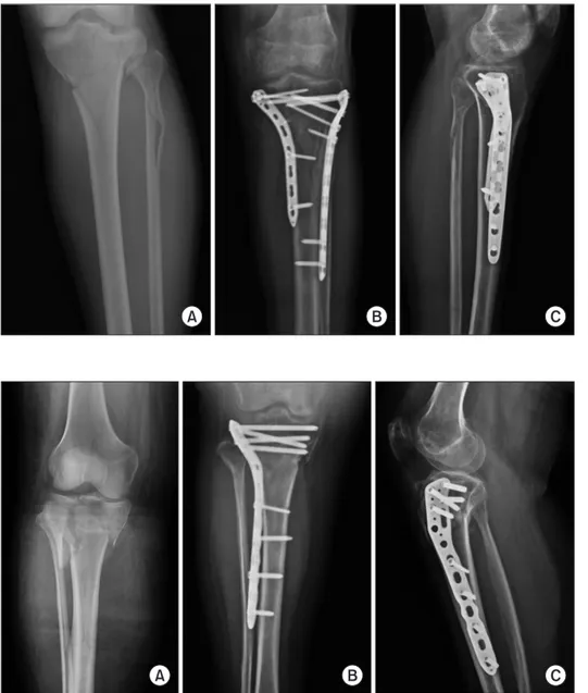
A B C
Fig. 1. (A) Preoperative radiograph showing left tibial plateau fracture in a 36-year-old male patient. (B, C) Radiographs taken 21 months after dual locking plate fixation.
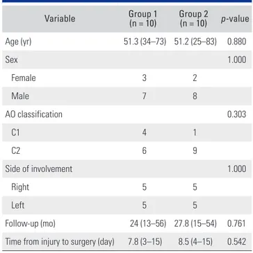
A B C
Fig. 2. (A) Preoperative radiograph showing right tibial plateau fracture in a 36-year- old female patient. (B, C) Radiographs taken 14 months after lateral locking plate fixation.
Statistical Analysis
Statistical analysis was performed using IBM SPSS ver.
22.0 (IBM Corp., Armonk, NY, USA). Fisher’s exact test and Pearson chi-square test were used to compare cat- egorical data between groups. Mann-Whitney U-test and Wilcoxon signed-rank test were used to compare continu- ous data. A p < 0.05 was considered statistically significant.
Ethical Approval
This study was conducted in accordance with the ethical guidelines of the Declaration of Helsinki and informed written consent was obtained from all the patients. The protocol was reviewed and approved by the Institutional Review Board of Izmir Bozyaka Education and Research Hospital (No. 29.12.2015-5).
RESULTS
Patient characteristics are presented in Table 2. The mean follow-up time was 24 months (range, 13 to 56 months) in group 1 and 27.8 months (range, 15 to 54 months) in group 2 (p = 0.761). The most common cause of injury was a traffic accident in 11 patients (55%), fall from height in five patients (25%), simple fall in three patients (15%) and crush injury in one patient (5%). No significant differ- ence was detected between group 1 and group 2 in terms of patient age, sex, fracture type, fracture side, time from
injury to operation (p > 0.05) (Table 2). Three of these fractures were open: two were Gustilo grade I and one was Gustilo grade II. Open fractures were initially managed with debridement and temporary fixation. The average delay in definitive surgery was 7.3 days (range, 6 to 9 days) in these patients. The bone defects were filled with allog- enous bone graft in one patient (10%) in group 2.
There were no significant difference between the groups with regard to KSS, RFS, RRS, VAS, and ROM at the final follow-up (Table 3). According to the KSS, we found four unsatisfactory results (two in group 1 and two in group 2, 20%); the remaining 16 cases were graded as good or excellent. Similarly, there were no significant in- tergroup differences in TFAA, MPTA, and PPTA at the final follow-up. However, there was a significant side-to- side difference in TFAA of group 2 (p = 0.046). Further- more, we detected significant side-to-side difference in PPTA of group 1 (p = 0.035). We attributed this difference to intraoperative malreduction measured on immediate postoperative X-rays (Table 4).
All fractures were healed. There were no late or mal- union cases. Partial and full weight-bearing were initiated 8 weeks and 12 weeks, respectively. The average time to ra- diological union was 14 weeks (range, 9 to 16 weeks). Late postoperative complications detected in group 1 were as follows: arthrosis in six patients (60%; Kellgren-Lawrence grade 1–3); flexion contracture in two patients (20%; 10°
and 15°); extension contracture in one patients (10%; 10°), which occurred at the 5 mm tibial medial metaphyseal de- pression and a total knee arthroplasty was performed at 15 months postoperatively; and superficial incision site infec- tion in one patient (10%), which was resolved with antibi- otic therapy and wound care. Late complications detected in group 2 include arthrosis in four patients (40%; grade 1–3), flexion contracture in five patients (50%; range, 10°
Table 2. Demographic Characteristics
Variable Group 1
(n = 10) Group 2
(n = 10) p-value
Age (yr) 51.3 (34–73) 51.2 (25–83) 0.880
Sex 1.000
Female 3 2
Male 7 8
AO classification 0.303
C1 4 1
C2 6 9
Side of involvement 1.000
Right 5 5
Left 5 5
Follow-up (mo) 24 (13–56) 27.8 (15–54) 0.761
Time from injury to surgery (day) 7.8 (3–15) 8.5 (4–15) 0.542 Values are presented as mean (range) or number.
Group 1: dual locking plate group, Group 2: lateral locking plate group.
Table 3. Functional Outcomes of Two Techniques
Variable Group 1 (n = 10) Group 2 (n = 10) p-value
KSS 79.1 (49–100) 72.9 (30–94) 0.496
RFS 24.3 (16–30) 22.9 (17–28) 0.238
RRS 15.8 (10–18) 15.2 (6–18) 0.903
VAS 4.5 (0–9) 5.5 (2–9) 0.517
ROM (°) 120 (100–130) 119 (110–130) 0.430 Values are presented as mean (range).
Group 1: dual locking plate group, Group 2: lateral locking plate group, KSS: Knee Society Knee Scoring System, RFS: Rasmussen functional score, RRS: Rasmussen radiological score, VAS: visual analog scale, ROM: range of motion.
to 15°) and extension contracture in two patients (20%; 5°
and 10°). There was no significant difference in terms of late complications between the groups (p = 0.582).
DISCUSSION
In this study, we compared the dual locking plate fixation technique and unilateral locking plate fixation technique in tibial bicondylar plateau fractures without posteromedi- al fragment. We did not observe significant differences in functional and clinical results between the groups, whereas the TFAA values of group 2 and the PPTA values of group 1 in the operated extremity showed significant difference when compared to those in the contralateral extremity.
The goals of operative treatment for the tibial plateau fractures include anatomic reduction with restoration of articular congruity and rigid fixation for recovery of previ- ous range of motion.1-4,7) There are many different surgical techniques that have been advocated for the management of bicondylar tibial plateau fractures such as dual buttress plate fixation, locking plate fixation, external fixation, and a combination of internal and external fixation.2-4,7,8,21-23)
The advantages of dual plate technique are visual reduction and maintenance of proximal tibial alignment,
but soft tissue complications and damage to the periosteal blood supply are major concerns in this method of inter- nal fixation.17,23) Some authors claimed a single lateral lock- ing plate can decrease the risk of soft tissue damage and wound infection, but it cannot provide sufficient stability for multipart fractures.3,9-13) The posteromedial fracture fragment has been reported to have a frequency of 33% in bicondylar tibial plateau fractures.24) A single lateral lock- ing plate fixation may not allow adequate stability in all bicondylar tibial plateau fractures.
Biomechanical and cadaver studies have shown that the dual locking plate fixation technique in bicondylar tibial plateau fractures allows less collapse when com- pared with the lateral locking plate fixation technique.25,26) Weaver et al.12) reported that fractures with medial coronal fracture lines had a higher rate of plateau collapse and loss of reduction in lateral locking plate fixations. They came to the conclusion that such fractures can be treated better with double plates. For this reason, complex fractures with posteromedial fragment were excluded from the current study; because of their different treatment strategy.
Different scoring systems have been recommended to evaluate functional results of tibial plateau fractures.
We used KSS and RFS in this study. With regard to these Table 4. Comparison of the Radiological Outcomes between Group 1 and Group 2 and between the Injured Side and the Uninjured Side in
Each Group
Variable Group 1 (n = 10) p-value* Group 2 (n = 10) p-value* p-value†
TFAA (°) 0.799 0.046 0.399
Fracture side, early postoperative 3.9 (2–6) 5.4 (2–13)
Fracture side, last visit 3.9 (2–6) 5.6 (2–15)
Normal side 4.0 (2–6) 3.3 (1–7)
MPTA (°) 0.221 0.779 0.545
Fracture side, early postoperative 86.1 (82–89) 86 (81–90)
Fracture side, last visit 86.8 (84–89) 86.3 (82–91)
Normal side 87.2 (85–88) 86.1 (83–88)
PPTA (°) 0.035 1.000 0.903
Fracture side, early postoperative 5.3 (2–7) 5.4 (4–9)
Fracture side, last visit 5.4 (3–7) 5.5 (4–9)
Normal side 6.3 (5–8) 5.4 (4–6)
Values are presented as mean (range).
Group 1: dual locking plate group, Group 2: lateral locking plate group, TFAA: tibiofemoral anatomic angle, MPTA: medial proximal tibial angle, PPTA:
proximal posterior tibial angle.
*Statistical comparison of the last visit values between the fracture side and the normal side. †Statistical comparison of the last visit values in the fracture side between group 1 and group 2.
systems, there were no difference between dual and single plating groups. In addition, in the literature, other series have reported good/excellent scores in 65%–89% of sub- jects.2,11) Our results are comparable with those of other published studies with 80% satisfaction rates.
Jiang et al.11) reported a higher incidence of postop- erative malalignment of the proximal tibia in the lateral Less Invasive Stabilization System group than in the dual plate group. Therefore, they suggested that the lateral lock- ing plate cannot replace the conventional double plate technique in bicondylar tibial plateau fractures.11) Similar- ly, another biomechanical study using the cadaveric model of the bicondylar tibial plateau fracture revealed that the double plate fixation resulted in less collapse in cyclic load- ing than isolated lateral locking plates did. This suggests that the lateral locking plate is not sufficient for bicondylar tibial plateau fractures.25)
In a biomechanical study by Mueller et al.,10) stability in a tibial plateau fracture model was compared between the unilateral locking plate fixation and the dual locking plate fixation. The results were similar to those in the study of Yao et al.9) who suggested that the shape of the medial condyle fracture plays a key role in single or double plate application. They reported there was no significant differ- ence in the incidence of early postoperative malalignment and malreduction between dual plates and lateral locking plates used to treat bicondylar tibial plateau fractures in patients with a relatively intact medial condyle.
Similarly, in our study, radiological parameters in- cluding TFAA, PPTA, and MPTA showed no significant difference between the groups. Two significant differences were noted only in the comparison with the uninjured extremities in each group. In the lateral plating group, the mean TFAA was changed from 5.6° to 3.3°. In the dual plating group, the mean PPTA was changed from 5.4° to 6.3°. These differences were attributed to intraoperative malreduction.
Soft tissue complications are a major concern in the treatment of bicondylar tibial plateau fractures with plates and the incidence is reported to range from 5% to 88%.27-29) Optimal timing for surgery, surgical time, soft tissue protec- tion, and prevention of infection in tibial plateau fractures are important factors.9) With the use of a single incision and less soft tissue damage, fracture healing may be improved as in the lateral locking plate fixation technique.7,9,13)
Deep infection with plates has been reported to occur in up to 22% in tibia plateau fractures.1,2,27,29) In our study, we
had no deep infection cases. Superficial tissue infection de- veloped in one patient in the dual plate group (10%), which was treated by oral antibiotics and wound care. In the lateral plating group, there was no sign of infection.
Studies have demonstrated that articular incongrui- ty and joint instability can lead to early posttraumatic joint degeneration.2,30) The incidence of osteoarthritis following tibial plateau fractures is 17%–83%.2) Since the follow-up period in this study was relatively short, the incidence of posttraumatic arthritis was not as high as that reported in other studies. In total, 10 cases had signs of arthrosis (grade I–III) when compared to the uninjured side (50%). We though our results are comparable to those in other pub- lished studies.
Nonunion of bicondylar tibial plateau fractures has an incidence of approximately 4%.3,7,13) Nutrient vascular injury, bone defect, and lack of adequate fixation can cause this complication.3,7,9,13,29) There was no nonunion in our study, which is related to the preservation of blood circu- lation of tissues during surgery. A spongious bone defect can easily occur in the tibial plateau fractures. The bone graft does protect the mechanical support and prevent late collapse.3,9,29,30) In this study, a bone graft was used in one patient in group 2.
One of the limitations of this study is the relatively small number of patients, for which we could not perform post-hoc analysis. In addition, the duration of follow-up was short, and the study was performed retrospectively.
Studies with a larger number of cases performed in a ran- domized controlled manner with a longer-term follow-up may allow more accurate assessment. However, a major advantage of this study is that we included only patients with bicondylar plateau fractures without posteromedial fracture fragment.
Dual locking plate fixation technique and lateral locking plate fixation technique are effective methods in the treatment of bicondylar tibial plateau fractures when used properly. The lateral locking plate fixation technique may provide comparable clinical and radiological out- comes with dual locking plate fixation technique for the treatment of bicondylar plateau fractures without postero- medial fragment.
CONFLICT OF INTEREST
No potential conflict of interest relevant to this article was reported.
REFERENCES
1. Chang H, Zhu Y, Zheng Z, et al. Meta-analysis shows that highly comminuted bicondylar tibial plateau fractures treated by single lateral locking plate give similar outcomes as dual plate fixation. Int Orthop. 2016;40(10):2129-41.
2. Manidakis N, Dosani A, Dimitriou R, Stengel D, Matthews S, Giannoudis P. Tibial plateau fractures: functional outcome and incidence of osteoarthritis in 125 cases. Int Orthop.
2010;34(4):565-70.
3. Watson JT. Tibia: proximal. In: Ruedi TP, Murphy WM, eds.
AO principles of fracture management. Stuttgart: Thieme;
2000. 499-517.
4. Lee TC, Huang HT, Lin YC, Chen CH, Cheng YM, Chen JC. Bicondylar tibial plateau fracture treated by open reduc- tion and fixation with unilateral locked plating. Kaohsiung J Med Sci. 2013;29(10):568-77.
5. Luo CF, Sun H, Zhang B, Zeng BF. Three-column fixa- tion for complex tibial plateau fractures. J Orthop Trauma.
2010;24(11):683-92.
6. Nikolaou VS, Tan HB, Haidukewych G, Kanakaris N, Giannoudis PV. Proximal tibial fractures: early experi- ence using polyaxial locking-plate technology. Int Orthop.
2011;35(8):1215-21.
7. Canadian Orthopaedic Trauma Society. Open reduction and internal fixation compared with circular fixator applica- tion for bicondylar tibial plateau fractures: results of a mul- ticenter, prospective, randomized clinical trial. J Bone Joint Surg Am. 2006;88(12):2613-23.
8. Moore TM, Patzakis MJ, Harvey JP. Tibial plateau fractures:
definition, demographics, treatment rationale, and long- term results of closed traction management or operative reduction. J Orthop Trauma. 1987;1(2):97-119.
9. Yao Y, Lv H, Zan J, et al. A comparison of lateral fixation versus dual plating for simple bicondylar fractures. Knee.
2015;22(3):225-9.
10. Mueller KL, Karunakar MA, Frankenburg EP, Scott DS. Bi- condylar tibial plateau fractures: a biomechanical study. Clin Orthop Relat Res. 2003;(412):189-95.
11. Jiang R, Luo CF, Wang MC, Yang TY, Zeng BF. A compara- tive study of Less Invasive Stabilization System (LISS) fixa- tion and two-incision double plating for the treatment of bicondylar tibial plateau fractures. Knee. 2008;15(2):139-43.
12. Weaver MJ, Harris MB, Strom AC, et al. Fracture pattern and fixation type related to loss of reduction in bicondylar tibial plateau fractures. Injury. 2012;43(6):864-9.
13. Egol KA, Su E, Tejwani NC, Sims SH, Kummer FJ, Koval KJ. Treatment of complex tibial plateau fractures using the
less invasive stabilization system plate: clinical experience and a laboratory comparison with double plating. J Trauma.
2004;57(2):340-6.
14. Lee MH, Hsu CJ, Lin KC, Renn JH. Comparison of outcome of unilateral locking plate and dual plating in the treatment of bicondylar tibial plateau fractures. J Orthop Surg Res.
2014;9:62.
15. Neogi DS, Trikha V, Mishra KK, Bandekar SM, Yadav CS.
Comparative study of single lateral locked plating versus double plating in type C bicondylar tibial plateau fractures.
Indian J Orthop. 2015;49(2):193-8.
16. Horwitz DS, Bachus KN, Craig MA, Peters CL. A biome- chanical analysis of internal fixation of complex tibial pla- teau fractures. J Orthop Trauma. 1999;13(8):545-9.
17. Gosling T, Schandelmaier P, Marti A, Hufner T, Parten- heimer A, Krettek C. Less invasive stabilization of complex tibial plateau fractures: a biomechanical evaluation of a unilateral locked screw plate and double plating. J Orthop Trauma. 2004;18(8):546-51.
18. Insall JN, Dorr LD, Scott RD, Scott WN. Rationale of the Knee Society clinical rating system. Clin Orthop Relat Res.
1989;(248):13-4.
19. Rasmussen PS. Tibial condylar fractures: impairment of knee joint stability as an indication for surgical treatment. J Bone Joint Surg Am. 1973;55(7):1331-50.
20. Whelan DB, Bhandari M, McKee MD, et al. Interobserver and intraobserver variation in the assessment of the healing of tibial fractures after intramedullary fixation. J Bone Joint Surg Br. 2002;84(1):15-8.
21. Ariffin HM, Mahdi NM, Rhani SA, Baharudin A, Shukur MH. Modified hybrid fixator for high-energy Schatzker V and VI tibial plateau fractures. Strategies Trauma Limb Re- constr. 2011;6(1):21-6.
22. El-Gafary K, El-adly W, Farouk O, Khaled M, Abdelaziz MM. Management of high-energy tibial plateau fractures by Ilizarov external fixator. Eur Orthop Traumatol. 2014;5(1):9- 14.
23. Musahl V, Tarkin I, Kobbe P, Tzioupis C, Siska PA, Pape HC.
New trends and techniques in open reduction and internal fixation of fractures of the tibial plateau. J Bone Joint Surg Br. 2009;91(4):426-33.
24. Barei DP, O’Mara TJ, Taitsman LA, Dunbar RP, Nork SE.
Frequency and fracture morphology of the posteromedial fragment in bicondylar tibial plateau fracture patterns. J Or- thop Trauma. 2008;22(3):176-82.
25. Higgins TF, Klatt J, Bachus KN. Biomechanical analysis of
bicondylar tibial plateau fixation: how does lateral lock- ing plate fixation compare to dual plate fixation? J Orthop Trauma. 2007;21(5):301-6.
26. Yoo BJ, Beingessner DM, Barei DP. Stabilization of the pos- teromedial fragment in bicondylar tibial plateau fractures:
a mechanical comparison of locking and nonlocking single and dual plating methods. J Trauma. 2010;69(1):148-55.
27. Phisitkul P, McKinley TO, Nepola JV, Marsh JL. Complica- tions of locking plate fixation in complex proximal tibia injuries. J Orthop Trauma. 2007;21(2):83-91.
28. Egol KA, Tejwani NC, Capla EL, Wolinsky PL, Koval KJ.
Staged management of high-energy proximal tibia fractures (OTA types 41): the results of a prospective, standardized protocol. J Orthop Trauma. 2005;19(7):448-55.
29. Spagnolo R, Pace F. Management of the Schatzker VI frac- tures with lateral locked screw plating. Musculoskelet Surg.
2012;96(2):75-80.
30. Kayali C, Citak C, Altay T, Kement Z. Subchondral raft con- struction with locking plates for the treatment of Schatzker type II fractures. Acta Ortop Bras. 2017;25(3):99-102.