INTRODUCTION
Glioblastoma multiforme (GBM) is the most common pri- mary malignant tumor in the central nervous system (CNS) and the most aggressive primary brain tumor in adults, with an incidence of 1.26/100,000 patient-years in Korea [1] and 3.19/100,000 patient-years in the world [2]. Among primary CNS tumors, GBM accounts for 5.3% of them in Korea [1]
and 14.7% of them in the Unites States [3]. GBM is defined as grade IV according to the World Health Organization (WHO) classification [4]. Despite treatment of choice currently con- sists of maximal safe surgical resection and postoperative con- comitant chemoradiotherapy followed by adjunctive chemo- therapy with temozolomide [5], its outcome is still dismal, with
Extensive Pachymeningeal Dissemination
of Glioblastoma Mimicking Chronic Subdural Hematoma:
A Case Report
Jiwook Lee1, Mee-Seon Kim2, Young Zoon Kim1
1Division of Neurooncology and Department of Neurosurgery, Samsung Changwon Hospital, Sungkyunkwan University School of Medicine, Changwon, Korea
2Department of Pathology, Samsung Changwon Hospital, Sungkyunkwan University School of Medicine, Changwon, Korea
Received November 13, 2018 Revised January 1, 2019 Accepted January 8, 2019 Correspondence Young Zoon Kim
Division of Neurooncology and Department of Neurosurgery, Samsung Changwon Hospital, Sungkyunkwan University School of Medicine,
158 Paryong-ro, Masanhoewon-gu, Changwon 51353, Korea Tel: +82-55-233-5241 Fax: +82-55-233-8040 E-mail: [email protected]
Meningeal dissemination (MDS) of glioblastoma is rare, although its incidence might have been un- derestimated. MDS of glioblastoma has a fatal course. Thus, rapid and precise diagnosis of MDS is important for further palliative treatment. Unfortunately, MDS of glioblastoma could be diagnosed at a delayed time, causing failure to treat patient optimally. Herein, we present a case of a 56-year-old male with MDS of glioblastoma mimicking chronic subdural hemorrhage (CSDH) after head trauma due to slip down. During treatment for CSDH, MDS of glioblastoma was not controlled appropriately.
The patient succumbed to MDS of glioblastoma at 9 weeks after the date of diagnosis of CSDH which could be an MDS.
Key Words Glioblastoma; Gliosarcoma; Meningeal dissemination; Mortality.
a median survival time of 14.6 months [6].
GBM often occurs in the supratentorial white matter in- cluding the corpus callosum. It usually spreads along white matter tracts [7]. Ependymal, subpial, and cerebrospinal fluid (CSF) metastases into the subarachnoid space are events of- ten seen in terminal patients. Leptomeningeal dissemination (LMD) of GBM is known to be rare. However, its incidence might have been underestimated in clinical practice. Clinical- ly, the reported incidence of LMS from malignant glioma had been 2% [8] and 3.1% [9]. However, autopsy studies have shown higher incidence of leptomeningeal seeding than those in the neurooncological practice [10,11]. One reason for such discrepancy may be that rapid disease progression can result in loss of chance to survey LMD of GBM. Since the prognosis of GBM patients with LMD is bleak often with fatal outcome, early and precise diagnosis of LMD is important to care for such patients.
Herein, we present a GBM patient with extensive meninge- al dissemination (MDS) mimicking chronic subdural hemor-
This is an Open Access article distributed under the terms of the Creative Commons Attribution Non-Commercial License (https://creativecommons.org/licenses/by-nc/4.0) which permits unrestricted non-commercial use, distribution, and reproduction in any medium, provided the original work is properly cited.
Copyright © 2019 The Korean Brain Tumor Society, The Korean Society for Neuro- Oncology, and The Korean Society for Pediatric Neuro-Oncology
rhage (CSDH) in CT scan performed after head trauma, which makes the best treatment delayed for the MDS of GBM. Also, we are reviewing the literature focused on GBM with MDS.
CASE REPORT
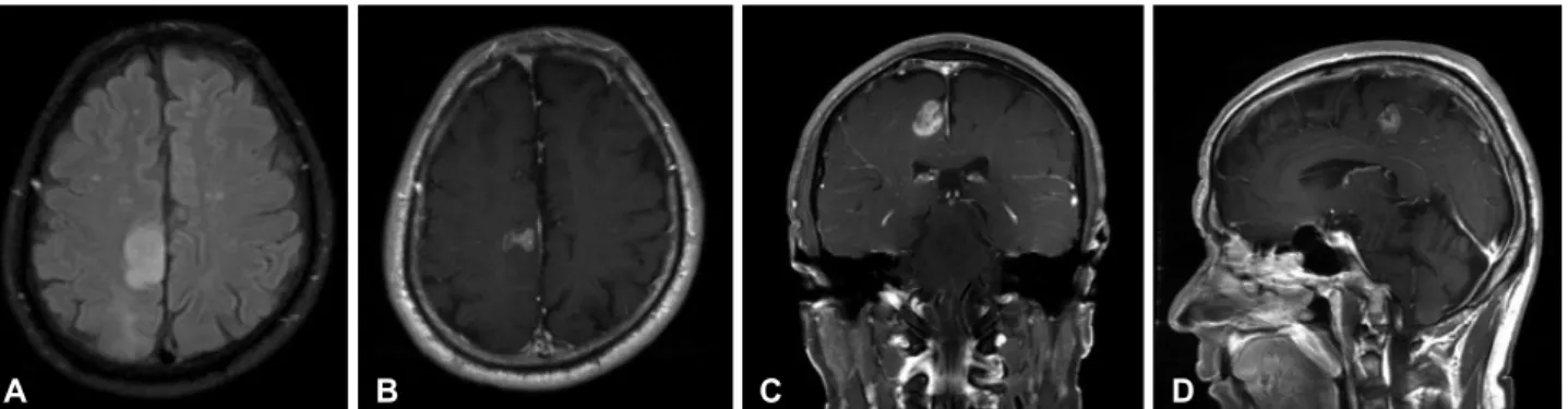
A 56-year-old male was admitted into the Neurosurgical Department via out-patient clinic due to progressive hemipa- resis of grade 3 weakness on the left side for two weeks. On a previous regular check for personal health performed four months earlier at our institute, magnetic resonance (MR) im- age showed a small focus with high signal intensity in fluid at- tenuated inversion recovery (FLAIR) image on the right pre- central gyrus of the frontal lobe. He had no focal neurological symptoms or signs. The lesion did not have enhancement with gadolinium in T1WI at that time. However, at the present ad- mission, MR image showed single nodule at size of 1 cm×1 cm with rim enhancement on the previous area showing high signal intensity in FLAIR image (Fig. 1).
GBM was diagnosed histopathologically based on naviga- tion-guided frameless stereotactic biopsy. Molecular and ge- netic analyses had the following results: O6-methyl guanine DNA methyltransferase (MGMT) promoter, unmethylated;
isocitrate dehydrogenase 1, wildtype; 1p19q, not deleted; al-
pha-thalassemia (ATRX), retained expression; amplification for p53 and epidermal growth factor receptor (EGFR), posi- tive; mitotic figures of 15/10 high power field (HPF); CD133, positive; phosphatase and tensin homolog (PTEN), negative;
glial fibrillary acidic protein (GFAP), positive; and Ki67 14%.
Then he underwent concurrent chemoradiotherapy with te- mozolomide for six weeks followed by six cycles of adjuvant treatment with temozolomide using Stupp’s regimen. There was no progression at the 12-months follow-up in MR imag- es. His neurological condition did not get worsen either. His physical condition was tolerable.
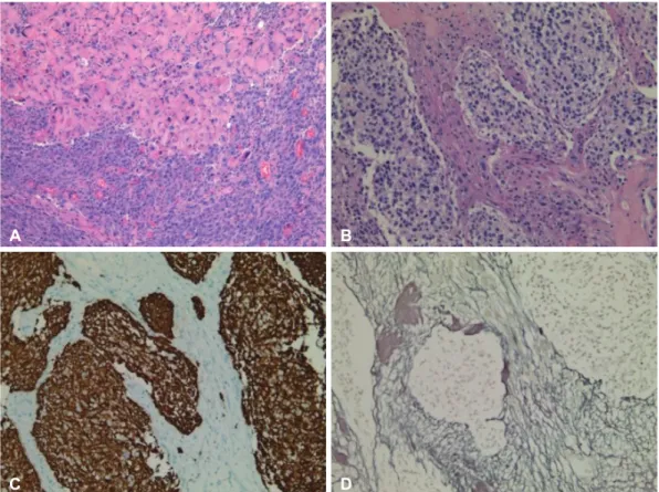
However, the MR image which was performed regularly by 6 months’ interval obtained at 2 years after the initial diagno- sis illustrated an aggressive progression with heterogeneous enhancement and extensive peritumoral edema (Fig. 2). His headache and motor weakness got worse gradually and did not respond to high dose corticosteroid. His motor function worsened to grade 1 on the left side. Therefore, craniotomy and gross total resection were performed to reduce the tumor burden and improve symptoms. Histopathological features showed two biphasic patterns in tumor of epithelioid glial com- ponent and cellular spindle mesenchymal component, leading to the diagnosis of gliosarcoma (Fig. 3). After surgical resec- tion, the headache was relieved and the hemiparesis on the
Fig. 1. The second MR image at the time of occurrence of progressive hemiparesis on the left side showing increased size of the previous area with high signal intensity in fluid attenuated inversion recovery image (A), and nodular rim enhancement at size of 1 cm×1 cm showing in T1WI axial (B) coronal (C), and sagittal images (D) in the right frontoparietal area.
Fig. 2. MR image for regular follow-up showing more extensive peritumoral edema in the fluid attenuated inversion recovery image (A), the low signal intensity suggesting necrosis, irregular shaped unevenly thicken walled mass with internal necrosis, and heterogeneous en- hancement on axial (B), coronal (C), and sagittal images (D) in the right frontoparietal area.
A B C D
A B C D
mal findings in the complete blood count, liver function test, bleeding times and coagulation test, but hypercholesterolemia (total cholesterol of 255 mg/dL and triglyceride of 455 mg/
dL) with hyperglycemia (HbA1c of 8.7%). In CT scan, fluid collection with low density under the dura mater on the right frontotemporoparietal area was shown (Fig. 4A). Straw-col- left side was improved to grade 4. He started to take metro-
nomic chemotherapy with daily treatment of temozolomide at dose of 50 mg/body surface area.
Four months later, he had a mild head trauma due to slip down which made him suffer from chronic headache and de- crease of mentality. The laboratory study showed no abnor-
Fig. 3. Histopathological features showing two biphasic patterns in tumor of epithelioid glial component (upper lane) and cellular spindle mesenchymal component (bottom lane); (H&E staining, ×20) (A). The glial component surrounded by elongated mesenchymal component and the glial area is composed of mild pleomorphic astrocyte (H&E staining, ×20) (B), leading to the diagnosis of gliosarcoma. Immunohis- tochemistry for GFAP demonstrates positivity in glial component (×20) (C), and the biphasic pattern shows strong reticulin positivity in mes- enchymal areas with reticulin negative glial areas (reticulin stain, ×20) (D). H&E: hematoxylin and eosin.
Fig. 4. The noncontrast CT scan showing a newly developed subdural fluid collection which is suggestive hematoma at the right hemi- sphere (A). Repeated CT scan without contrast showing the lining pattern of soft tissue density as well as fluid collection at the right hemi- sphere (B). Intraoperative findings (C) show that thickened dura matter with attachment of the tumors ( 1 ), whitish colored tumor with a fragile texture and relatively poor vascularity ( 2 ), and the relatively well preserved arachnoid membrane beneath the tumor without de- struction nor invasion by the tumor ( 3 ).
A
C
B
D
A B C
ored fluid was drained via the burrhole trephination. The fluid had high viscosity and looked like mucoid fluid. After drain- age, the patient’s headache subsided moderately. However, the low density remained minimally in the follow-up CT scan which was checked 4 weeks after the first burrhole drainage.
And progressive decrease of mentality to stupor with thickened low-density area in the CT scan. Thus, he underwent a sec- ond burrhole trephination for drainage of CSDH. However, no fluid was drained. Therefore, craniotomy was performed to remove the subdural fluid collection (Fig. 4B). During op- eration, there was no fluid collection but a mass covering the whole hemisphere on the right side of frontotemporoparietal lobe. The whitish-colored tumor had a fragile texture and rel- atively poor vascularity, was attached at the whole pachyme- ninges of the right hemisphere (Fig. 4C), and easily detached by bipolar coagulators. However, the arachnoid membrane beneath the tumor was not destructed nor invaded by the tu- mor but relatively well preserved. The mass was removed as much as possible, and histological characteristics of the glio- blastoma were changed into more aggressive and more malig- nant biology as follows: 1p19q non-codeleted; MGMT non- methylated; CD133, strong positive; PTEN, negative; GFAP, strong positive; EGFR, 3+; ATRX, retained expression; p53 with about 35%; Ki67 with about 50%; mitotic figures of 85/10 HPF.
His family members were reluctant to let him undergo fur- ther palliative therapies such as re-irradiation or chemothera- py despite worsening features in patient’s condition and CT scan. He was succumbed to the disease at 4 weeks after the last craniotomy. His overall survival was 32 months. He died at 9 weeks after diagnosis of LMD based on the CT scan. In- formed consent was waived due to its retrospective nature and minimal risk for harm to patient. And this report was conduct- ed according to guidelines of the Declaration of Helsinki for biomedical research.
DISCUSSION
Meningeal spread including ependymal, subpial, and CSF metastases into the subarachnoid and subdural space is not commonly encountered in the clinical field of treating GBM and even events seen in terminal patients. Symptomatic CSF seeding of intracranial GBM is also rare, accounting for only 2% of cases in the largest study of 600 patients [12]. That study also showed that CSF seeding occurred mostly in the late stage of GBM [12]. However, it is still unknown whether lengthier survival may lead to greater chance of CSF seeding.
In fact, glioma dissemination, especially invasion into the leptomeninges, has been largely studied. There are growing studies reporting potential risk factors and possible mecha-
nisms for leptomeningeal spread of GBMs. Among them, the anatomical location of GBM is the most important risk factor.
It is well-known that there is a particular relationship between GBM and the subventricular zone (SVZ) neural stem cells re- gion [13]. GBMs in contact with the SVZ are very likely to be multifocal at diagnosis and recur a distance from the initial lesion [13]. Additionally, ventricular entry at operation, re- peated tumor resection, male sex, ependymal invasion, fissur- ing of the ependyma due to hydrocephalus, depressed immune function after radiotherapy and chemotherapy, and fragmen- tation of the tumor in contact with CSF are significant factors associated with increased incidence of MDS [14].
In terms of molecular biology, a number of factors and mechanisms for MDS of GBM have been implicated, includ- ing CD133-postive stem cells as possible initiators of metasta- sis. Matrix metalloproteinases involved in enzymatic degrada- tion of the extracellular matrix also appear to play a key role in the progression of various tumors. Mutations in tumor sup- pressor gene PTEN as well as gains at the 1p36 chromosomal region have also been preferentially found in disseminated gli- oma [15-17]. For histopathological diagnosis, some studies have suggested that the degree of astrocytic differentiation of tumor cells seen in GFAP-immunohistochemistry and MIB-1 labelling indexes are potential markers of invasiveness [15,18, 19]. However, the present case did not show CD133 positivity nor loss of PTEN, although he showed low expression level of GFAP in immunohistochemical staining with high Ki67 in- dex (about 40%). Since these molecular features are from a single case, they may not reflect all cases. Further comprehen- sive studies are essential for establishment of predictive role of molecular studies.
Treatment for MDS of GBM is chiefly palliative due to its fatal prognosis. The mean survival time between diagnosis of MDS and death is approximately 2 to 3 months [20,21]. Ra- diotherapy is the most common treatment of choice, with a total dose of 25–40 Gy. It may provide pain relief and some improvements in neurological function. However, it may not offer survival benefits [22]. Intravenous or intrathecal chemo- therapy has not found to be very useful for improving the over- all survival of such patients either. Repeated surgical resection may be attempted if there is a symptomatic, large metastatic deposit causing cord compression. However, usually MDS is not amenable to surgery due to diffuse nature of the disease [22]. In the present case, we performed surgical resection for the subdural located large mass covering the right hemisphere.
However, the patient’s neurological status or survival was not improved at all.
In the present case, we misdiagnosed MDS as CSDH due to obvious history of head trauma and clinical manifestation.
However, several diseases must be differentiated from MDS
in CT scan and MR image. The first of such diseases that need to be differentiated is meningeal metastasis of systemic cancer such as breast cancer and prostate cancer [23]. On non-con- trast-enhanced CT images and MR image, it is difficult to dif- ferentiate CSDH from meningeal metastasis of systemic can- cer. Dural metastases appearing as relatively dense lesions and hemorrhagic hyperdensities may be found. On MR, most masses are hypointense to grey matter in T1WI and hyperin- tense in T2WI with possible hemorrhagic signal as in any me- tastasis. The second of such diseases that need to be differenti- ated from MDS is meningiomas, especially the en plaque type which is a common primary adult intracranial tumor [23]. In nonenhanced CT, the mass is sharply circumscribed in most cases (70–75%) while it is hyperdense with calcification in 20–
25% of cases. Necrosis, cysts, and hemorrhage occur in 8–23%
of cases. The third is neurosarcoidosis, a multisystem inflam- matory disease characterized by non-caseating epitheloid-cell granulomas of unknown etiology with granulomatous lepto- meningitis or dural-based mass as the most common gross pathological features [23]. Therefore, physicians must differen- tiate MDS from other pathologic conditions even if the cause is obvious such as the presenting case. Despite the MR image is superior to CT scan for the differentiation of these patholog- ical condition, we did not check the MR image unfortunately.
The omitting check of MR image could be one of reasons do- ing our misdiagnosis.
In summary, we experienced a case of extensive MDS of GBM misdiagnosed as CSDH due to obvious history of head trauma. Here we report this case with literature review. Al- though the dismal outcome of MDS of GBM makes physician and patients difficult to treat this disease, further comprehen- sive study is needed to improve the outcome of patients with MDS of GBM.
Conflicts of Interest
The authors have no potential conflicts of interest.
Acknowledgments
We appreciate the two radiologist, Young Min Kim, M.D., and Sunwoo Mi-Ok, M.D. for the review of radiographic features of the patient.
REFERENCES
1. Dho YS, Jung KW, Ha J, et al. An updated nationwide epidemiology of primary brain tumors in Republic of Korea, 2013. Brain Tumor Res Treat 2017;5:16-23.
2. Louis DN, Perry A, Burger P, et al. International Society of Neuropa- thology--Haarlem consensus guidelines for nervous system tumor classification and grading. Brain Pathol 2014;24:429-35.
3. Ostrom QT, Gittleman H, Truitt G, et al. CBTRUS statistical report:
primary brain and other central nervous system tumors diagnosed in the United States in 2011-2015. Neuro Oncol 2018;20(suppl_4):iv1-86.
4. Louis DN, Perry A, Reifenberger G, et al. The 2016 World Health Or-
ganization Classification of Tumors of the Central Nervous System: a summary. Acta Neuropathol 2016;131:803-20.
5. Preusser M, de Ribaupierre S, Wöhrer A, et al. Current concepts and management of glioblastoma. Ann Neurol 2011;70:9-21.
6. Stupp R, Hegi ME, Mason WP, et al. Effects of radiotherapy with con- comitant and adjuvant temozolomide versus radiotherapy alone on survival in glioblastoma in a randomised phase III study: 5-year analy- sis of the EORTC-NCIC trial. Lancet Oncol 2009;10:459-66.
7. Kumar R, Jain R, Tandon V. Thalamic glioblastoma with cerebrospinal fluid dissemination in the peritoneal cavity. Pediatr Neurosurg 1999;31:
242-5.
8. Salazar OM, Rubin P. The spread of glioblastoma multiforme as a deter- mining factor in the radiation treated volume. Int J Radiat Oncol Biol Phys 1976;1:627-37.
9. Macdonald DR, Cascino TL, Schold SC Jr, Cairncross JG. Response criteria for phase II studies of supratentorial malignant glioma. J Clin Oncol 1990;8:1277-80.
10. Bryan P. CSF seeding of intra-cranial tumours: a study of 96 cases.
Clin Radiol 1974;25:355-60.
11. Saito R, Kumabe T, Jokura H, Shirane R, Yoshimoto T. Symptomatic spinal dissemination of malignant astrocytoma. J Neurooncol 2003;61:
227-35.
12. Vertosick FT Jr, Selker RG. Brain stem and spinal metastases of supra- tentorial glioblastoma multiforme: a clinical series. Neurosurgery 1990;
27:516-21.
13. Lim DA, Cha S, Mayo MC, et al. Relationship of glioblastoma multi- forme to neural stem cell regions predicts invasive and multifocal tu- mor phenotype. Neuro Oncol 2007;9:424-9.
14. Lawton CD, Nagasawa DT, Yang I, Fessler RG, Smith ZA. Leptomenin- geal spinal metastases from glioblastoma multiforme: treatment and management of an uncommon manifestation of disease. J Neurosurg Spine 2012;17:438-48.
15. Kato H, Fujimura M, Kumabe T, Ishioka C, Kanamaru R, Yoshimoto T.
PTEN gene mutation and high MIB-1 labeling index may contribute to dissemination in patients with glioblastoma. J Clin Neurosci 2004;11:
37-41.
16. Korshunov A, Sycheva R, Golanov A, Pronin I. Gains at the 1p36 chro- mosomal region are associated with symptomatic leptomeningeal dis- semination of supratentorial glioblastomas. Am J Clin Pathol 2007;127:
585-90.
17. Sato A, Sakurada K, Kumabe T, et al. Association of stem cell marker CD133 expression with dissemination of glioblastomas. Neurosurg Rev 2010;33:175-83.
18. Arita N, Taneda M, Hayakawa T. Leptomeningeal dissemination of malignant gliomas. Incidence, diagnosis and outcome. Acta Neurochir (Wien) 1994;126:84-92.
19. Onda K, Tanaka R, Takahashi H, Takeda N, Ikuta F. Cerebral glioblasto- ma with cerebrospinal fluid dissemination: a clinicopathological study of 14 cases examined by complete autopsy. Neurosurgery 1989;25:533- 20. Lomax AJ, Yannakou CK, Rosenthal MA. Spinal cord metastasis in a 40.
patient treated with bevacizumab for glioblastoma. Target Oncol 2013;
8:153-5.
21. Bae JS, Yang SH, Yoon WS, Kang SG, Hong YK, Jeun SS. The clinical features of spinal leptomeningeal dissemination from malignant glio- mas. J Korean Neurosurg Soc 2011;49:334-8.
22. Grah JJ, Katalinic D, Stern-Padovan R, et al. Leptomeningeal and intra- medullary metastases of glioblastoma multiforme in a patient reoperated during adjuvant radiochemotherapy. World J Surg Oncol 2013;11:55.
23. Taschner CA, Brendecke S, Weyerbrock A, Egger K, Prinz M. Freiburg neuropathology case conference: widespread mass lesions after resec- tion of a glioblastoma multiforme. Clin Neuroradiol 2012;22:375-80.