ABSTRACT
Purpose: Common human coronaviruses (HCoVs) are relatively understudied due to the mild nature of HCoV infection. Given the lack of local epidemiology data on common HCoVs, we aimed to describe clinical and epidemiological characteristics of common HCoVs in children.
Methods: Respiratory viral test results from 9,589 respiratory samples from Seoul National University Children's Hospital were analyzed from January 2015 to December 2019. Viral detection was done by the multiplex reverse transcription polymerase chain reaction.
Demographics and clinical diagnosis were collected for previously healthy children tested positive for HCoVs.
Results: Of the 9,589 samples tested, 1 or more respiratory viruses were detected from 5,017 (52.3%) samples and 463 (4.8%) samples were positive for HCoVs (OC43 2.8%, NL63 1.4%, 229E 0.7%). All 3 types co-circulated during winter months (November to February) with some variation by type. HCoV-OC43 was the most prevalent every winter season. HCoV- NL63 showed alternate peaks in late winter (January to March) and early winter (November to February). HCoV-229E had smaller peaks every other winter. Forty-one percent of HCoV-positive samples were co-detected with additional viruses; human rhinovirus 13.2%, respiratory syncytial virus 13.0%, influenza virus 4.3%. Common clinical diagnosis was upper respiratory tract infection (60.0%) followed by pneumonia (14.8%), croup (8.1%), and bronchiolitis (6.7%). Croup accounted for 17.0% of HCoV-NL63-positive children.
Conclusions: This study described clinical and epidemiological characteristics of common HCoVs (OC43, NL63, 229E) in children. Continuing surveillance, perhaps by adding HKU1 in the diagnostic panel can further elucidate the spectrum of common HCoV infections in children.
Keywords: Coronavirus; Children; Epidemiology
INTRODUCTION
Since the first report of human coronaviruses (HCoVs) in the 1960s, more HCoVs have been discovered, including the 4 common HCoVs; HCoV-229E, HCoV-NL63 in alpha genus and HCoV-OC43, HCoV-HKU1 in beta genus. Common HCoVs circulate worldwide as contributors to a range of respiratory outcomes. Infections generally cause mild to moderate
Original Article
Received: Mar 8, 2021 Revised: Aug 11, 2021 Accepted: Aug 12, 2021 Correspondence to Eun Hwa Choi
Department of Pediatrics, Seoul National University College of Medicine, 103 Daehak-ro, Jongno-gu, Seoul 03080, the Republic of Korea.
E-mail: [email protected]
Copyright © 2021 The Korean Society of Pediatric Infectious Diseases
This is an Open Access article distributed under the terms of the Creative Commons Attribution Non-Commercial License (https://
creativecommons.org/licenses/by-nc/4.0/) which permits unrestricted non-commercial use, distribution, and reproduction in any medium, provided the original work is properly cited.
ORCID iDs Youn Young Choi
https://orcid.org/0000-0001-8749-0951 Ye Kyung Kim
https://orcid.org/0000-0001-7145-8738 Eun Hwa Choi
https://orcid.org/0000-0002-5857-0749 Conflict of Interest
No potential conflict of interest relevant to this article was reported.
Youn Young Choi ,1 Ye Kyung Kim ,1 Eun Hwa Choi 1,2
1Department of Pediatrics, Seoul National University Children's Hospital, Seoul, the Republic of Korea
2Department of Pediatrics, Seoul National University College of Medicine, Seoul, the Republic of Korea
Clinical and Epidemiological
Characteristics of Common Human
Coronaviruses in Children: A Single
Center Study, 2015–2019
Author Contributions
Conceptualization: Choi EH; Data curation:
Choi YY, Kim YK; Formal analysis: Choi YY, Kim YK, Choi EH; Investigation: Choi YY, Kim YK; Methodology: Choi YY, Kim YK, Choi EH; Supervision: Choi EH; Validation: Choi EH; Writing - original draft: Choi YY, Kim YK;
Writing - review & editing: Choi EH.
upper respiratory tract illness in children and HCoVs are considered the second most common cause of the common cold.1,2) HCoV infections can also result in lower respiratory tract illness including bronchiolitis and pneumonia, especially in infants.
Different HCoV types may be detected more frequently in different age groups and associated with different clinical manifestation.3) Also, the predominant HCoV type may vary by region or year. However, due to the mild nature of HCoV infection, common HCoVs are understudied compared to influenza virus and respiratory syncytial virus. In Korea, only a few reports have analyzed clinical or epidemiological characteristics of the single HCoV type, mostly during a short period.4-6) Because the data on the circulation pattern of each common HCoVs has been limited in Korea, we aimed to characterize detailed epidemiology and clinical characteristics of HCoVs in children from 2015 to 2019 in a single hospital.
MATERIALS AND METHODS
Respiratory viral test results from respiratory samples from the Seoul National University Children's Hospital (SNUCH) were analyzed from January 2015 to December 2019.
SNUCH is a 300-bed pediatric referral center located in Seoul, Korea. In SNUCH, respiratory viruses were tested for children and adolescents under 20 years of age who presented with fever and/or respiratory symptoms as a part of standard patient care. Respiratory samples were obtained by nasopharyngeal aspiration, nasopharyngeal swab, and/or sputum collection, transtracheal aspiration, or bronchoalveolar lavage. Samples collected within 3 days of admission were included. When samples from a single person were consecutively positive for the same virus within 1 month, only the initial result was included.
The multiplex reverse transcription polymerase chain reaction (RT-PCR) detects 3 types of HCoVs; HCoV-229E, HCoV-NL63, and HCoV-OC43 along with other common respiratory viruses (influenza A virus, influenza B virus, respiratory syncytial virus, human adenovirus, human metapneumovirus, human rhinovirus, parainfluenza virus types 1–4, and human bocavirus). HCoV-HKU1 was not included in the assays. The RT-PCR was performed using the Anyplex™ II RV16 Detection (Seegene Inc., Seoul, Korea) or Allplex™ Respiratory Panel Assays (Seegene Inc.).
We assessed the detection rate of HCoVs for each type, viral co-detections, and demographics of patients for all samples tested positive for HCoV. According to the circulation pattern of HCoVs, which reached a nadir in August and rose from September, one epidemic year was set between September 1 of the year and the end of August of next year. Medical records of the previously healthy children with a single HCoV detection were retrospectively reviewed to analyze the clinical features by each type.
Statistical analyses were conducted using R 4.0 (R Foundation for Statistical Computing, Vienna, Austria). To compare the clinical manifestations of HCoV infection by type, Kruskal-Wallis and Fisher's exact test was used. P-value <0.05 in the analyses was considered significant.
Seoul National University Hospital Institutional Review Board approved this study (No. 2003- 194-1112) and written consent was waived.
RESULTS
1. Prevalence of respiratory viruses
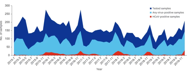
A total of the 9,589 respiratory samples were tested in SNUCH. The rate of detection for all respiratory viruses was 52.3% (5,017/9,589). 463 (4.8%) samples were positive for HCoVs (Fig. 1). Of these samples positive for HCoVs, 67 (0.7%) were positive for HCoV-229E, 139 (1.4%) for HCoV-NL63, 268 (2.8%) for HCoV-OC43 (Table 1). For comparison, human rhinoviruses were detected in 2,132 samples (22.2%), respiratory syncytial viruses in 974 (10.2%), adenoviruses in 668 (7.0%), parainfluenza viruses in 592 (6.2%), influenza viruses in 472 (4.9%).
Simultaneous detection of more than one respiratory virus was observed in 1,038 (10.8%) samples and the total number of detected viruses was 6,268. The proportion positive for each virus of the 6,268 test-positive viruses was shown in Fig. 2. The average proportion of HCoVs of the test-positive viruses during the study period was 7.6%. Human rhinovirus was the most commonly detected virus (34.0%). The second most prevalent virus was respiratory syncytial virus (15.5%).
No. of samples
Year 0
2015-12015-32015-52015-72015-92015-112016-12016-32016-52016-72016-92016-112017-12017-32017-52017-72017-92017-112018-12018-32018-52018-72018-92018-112019-12019-32019-52019-72019-92019-11 300
250 200 150 100 50
Tested samples
Any virus-positive samples HCoV-positive samples
Fig. 1. Monthly number of tested samples, any respiratory virus-positive samples, and HCoV positive samples, 2015–2019.
Abbreviations: HCoV, human coronavirus.
Table 1. The number of samples tested positive for HCoVs in Seoul National University Children's Hospital, 2015–2019
Variables No. of samples (%, yearly detection rate)
2015 Jan–Aug 2015/16 2016/17 2017/18 2018/19 2019 Sep–Dec Total
No. of sample tested 1,409 2,382 1,954 1,717 1,461 666 9,589
All HCoVs 21 (1.5) 122 (5.1) 103 (5.3) 99 (5.8) 72 (4.9) 46 (6.9) 463 (4.8)
HCoV-229E 1 (0.1) 28 (1.2) 4 (0.2) 25 (1.5) 5 (0.3) 4 (0.6) 67 (0.7)
HCoV-NL63 1 (0.1) 33 (1.4) 43 (2.2) 38 (2.2) 14 (1.0) 10 (1.5) 139 (1.4)
HCoV-OC43 20 (1.4) 64 (2.7) 58 (3.0) 41 (2.4) 53 (3.6) 32 (4.8) 268 (2.8)
Data presented as number of samples (%). One epidemic year was set between September 1 of the year and the end of August of next year. Co-detection with 2 different types of HCoVs was observed in 11 samples.
Abbreviations: HCoV, human coronavirus.
2. Demographics
The median age of patients tested was 2.8 years (interquartile range [IQR], 0.8–7.2 years) and 57.5% were male. In 463 children with HCoV detection, the median age was 2.3 years (IQR, 0.9–4.9 years) and 54.9% were male. The detection rate of HCoV was highest in 2–4 year old group with 6.4%, followed by 5.2% in 0–1 year old group and 3.4% in 5–19 year old group.
3. Annual and seasonal distribution of HCoVs
Annual detection rates of HCoV were ranged from 4.9% in 2018/2019 to 5.8% in 2017/2018 (Table 1). The detection rate of HCoV was high during winter season (November to February) each year, showing the highest monthly detection rates up to 16.1% in November 2018 (Fig. 3).
HCoV-OC43 was most prevalent among 3 types and mostly peaked during winter season.
HCoV-NL63 was identified with alternate peaks occurring late winter (from January to March) or early winter (from November to January). Meanwhile, HCoV-229E had smaller peaks every other winter in 2015/16 and 2017/18.
4. Co-detection with respiratory viruses
Of the 463 HCoV-positive samples, co-detection of HCoV and/or non-HCoV respiratory viruses occurred in 197 (42.5%) samples. Co-detection with 2 different types of HCoVs was observed in 11 samples. Therefore, the total number of HCoVs detected was 474. 189 (40.8%) samples were co-detected with one or more respiratory viruses other than HCoVs. Of these, 2 non-HCoV respiratory viruses were co-detected in 29 samples, 3 non-HCoV viruses in 6 samples, and 4 non-HCoV viruses in 2 samples. Frequently co-detected viruses with HCoVs were human rhinovirus (13.2%), respiratory syncytial virus (13.0%), and adenovirus (10.4%).
Influenza virus (4.3%) was less commonly detected with HCoVs (Table 2).
5. Clinical diagnosis of previously healthy children
Medical records of 135 previously healthy children with a single HCoV detection were retrospectively reviewed. The median age was 1.1 year (IQR, 0.3–2.95 years) and 54.1% were male. The clinical diagnoses by type are summarized in Table 3. There were no significant differences in the median age and age distribution by type (P=0.447, P=0.460). Common
Detection rate (%)
Year
0 15/16
100
80
60
40
20
16/17 17/18 18/19
Others HRV PIV HAdV RSV IFV HCoV
Fig. 2. Respiratory viruses identified in children, 2015–2019. One epidemic year was set between September 1 of the year and August 31 of next year. Others indicated human metapneumovirus, bocavirus, and enterovirus.
Abbreviations: HRV, human rhinovirus; PIV, parainfluenza virus; HAdV, human adenovirus; RSV, respiratory syncytial virus; IFV, influenza virus; HCoV, human coronavirus.
clinical diagnoses in overall were upper respiratory tract infection (81, 60.0%) followed by pneumonia (20, 14.8%), febrile illness (14, 10.4%), croup (11, 8.1%), and bronchiolitis (9, 6.7%). Upper respiratory infection was the most common clinical diagnosis of all 3 types of HCoV. HCoV-NL63 was more frequently associated with croup (P=0.029). Simple febrile illness was less frequently observed in HCoV-NL63 (P=0.044).
Detection rate (%)
Year 0
2015-12015-32015-52015-72015-92015-112016-12016-32016-52016-72016-92016-112017-12017-32017-52017-72017-92017-112018-12018-32018-52018-72018-92018-112019-12019-32019-52019-72019-92019-11 15
10
5
Detection rate (%)
Year 0
2015-12015-32015-52015-72015-92015-112016-12016-32016-52016-72016-92016-112017-12017-32017-52017-72017-92017-112018-12018-32018-52018-72018-92018-112019-12019-32019-52019-72019-92019-11 15
10
5
Detection rate (%)
Year 0
2015-12015-32015-52015-72015-92015-112016-12016-32016-52016-72016-92016-112017-12017-32017-52017-72017-92017-112018-12018-32018-52018-72018-92018-112019-12019-32019-52019-72019-92019-11 15
10
5
HCoV-Total HCoV-OC43 HCoV-Total HCoV-NL63 HCoV-Total HCoV-229E
Fig. 3. The percentage of tests positive for HCoVs -229E, -NL63, and -OC43 in children, 2015–2019.
Abbreviations: HCoV, human coronavirus.
Table 2. Frequency of respiratory viral codetection with HCoV in children, 2015–2019
Variables No. of samples positive for each virus (%, codetection rate)
HCoV-229E HCoV-NL63 HCoV-OC43 HAdV PIV RSV IFV-A IFV-B HRV HBoV HMPV EV Any non-HCoV
HCoV-229E (n=67) - 2 (3.0) 5 (7.5) 6 (9.0) 2 (3.0) 10 (14.9) 5 (7.5) 0 (0) 13 (19.4) 2 (3.0) 0 (0) 1 (1.5) 32* (47.8) HCoV-NL63 (n=139) 2 (1.4) - 4 (2.9) 9 (6.5) 0 (0) 16 (11.5) 4 (2.9) 1 (0.7) 10 (7.2) 5 (3.6) 2 (1.4) 1 (0.7) 44* (31.7) HCoV-OC43 (n=268) 5 (1.9) 4 (1.5) - 33 (12.3) 11 (4.1) 36 (13.4) 10 (3.7) 1 (0.4) 38 (14.2) 16 (6.0) 3 (1.1) 4 (1.5) 116* (43.3) All HCoV (n=463†) - - - 48 (10.4) 13 (2.8) 60‡ (13.0) 18‡ (3.9) 2 (0.4) 61 (13.2) 23 (5.0) 5 (1.1) 6 (1.3) 189* (40.8) Data presented as number of samples (%).
Abbreviations: HCoV, human coronavirus; HAdV, human adenovirus; PIV, parainfluenza virus; RSV, respiratory syncytial virus; IFV-A, influenza A; IFV-B, influenza B; HRV, human rhinovirus; HBoV, human bocavirus; HMPV, human metapneumovirus; EV, enterovirus.
*Due to the samples with ≥2 non-HCoV co-detections, the sum of the number of samples positive for each non-HCoV is greater than the number of samples with any non-HCoV co-detection; †Two different types of HCoVs were co-detected in 11 samples; ‡Two different types of HCoVs were co-detected with a non-HCoV.
DISCUSSION
In this study, we investigated the prevalence and circulation pattern of HCoVs using 9,589 respiratory samples during 5 consecutive years, from 2015 to 2019. All 3 types of HCoVs showed a marked seasonality with peaks from winter to early spring and were rarely detected in summer.
Our findings that HCoV mainly circulated from November to February with some variations are consistent with earlier studies.3,6,7) By types, HCoV-OC43 mainly contributed to the overall HCoV infection annually, followed by HCoV-NL63 and HCoV-229E. While HCoV-OC43 and HCoV-NL63 were detected with distinct prevalence every year, biennial pattern was seen in HCoV-229E that peaked in 2015/16 and 2017/18. Although the circulation peaks were not as high as influenza virus, there was a unique seasonality for all 3 types of HCoVs with a minimal prevalence in summer.
The current real-time RT-PCR tests in the study site do not include tests for HCoV-HKU1. In Korea, only a single study has looked a prevalence and epidemiology of HCoV-HKU1 from January 2007 to May 2008.5) In the study, HCoV-HKU1 was detected in 50 of 1,985 specimens with the detection rate of 2.5% which was similar to the recent 5-year average detection rate (2.8%) of HCoV-OC43, the most commonly detected HCoV in this study. The nationwide surveillance through Korea Influenza and Respiratory Viruses Surveillance System
(KINRESS) does not include a test for HCoV-HKU1 either.8) Considering the surveillance data from the U.S. Centers for Disease Control and Prevention that showed HCoV-HKU1 was the most commonly detected HCoV (4.8% in January 2020) in the 2019-2020 season,9) new panel with an inclusion of HCoV-HKU1 should be launched.
Since the emergence of the pandemic coronavirus, severe acute respiratory syndrome coronavirus-2 (SARS-CoV-2), interest in the epidemiology of common HCoVs, which has not been appreciated in the past, is now growing. Although effective and safe vaccines have become available, there are still uncertainties about how the pandemic may progress. A study which predicted how transmission of SARS-CoV-2 will develop proposed that seasonality, Table 3. Clinical features of previously healthy children with a single HCoV detection in Seoul National University Children's Hospital, 2015–2019
Variables Total tested (n=9,589) HCoV-229E (n=12) HCoV-NL63 (n=47) HCoV-OC43 (n=76) P-value
Age
Median age 2.8 (0.8–7.2) 2.35 (0.525–5.5) 0.9 (0.3–2.9) 1.15 (0.2–2.825) 0.447
0–<2 yr 4,054 (42.3) 6 (50.0) 32 (68.1) 49 (64.5) 0.460
2–<5 yr 2,141 (22.3) 3 (25.0) 12 (25.5) 19 (25.0)
5–19 yr 3,394 (35.4) 3 (25.0) 3 (6.4) 8 (10.5)
Sex 0.336
Male 5,512 (57.5) 9 (75.0) 24 (51.1) 40 (52.6)
Female 4,077 (42.5) 3 (25.0) 23 (48.9) 36 (47.4)
Clinical diagnosis
Upper respiratory infection - 7 (58.3) 29 (61.7) 45 (59.2) 0.965
Croup - 0 (0.0) 8 (17.0) 3 (3.9) 0.029
Pneumonia - 2 (16.7) 6 (12.8) 12 (15.8) 0.875
Bronchiolitis - 1 (8.3) 3 (6.4) 5 (6.6) 1.000
Febrile illness* - 2 (16.7) 1 (2.1) 11 (14.5) 0.044
Data presented as number of cases (%) or median values (interquartile range). HCoV-positive cases with other co-detected viruses were excluded. Bold-faced values indicate P-value <0.05.
Abbreviations: HCoV, human coronavirus.
*Febrile illness did not accompany any respiratory symptoms.
the duration of immunity, and cross-immunity from other common HCoVs would be the determining factors.10) Changes in temperature and humidity by season could be a major determinant. Some experts suggested SARS-CoV-2 might decline during summertime and rebound with the start of epidemics of wintertime respiratory viruses such as influenza virus, respiratory syncytial virus, and common HCoVs in the Northern Hemisphere. However, it is too early to predict future seasonality of SARS-CoV-2 based on the seasonality characteristics of common HCoVs. Many places experienced a resurgence of new confirmed cases following a loosening of social distancing regardless of the season. While the prediction of the
COVID-19 outbreak in coming years remains uncertain, decline in the summer does not seem correct at this point. This strongly suggests that human contact patterns among population contribute to occurrence and seasonality of respiratory viruses along with the climate characteristics in the geographic region.11)
Each HCoV type may also independently affect circulation patterns of SARS-CoV-2. Degree of immunity to SARS-CoV-2 along with the cross-immunity within or between the different genera may contribute to the fate of SARS-CoV-2.11,12) Although the existence of cross- immunity is controversial, some expectation remains that immune responses developed after infections by the closest betacoronaviruses, HCoV-OC43 and HCoV-HKU1, may induce cross- immunity with SARS-CoV-2.13,14) Currently, we expect spread of the pandemic virus will stop if the herd immunity threshold of 60–67% is achieved.15,16) Two main caveats are the duration of the immunity and the emergence of the variants of concern that influence the transmission efficiency.
This study has several limitations. Epidemiological characteristics of HCoVs were analyzed in a single center and clinical features were evaluated for previously healthy children only.
There were no available studies that looked at HCoV infections in adults in Korea, while there was only a single study that compared the prevalence of common respiratory viruses between children and adults.17)
Despite these limitations, this study provides the current epidemiology of common HCoVs in children and suggests that there is a room for improvement with the inclusion of HCoV- HKU1 in the nationwide KINRESS and in hospital-based laboratories. As interaction and contact patterns between people in diverse age groups are important in shaping seasonality and epidemiology of respiratory viruses in community, further studies need to establish data on the epidemiology and clinical characteristics of HCoV infections including the adult population in Korea.
REFERENCES
1. van der Hoek L. Human coronaviruses: what do they cause? Antivir Ther 2007;12:651-8.
PUBMED
2. Mäkelä MJ, Puhakka T, Ruuskanen O, Leinonen M, Saikku P, Kimpimäki M, et al. Viruses and bacteria in the etiology of the common cold. J Clin Microbiol 1998;36:539-42.
PUBMED | CROSSREF
3. Killerby ME, Biggs HM, Haynes A, Dahl RM, Mustaquim D, Gerber SI, et al. Human coronavirus circulation in the United States 2014–2017. J Clin Virol 2018;101:52-6.
PUBMED | CROSSREF
4. Sung JY, Lee HJ, Eun BW, Kim SH, Lee SY, Lee JY, et al. Role of human coronavirus NL63 in hospitalized children with croup. Pediatr Infect Dis J 2010;29:822-6.
PUBMED | CROSSREF
5. Lee WJ, Chung YS, Yoon HS, Kang C, Kim K. Prevalence and molecular epidemiology of human coronavirus HKU1 in patients with acute respiratory illness. J Med Virol 2013;85:309-14.
PUBMED | CROSSREF
6. Kim KH, Lee JH, Sun DS, Kim YB, Choi YJ, Park JS, et al. Detection and clinical manifestations of twelve respiratory viruses in hospitalized children with acute lower respiratory tract infections: focus on human metapneumovirus, human rhinovirus and human coronavirus. Korean J Pediatr 2008;51:834-41.
CROSSREF
7. Dare RK, Fry AM, Chittaganpitch M, Sawanpanyalert P, Olsen SJ, Erdman DD. Human coronavirus infections in rural Thailand: a comprehensive study using real-time reverse-transcription polymerase chain reaction assays. J Infect Dis 2007;196:1321-8.
PUBMED | CROSSREF
8. Korea Centers for Disease Control and Prevention (KCDC). Public Health Weekly Report 2015–2019 [Internet]. Cheongju: KCDC; 2015–2019 [cited 2021 Apr 30]. Available from: https://www.kdca.go.kr/
board/board.es?mid=a30501000000&bid=0031&cg_code=C04.
9. U.S. Centers for Disease Control and Prevention (CDC). The National Respiratory and Enteric Virus Surveillance System (NREVSS) 2019–2020 [Internet]. Atlanta: CDC; 2019–2020 [cited 2021 Apr 30].
Available from: https://www.cdc.gov/surveillance/nrevss/coronavirus/natl-trends.html.
10. Kissler SM, Tedijanto C, Goldstein E, Grad YH, Lipsitch M. Projecting the transmission dynamics of SARS-CoV-2 through the postpandemic period. Science 2020;368:860-8.
PUBMED | CROSSREF
11. Sette A, Crotty S. Pre-existing immunity to SARS-CoV-2: the knowns and unknowns. Nat Rev Immunol 2020;20:457-8.
PUBMED | CROSSREF
12. Ma Z, Li P, Ji Y, Ikram A, Pan Q. Cross-reactivity towards SARS-CoV-2: the potential role of low- pathogenic human coronaviruses. Lancet Microbe 2020;1:e151.
PUBMED | CROSSREF
13. Hicks J, Klumpp-Thomas C, Kalish H, Shunmugavel A, Mehalko J, Denson JP, et al. Serologic cross- reactivity of SARS-CoV-2 with endemic and seasonal Betacoronaviruses. J Clin Immunol 2021;41:906-13.
PUBMED | CROSSREF
14. Grifoni A, Weiskopf D, Ramirez SI, Mateus J, Dan JM, Moderbacher CR, et al. Targets of T cell responses to SARS-CoV-2 coronavirus in humans with COVID-19 disease and unexposed individuals. Cell 2020;181:1489-501.
PUBMED | CROSSREF
15. Britton T, Ball F, Trapman P. A mathematical model reveals the influence of population heterogeneity on herd immunity to SARS-CoV-2. Science 2020;369:846-9.
PUBMED | CROSSREF
16. Randolph HE, Barreiro LB. Herd immunity: understanding COVID-19. Immunity 2020;52:737-41.
PUBMED | CROSSREF
17. Seo YB, Song JY, Choi MJ, Kim IS, Yang TU, Hong KW, et al. Etiology and clinical outcomes of acute respiratory virus infection in hospitalized adults. Infect Chemother 2014;46:67-76.
PUBMED | CROSSREF
요약
목적: 사람 코로나바이러스(human coronavirus [HCoV]) 감염은 보통 경미한 임상 양상을 보이는 경우가 많으므로,
HCoV에 대한 연구가 다른 호흡기 바이러스들에 비해 상대적으로 잘 이루어지지 않고 있다. 본 연구는 국내 소아청소 년에서의 HCoV 역학 및 임상적 특성을 기술하고자 하였다.
방법: 2015 년 1 월부터 2019년 12월까지 서울대학교 어린이병원에서 수집한 호흡기 검체의 호흡기바이러스 검사 결과
를 분석하였다. 다중 reverse transcription polymerase chain reaction을 이용하여 바이러스를 검출하여 역학을 조 사하였고, 이전 건강했던 어린이에게서 HCoV가 검출된 경우에 한하여 임상 진단을 분석하였다.
결과: 총 9,589 개 검체 중 5,017개(52.3%)에서 1개 이상의 호흡기바이러스가 검출되었고 463개(4.8%)가 HCoV 양성이 었다(OC43 2.8%, NL63 1.4%, 229E 0.7%). 3가지 유형 모두 겨울철(11월–2월)에 유행하였으며 유형별로 역학적 차 이를 보였다. HCoV-OC43이 가장 흔하였으며 매년 겨울에 유행하였다. HCoV-NL63은 1월–3월과 11월–2월 사이에 유 행하였다. 한편, HCoV-229E는 2015/2016 및 2017/2018 겨울에 격년으로 유행하였다. 연령군을 0–1세, 2–4세, 5–19세 로 분류하였을 때, HCoV 감염의 빈도는 2–4세에서 가장 높았고, 5–19세에서 가장 낮았다. HCoV 양성 검체의 40.8%
에서 다른 호흡기바이러스가 함께 검출되었다. 리노바이러스 및 호흡기세포융합바이러스의 동시검출률이 각각 13.2%, 13.0%로 높았고, 인플루엔자 바이러스는 4.3%로 낮았다. 이전에 건강했던 135 명의 어린이에게서 가장 흔한 임상 진 단은 상기도감염(60.0%)이었고, 폐렴(14.8%), 크루프(8.1%), 세기관지염(6.7%) 순이었다. 크루프는 NL63 양성인 어 린이의 임상 진단 중 17.0%를 차지했다.
결론: 본 연구는 소아청소년에서의 HCoV (OC43, NL63, 229E) 감염의 역학적, 임상적 특성을 기술하였다. 소아청소년
에서의 HCoV 감염에 대한 지속적인 감시가 필요하며, 추가적으로 HCoV-HKU1을 진단 패널에 포함한다면 HCoV 감 염의 임상역학적 특징을 파악하는 데 더 도움이 될 것이다.