Received: March 5, 2019 Revised: May 29, 2019 Accepted: June 24, 2019
Address for Correspondence: Sun Yi Choe, Department of Obstetrics & Gynecology, Dong-A University Hospital, 26 Daesingongwon-ro, Seo- gu, Busan 49201, Korea
Tel: 82-51-240-2864, E-mail: [email protected], ORCID: https://orcid.org/0000-0002-6890-1329
ORIGINAL ARTICLE
INTRODUCTION
Osteoporosis is a skeletal disorder characterized by a decrease in bone mineral density. With the growing el- derly population, morbidity and mortality rates associ- ated with osteoporotic fractures are also increasing [1].
According to a study that analyzed the results of 2008 to 2011 Korea National Health and Nutrition Examina- tion Survey (KNHANES) [2], the prevalence of osteo- porosis among Korean women aged 50 years or older was high: 30.1% in the lumbar spine and 23.1% in the femoral neck. Therefore, early detection is important for reducing the fracture-related mortality rate among
people with a risk of osteoporosis.
Bone mass is determined by the peak bone mass and extent of subsequent bone loss. Peak bone mass is not only affected by genetic factors, but also by nutritional status, physical activities, and hormonal changes [3].
Childbirth and breast feeding usually take place around the age of 30 to 39 years when the peak bone mass is achieved; thus, investigating the effects of parity and duration of breast feeding on the risk of osteoporosis among postmenopausal women is very important for the prevention of osteoporosis [4].
Childbirth and breast feeding affect calcium metabo- lism in women, which in turn, affects bone density [5].
Effects of Parity and Breast Feeding Duration on the Risk of Osteoporosis in Postmenopausal Korean Women:
A Systematic Review and Meta-Analysis
Eun Nam Lee1, Sun Yi Choe2, Eun Hui Choi3, Min Ju Lee1
1College of Nursing, Dong-A University, Busan, Korea, 2Department of Obstetrics & Gynecology, Dong-A University Hospital, Busan, Korea, 3Department of Nursing, Masan University, Masan, Korea
Objectives: To summarize the evidence regarding the association of parity and breast feeding duration with the risk of osteoporosis in postmenopausal Korean women. This was because studies have been inconsistent regarding the effect of parity and breast feeding duration on the risk of osteoporosis.
Methods: A systematic literature search of relevant studies published by December 26, 2018 was conducted in PubMed, EMBASE, the Cochrane Library, CINAHL, RISS, KISS, KMbase, and KoreaMed. Outcome estimates of odds ratio (OR) or standardized mean difference were pooled with fixed or random-effect model. In case of heterogeneity, subgroup analysis was conducted.
Results: Seven cross-sectional studies (with 3,813 subjects) were included in the analysis. OR for osteoporosis was 1.43 (95%
confidence interval [CI] = 1.09–1.88, P = 0.010) in postmenopausal women with higher parity compared to those with less parity.
Moreover, OR for osteoporosis was 1.93 (95% CI = 1.28–2.93, P = 0.002) in postmenopausal women with longer durations of breast feeding than in those with shorter durations of breast feeding.
Conclusions: This study revealed that duration of breast feeding increased the risk of osteoporosis in postmenopausal Korean women.
More cohort studies with high quality research designs are needed to confirm our results.
Key Words: Bone density, Breast feeding, Parity, Postmenopause
While some studies have reported reduction in femoral bone density due to childbirth [3,6,7], other studies reported that parity may have a protective effect on the bones in multiparous women [8,9]. In addition, some authors claim that the number of childbirths does not have a significant effect on bone density in postmeno- pausal women since bone loss due to childbirth is re- covered after delivery [10-12]. Although some studies have reported that estrogen levels in mothers decrease during the breast feeding period and that the reduction is caused by calcium loss through breast milk [13-15], other studies indicate that the duration of breast feed- ing and postmenopausal bone density are unrelated since bone loss stops once breast feeding is stopped and bones are restored to their original state [3,16,17].
As shown, reduction in bone mass may be caused by calcium loss and hormonal change during preg- nancy and breast feeding, but a consensus has not been reached on the long-term effects of such changes on the bone density of postmenopausal women. The reason for such conflicting findings between studies may be attributed to differences in ethnicity, age, menopausal status, parity, and/or duration of breast feeding of women in these studies, and the study designs. In par- ticular, the average number of childbirths or duration of breast feeding among women in Korea differs from that in other Asian or Western countries. Therefore, results from studies outside Korea may not be directly applicable.
A recently published meta-analysis [18] showed that an increase in parity reduced the risk of hip fracture among postmenopausal women. However, another me- ta-analysis on the effects of parity on the bone density of pre-menopausal and post-menopausal women [19]
reported that parity did not affect bone density in the lumbar spine and femoral neck, although primiparous or multiparous women showed significantly higher total femoral bone density than nulliparous women, indicating that the effect of parity on bone density may differ depending on the site of measurement.
Thus, even meta-analyses have failed to reach a con- sensus on the relationship between parity or duration of breast feeding and bone density. Particularly, since such correlations may appear differently depending on the ethnicity and menopausal status of the subjects and the site of bone density measurement, the present study included studies that investigated the relationship between parity or duration of breast feeding and bone density in the lumbar spine or femoral neck in post-
menopausal Korean women alone. A meta-analysis was also performed to derive integrative results regarding these studies.
MATERIALS AND METHODS
Search strategy and study selection
The following terms were used to search Korean data- bases (RISS, KISS, KMbase, and KoreaMed) and non- Korean databases (PubMed, EMBASE, the Cochrane Library, and CINAHL) up to December 26, 2018. The search terms were “parity”, “bone mineral density”,
“BMD”, “osteoporosis”, “bone mass”, “birth”, “labor”, “de- livery”, “pregnancy”, “breast feeding”, “feeding”, and “lac- tation”.
Study quality assessment
The quality of individual studies was assessed using the modified form of the New Castle–Ottawa qual- ity assessment scale (NOS) [20]. This scale consists of three major categories: selection, comparability, and outcome. Using this assessment tool, items with high quality of evidence were marked with an asterisk (*) and scores were assigned according to the total num- ber of asterisks. The maximum possible score for each study was 8 points.
Inclusion and exclusion criteria
Studies that compared the relationship between parity or duration of breast feeding and bone density among postmenopausal Korean women were considered to have satisfied the inclusion criteria but only studies that measured bone density in the lumbar spine or femoral neck using dual energy X-ray absorptiometry were included. Only articles published in English or Korean were included. Among studies that used the same data, articles that presented statistics appropriate for meta- analysis were selected first, after which, articles with the largest sample sizes were selected. Moreover, stud- ies that did not apply the World Health Organization (WHO) criteria for osteoporosis (T-score < –2.5 stan- dard deviation [SD]) were excluded.
Data extraction
Three researchers independently assessed the suitabil- ity of the selected studies and extracted the data. The extracted data included the name(s) of the author(s), sample size, age of subjects, outcomes (parity and dura- tion of breast feeding: mean, SD, odds ratio, and 95%
confidence interval [CI]), and quality of study scores.
Statistical methods
Data analysis was performed using Comprehensive Meta-Analysis (CMA) version 2.2 (Biostat, Englewood, NJ, USA). The outcome variable in the present study was osteoporosis status. Meta-analysis was performed with fixed and random effects, where analysis was performed with random effects in cases with high het- erogeneity. Heterogeneity was assessed using Higgins I2 and χ2, where, if I2 was ≥ 50% and χ2 was < 0.10, the case was determined to show heterogeneity.
Publication bias was assessed using a funnel plot and Egger’s test, while sub-analyses were performed accord- ing to the characteristics of the subjects and studies (quality of study).
RESULTS
Search for studies
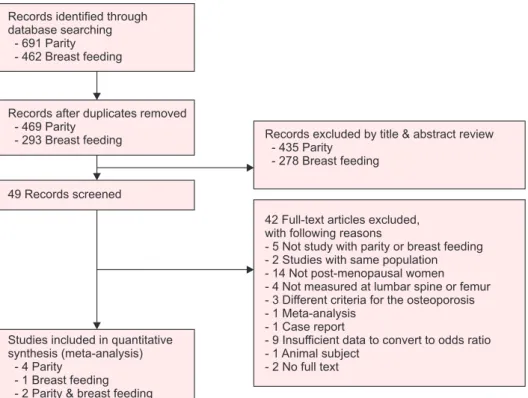
Based on the literature search, a total of 1,153 studies (691 studies on parity and 462 studies on breast feed- ing) were found. After excluding duplicate studies, 762 articles were extracted. An additional 713 articles (435 on parity and 278 on breast feeding) were excluded based on a review of the title and abstract, after which,
the full text of 49 articles were reviewed. Ultimately, a total of 7 articles (4 on parity, 1 on breast feeding, and 2 on both parity and breast feeding) [21-27] were consid- ered suitable for the present study, and a meta-analysis was performed on the studies (Fig. 1).
Study characteristics
The characteristics of the 7 studies included in the fi- nal analysis are shown in Tables 1 and 2. The total sam- ple size was 3,813 patients, including 1,052 patients in the osteoporosis group and 2,761 patients in the non- osteoporosis group (including osteopenia and healthy patients). The mean ages of the osteoporosis and non- osteoporosis groups were 62.49 and 61.98 years, respec- tively. In 6 out of the 7 articles, the relationship between parity and risk of osteoporosis was analyzed: 3 studies [21-23] presented parity as ranges of values while 3 studies [24-26] used mean parity values. Moreover, 3 studies analyzed the relationship between duration of breast feeding and risk of osteoporosis: 2 studies [21,27]
presented duration as ranges of values and 1 study [24]
used mean values.
The quality scores of the included studies ranged between 2 and 8 points. Among the studies on parity, 4 out of 6 studies showed a low quality with a score of 2 points, whereas, the other 2 studies showed a very
Records identified through database searching
- 691 Parity - 462 Breast feeding
Records after duplicates removed - 469 Parity
- 293 Breast feeding
49 Records screened
Studies included in quantitative synthesis (meta-analysis)
- 4 Parity - 1 Breast feeding - 2 Parity & breast feeding
Records excluded by title & abstract review - 435 Parity
- 278 Breast feeding
42 Full-text articles excluded, with following reasons
- 5 Not study with parity or breast feeding - 2 Studies with same population - 14 Not post-menopausal women - 4 Not measured at lumbar spine or femur - 3 Different criteria for the osteoporosis - 1 Meta-analysis
- 1 Case report
- 9 Insufficient data to convert to odds ratio - 1 Animal subject
- 2 No full text
Fig. 1. Flow diagram of the study selec- tion process.
high score of 8 points. Among the studies on dura- tion of breast feeding, 2 out of 3 studies showed a very high score of 8 points, whereas, the remaining 1 study showed a low score of 2 points.
Estimation of the effect size of parity on the risk of osteoporosis
Among the 7 studies included in the meta-analysis,
6 studies analyzed the risk of osteoporosis according to parity, of which, 3 studies presented parity as mean values and the remaining 3 studies presented parity as ranges of values. The range of values varied between studies, including ranges of 0–2, 3–5, ≥ 6, ≤ 4, ≥ 5, 1–2, and ≥ 3. In 6 studies that analyzed the effect of parity on osteoporosis in postmenopausal Korean women, the effect size was derived by a fixed-effect model since Table 1. Characteristics of included studies: parity and osteoporosis risk
Study
Mean age of non- osteoporosis/
osteoporosis (y) Cases Controls
Parity
OR (95% CI) Study
quality Comments
Range Osteoporosis Non- osteoporosis
Jang et al. [21] 65.5/70.6 182 180 0–2
3–5≥ 6
1.00 (reference) 1.77 (0.57–2.41) 2.89 (1.12–7.42)
8 Study quality (selection 4, comparability 2, outcome 2) Ju and Nam
[22] 44 66 ≤ 4
≥ 5 1.00 (reference)
1.52 (0.62–3.75) 2 Study quality (selection 2, comparability 0, outcome 0) Choi and Kim
[23]
63.93a 340 1,358 1–2
≥ 3 1.00 (reference)
0.51 (0.21–1.23) 8 Study quality (selection 4, comparability 2, outcome 2) Choi and Sung
[24] 64.65/60.77 20 44 4.05 ± 1.85 4.07 ± 1.91 2 Study quality (selection
1, comparability 0, outcome 1)
Doh et al. [25] 57.4/58.4 102 51 4.1 ± 1.1 3.7 ± 1.4 2 Study quality (selection
1, comparability 0, outcome 1)
Park [26] 55.6/52.5 60 135 3.60 ± 1.92 2.92 ± 1.52 2 Study quality (selection
1, comparability 0, outcome 1) Data are presented as mean only, number only, or mean ± standard deviation.
OR: odds ratio, CI: confidence interval.
aThis is an average age of all subjects without distinction between the average age of osteoporosis and non-osteoporosis groups.
Table 2. Characteristics of included studies: breast feeding duration and osteoporosis risk Study Mean age of non-
osteoporosis/
osteoporosis (y) Cases Controls
Breast feeding
OR (95% CI) Study
quality Comments
Month Osteoporosis Non- osteoporosis Jang et al. [21] 65.5/70.6 182 180 < 12
12–23
≥ 24
1.00 (reference) 1.40 (0.46–4.28) 1.08 (0.36–3.26)
8 Study quality (selection 4, comparability 2, outcome 2)
Yun et al. [27] 62.7/58.3 304 927 0
< 24
≥ 24
1.00 (reference) 1.99 (0.90–4.42) 2.49 (1.11–5.58)
8 Study quality (selection 4, comparability 2, outcome 2) Choi and Sung
[24] 64.65/60.77 20 44 19.64 ± 11.35 15.10 ± 7.36 2 Study quality (selection
1, comparability 0, outcome 1) Data are presented as mean only, number only, or mean ± standard deviation.
OR: odds ratio, CI: confidence interval.
heterogeneity was not high [χ2 = 8.193, degree of free- dom (df) = 5, P = 0.146; I2 = 38.97%]. In these stud- ies, the effect size of parity on the risk of osteoporosis was 1.43 (95% CI = 1.09–1.88), which was statistically significant (Z = 2.583, P = 0.010) (Fig. 2). The funnel plot did not show any publication bias, which was sup- ported by Egger test results.
However, as shown in Figure 2, the weight value of one of the six studies that examined the effects of parity on the risk of osteoporosis was significantly higher than that of the other studies. Therefore, as a result of the sensitivity test for two high quality studies, the effect of
parity on the risk of osteoporosis was not significant.
Estimation of the effect size of breast feeding du- ration on the risk of osteoporosis
Among the 7 studies included in the meta-analysis, 3 studies analyzed the risk of osteoporosis according to duration of breast feeding, of which, 1 study pre- sented the duration of breast feeding as mean values and the other 2 studies presented it as ranges of values (< 12, 12–23, and ≥ 24 months or none, < 24, and ≥ 24 months). All 3 studies that analyzed the effect of breast feeding duration on osteoporosis in postmenopausal
Fig. 3. Risk of osteoporosis by breast feeding duration. (A) Overall effect size. (B) Sensitivity analysis (quality assessment result good only). CI:
confidence interval, df: degree of freedom.
Fig. 2. Risk of osteoporosis by parity.
(A) Overall effect size. (B) Sensitivity analysis (quality assessment result good only). CI: confidence interval, df:
degree of freedom.
Korean women were homogeneous (χ2 = 1.843, df = 2, P = 0.398; I2 = 0%); thus, the effect size was derived by a fixed-effect model. In these studies, the effect size of breast feeding duration on the risk of osteoporosis was 1.93 (95% CI = 1.28–2.93), which was statistically significant (Z = 3.108, P = 0.002) (Fig. 3). The funnel plot did not show any publication bias, which was sup- ported by Egger test results.
DISCUSSION
Although some systematic review studies [18-20,28- 31] have investigated the relationship between parity or duration of breast feeding and the risk of osteoporosis, no consensus has been reached. The present study is important as it is the first meta-analysis to investigate the relationship between parity or duration of breast feeding and the risk of osteoporosis in postmenopausal Korean women alone.
Although our meta-analysis was limited by the fact that the studies we analyzed used different ranges of parity, the results showed that postmenopausal women with higher parity had a higher risk of osteoporosis compared to those with lower parity. Generally, the loss of calcium required for fetal growth and develop- ment during pregnancy causes a 3% reduction in bone density in the mother [32] and estrogen deficiency dur- ing the period of postpartum amenorrhea. Therefore, it is suspected that multiple childbirths may result in reduction in bone mass due to conception and delivery before bone mass has sufficiently recovered from the previous childbirth.
However, a meta-analysis on the effect of parity on bone density [19], conducted outside Korea, reported that parity did not have a significant effect on the femo- ral neck and lumbar spine, but primiparous or multipa- rous women showed significantly higher total femoral bone density compared to nulliparous women, which is contrary to the findings of the present study. How- ever, the study population of all the studies included in that meta-analysis, except for one study, consisted of Caucasians, and included both premenopausal and postmenopausal women. Moreover, parity status was analyzed without categorization. Therefore, it would be difficult to make a direct comparison between the above study and the present study, which analyzed only postmenopausal Korean women. Bone mass may decrease due to increased calcium demand during pregnancy, and calcium absorption by the intestines in-
creases due to increased estrogen concentration during the latter part of pregnancy. Increase in bone density may be caused by increased load on the bones due to increased body weight during pregnancy and increased physical activities during childrearing.
However, only a few studies in Korea have system- atically investigated the effect of parity on the risk of osteoporosis, and they used different ranges of parity.
Therefore, the threshold of parity with respect to the risk of osteoporosis could not be analyzed. Moreover, among the 7 studies included in the meta-analysis, only 2 showed a high-quality score. Sensitivity tests conduct- ed with only these two studies of high quality showed that parity had not affected the risk of osteoporosis in the femur or lumbar vertebrae. Therefore, it is difficult to reach a definite conclusion on the impact of parity on the risk of osteoporosis in postmenopausal Korean women.
Meanwhile, WHO recommends exclusive breast feed- ing for the first 6 months after childbirth, and continu- ing breast feeding for 2 years or more [33]. However, there have been conflicting results on the effect of breast feeding duration on the risk of osteoporosis to date.
In the present study, postmenopausal women with longer durations of breast feeding showed a higher risk of osteoporosis in the lumbar spine or femur. Although the mechanism for regulating calcium metabolism during breast feeding is not well known, it has been reported to be mediated by a reduction in parathyroid hormone-related peptide and estrogen concentrations [34]. Because an average of 300–400 mg of calcium is lost daily to breast milk during breast feeding, the bone mass of mothers may decrease when the duration breast feeding is prolonged, despite the actions of regu- lating mechanisms that increase calcium retention in the kidneys and calcium reabsorption by the intestines [34]. In a study that analyzed data from 2010–2011 KNHANES [35], prolonged breast feeding lasting ≥ 37 months, increased the risk of osteoporosis in the lum- bar spine of postmenopausal women, which supports the findings of the present study. However, a meta- analysis on the effect of breast feeding on the health of women reported that the duration of breast feeding did not have a significant effect on bone density [30]. In particular, the findings of the present study contradicts another study, which reported that a 4%–7% bone loss occurs in the lumbar spine and femur during breast feeding but that bone density returns to the previous
state within 1 year from discontinuing breast feeding [36]. One study reported that breast feeding for only 6 months resulted in a reduction in bone density, which stopped after 6 months and returned to previous levels after another 6 months, whereas bone density did not return to original levels when breast feeding lasted ≥ 12 months [36]. In view of that study, identifying the threshold duration of breast feeding with respect to the risk of osteoporosis seems important. Therefore, it is necessary to longitudinally study changes in bone den- sity according to the duration of breast feeding through cohort or prospective studies.
One of the strengths of the present meta-analysis is that homogeneity was assured since it integrated only results from studies that measured bone density by the same method in the same area among postmenopausal women alone. Even among menopausal women in the same population, bone density may be affected by the area measured, age of the subjects, and measurement method.
There were some limitations to this study. First, the cross-sectional studies included in the meta-analysis did not control for factors that can affect calcium me- tabolism during breast feeding, such as nutritional sta- tus, activity level, and change in body weight, and thus, their effects cannot be disregarded. Moreover, because only 3 studies were included in the meta-analysis, the threshold duration of breast feeding with respect to the risk of osteoporosis could not be analyzed. Further- more, the joint effect of parity and duration of breast feeding could not be analyzed. Moreover, the small number of studies with different scale such as mean value, ranges included in the meta-analysis and low- quality studies may also have limited the generalization of the results.
However, it may be considered that women with lon- ger duration of breast feeding are exposed to a greater risk of osteoporosis. Therefore, countermeasures for the prevention of hip fracture are needed for women with prolonged breast feeding.
In conclusion, the findings in the present meta- analysis showed that longer duration of breast feeding increased the risk of osteoporosis in the lumbar spine or femoral neck. On the other hand, the impact of par- ity on the risk of osteoporosis needs to be explored in future studies with high quality research designs.
ACKNOWLEDGMENTS
This work was supported by the 2019 Dong-A Uni- versity research fund.
CONFLICT OF INTEREST
No potential conflict of interest relevant to this article was reported.
REFERENCES
1. NIH Consensus Development Panel on Osteoporosis Prevention, Diagnosis, and Therapy. Osteoporosis prevention, diagnosis, and therapy. JAMA 2001; 285: 785-95.
2. Park EJ, Joo IW, Jang MJ, Kim YT, Oh K, Oh HJ. Prevalence of osteoporosis in the Korean population based on Korea National Health and Nutrition Examination Survey (KNHANES), 2008- 2011. Yonsei Med J 2014; 55: 1049-57.
3. Demir B, Haberal A, Geyik P, Baskan B, Ozturkoglu E, Karacay O, et al. Identification of the risk factors for osteoporosis among post- menopausal women. Maturitas 2008; 60: 253-6.
4. Salari P, Abdollahi M. The influence of pregnancy and lactation on maternal bone health: a systematic review. J Family Reprod Health 2014; 8: 135-48.
5. Oliveri B, Parisi MS, Zeni S, Mautalen C. Mineral and bone mass changes during pregnancy and lactation. Nutrition 2004; 20: 235- 40.
6. Tsvetov G, Levy S, Benbassat C, Shraga-Slutzky I, Hirsch D. Influ- ence of number of deliveries and total breast-feeding time on bone mineral density in premenopausal and young postmenopausal women. Maturitas 2014; 77: 249-54.
7. Sharma N, Natung T, Barooah R, Ahanthem SS. Effect of multi- parity and prolonged lactation on bone mineral density. J Meno- pausal Med 2016; 22: 161-6.
8. To WW, Wong MW. Changes in bone mineral density of the os calcis as measured by quantitative ultrasound during pregnancy and 24 months after delivery. Aust N Z J Obstet Gynaecol 2011;
51: 166-71.
9. Streeten EA, Ryan KA, McBride DJ, Pollin TI, Shuldiner AR, Mitchell BD. The relationship between parity and bone mineral density in women characterized by a homogeneous lifestyle and high parity. J Clin Endocrinol Metab 2005; 90: 4536-41.
10. Hiz O, Ediz L, Tekeoglu I. Effect of number of pregnancies on bone mineral density. J Int Med Res 2010; 38: 1816-23.
11. Lenora J, Lekamwasam S, Karlsson MK. Effects of multiparity and prolonged breast-feeding on maternal bone mineral density:
a community-based cross-sectional study. BMC Womens Health 2009; 9: 19.
12. Kojima N, Douchi T, Kosha S, Nagata Y. Cross-sectional study of the effects of parturition and lactation on bone mineral density later in life. Maturitas 2002; 41: 203-9.
13. Hopkinson JM, Butte NF, Ellis K, Smith EO. Lactation delays post- partum bone mineral accretion and temporarily alters its regional distribution in women. J Nutr 2000; 130: 777-83.
14. Okyay DO, Okyay E, Dogan E, Kurtulmus S, Acet F, Taner CE.
Prolonged breast-feeding is an independent risk factor for post- menopausal osteoporosis. Maturitas 2013; 74: 270-5.
15. Yilmaz H, Erkin G, Polat HAD, Küçüksen S, Sallı A, Uğurlu H. Ef- fects of reproductive factors on bone mineral densitometry. Turk J Osteoporos 2012; 18: 8-12.
16. Hadji P, Ziller V, Kalder M, Gottschalk M, Hellmeyer L, Hars O, et al. Influence of pregnancy and breast-feeding on quantitative ultrasonometry of bone in postmenopausal women. Climacteric 2012; 5: 277-85.
17. Yazici S, Korkmaz U, Erkan M, Korkmaz N, Erdem Baki A, Alçe- lik A, et al. The effect of breast-feeding duration on bone mineral density in postmenopausal Turkish women: a population-based study. Arch Med Sci 2011; 7: 486-92.
18. Wang Q, Huang Q, Zeng Y, Liang JJ, Liu SY, Gu X, et al. Parity and osteoporotic fracture risk in postmenopausal women: a dose-re- sponse meta-analysis of prospective studies. Osteoporos Int 2016;
27: 319-30.
19. Song SY, Kim Y, Park H, Kim YJ, Kang W, Kim EY. Effect of parity on bone mineral density: a systematic review and meta-analysis.
Bone 2017; 101: 70-6.
20. Wells GA, Shea B, O’Connell D, Peterson J, Welch V, Losos M, et al. The Newcastle-Ottawa Scale (NOS) for assessing the quality of nonrandomised studies in meta-analyses. Ottawa (ON): Ottawa Hospital Research Institute [cited 2019 Jan 5]. Available from:
http://www.ohri.ca/programs/clinical_epidemiology/oxford.asp.
21. Jang SN, Choi YH, Choi MG, Kang SH, Jeong JY, Choi YJ, et al.
Prevalence and associated factors of osteoporosis among post- menopausal women in Chuncheon: Hallym Aging Study (HAS). J Prev Med Public Health 2006; 39: 389-96.
22. Ju MS, Nam SL. A study on risk factors of osteoporosis. J Rheuma- tol Health 1999; 6: 37-50.
23. Choi KJ, Kim KH. Factors influencing bone mineral density by
postmenopausal ages. Korean J Heal Serv Manag 2017; 11: 145-55.
24. Choi YH, Sung CJ. Effects of physiological factors and lifestyles on bone mineral density in postmenopausal women. Korean J Nutr 2007; 40: 517-25.
25. Doh JH, Kang PS, Joo R, Kim SB, Kim SK. Determinants of bone mineral density in adult women. J Korean Soc Matern Child Health 2000; 4: 189-98.
26. Park MH. Risk factors on osteoporosis in the menopausal women [Master’s thesis]. Seoul: Ewha Womans University; 1995.
27. Yun BH, Chon SJ, Choi YS, Cho S, Lee BS, Seo SK. The effect of prolonged breast-feeding on the development of postmenopausal osteoporosis in population with insufficient calcium intake and vitamin D level. Osteoporos Int 2016; 27: 2745-53.
28. Duan X, Wang J, Jiang X. A meta-analysis of breastfeeding and osteoporotic fracture risk in the females. Osteoporos Int 2017; 28:
495-503.
29. Bayray A, Enquselassie F. The effect of parity on bone mineral density in postmenopausal women: a systematic review. J Osteo- por Phys Act 2013; 1: 1-6.
30. Chowdhury R, Sinha B, Sankar MJ, Taneja S, Bhandari N, Rollins N, et al. Breastfeeding and maternal health outcomes: a systematic review and meta-analysis. Acta Paediatr 2015; 104: 96-113.
31. Naz MSG, Ghasemi V, Kiani Z, Fakari FR, Ozgoli G. The effect of breastfeeding duration on bone mineral density (BMD): a system- atic review and meta-analysis. Int J Pediatr 2019; 7: 8831-43.
32. Kovacs CS. Calcium and bone metabolism during pregnancy and lactation. J Mammary Gland Biol Neoplasia 2005; 10: 105-18.
33. World Health Organization. Infant and young child feeding:
model chapter for textbooks for medical students and allied health professionals. Geneva: World Health Organization; 2009.
34. Kovacs CS. Calcium and bone metabolism in pregnancy and lac- tation. J Clin Endocrinol Metab 2001; 86: 2344-8.
35. Hwang IR, Choi YK, Lee WK, Kim JG, Lee IK, Kim SW, et al.
Association between prolonged breastfeeding and bone mineral density and osteoporosis in postmenopausal women: KNHANES 2010-2011. Osteoporos Int 2016; 27: 257-65.
36. More C, Bettembuk P, Bhattoa HP, Balogh A. The effects of preg- nancy and lactation on bone mineral density. Osteoporos Int 2001;
12: 732-7.