Solo single incision laparoscopic cholecystectomy using the parallel method; Surgical technique reducing a steep learning curve
Boram Lee1, Suk-Won Suh2, YoungRok Choi1, Ho-Seong Han1, Yoo-Seok Yoon1, Jai Young Cho1, Kil Hwan Kim1, In Gun Hyun1, and Sun Jong Han1
Department of Surgery, 1Seoul National University, College of Medicine, Seoul National University Bundang Hospital, Seongnam, 2Chung-Ang University, College of Medicine, Chung-Ang University Hospital, Seoul, Korea
Backgrounds/Aims: To describe the techniques, short-term outcomes, and learning curve of solo single-incision laparo- scopic cholecystectomy (Solo-SILC) using a laparoscopic scope holder. Methods: A total of 591 patients who underwent Solo-SILC from July 2014 to December 2016 performed by four experienced hepatobiliary surgeons were retro- spectively assessed. Solo-SILC was performed using the parallel method using a scope holder. The moving average method was used to investigate the learning curve in terms of operative time. Results: In total, 590 Solo-SILC proce- dures were performed. Very few procedures were converted to multi-port laparoscopic cholecystectomy. There was one case of bile duct injury. The mean operative time (59.93±25.77 min) was shorter than that in other studies of SILC. Three postoperative complications, delaying bile leakage, occurred in the patients treated by one surgeon. These cases were resolved by ultrasound-guided puncture and drainage. The learning curve for surgeons A, B, and C was overcome after 14, 12, and 12 cases. Surgeon D, who had the most experience with SILC, had no obvious learning curve. Conclusions: Hepatobiliary surgeons experienced in LC can perform Solo-SILC almost immediately. Solo-SILC using the parallel technique represents a more stable option and is a promising treatment for gallbladder disease.
(Ann Hepatobiliary Pancreat Surg 2019;23:344-352)
Key Words: Cholecystectomy; Laparoscopy; Minimally invasive surgical procedures; Learning curve
Received: May 30, 2019; Revised: July 14, 2019; Accepted: July 25, 2019 Corresponding author: YoungRok Choi
Department of Surgery, Seoul National University Bundang Hospital, 82 Gumi-ro, 173 Beon-gil, Bundang-gu, Seongnam 13620, Korea Tel: +82-31-787-7111, Fax: +82-31-787-4055, E-mail: [email protected]
Copyright Ⓒ 2019 by The Korean Association of Hepato-Biliary-Pancreatic Surgery
This is an Open Access article distributed under the terms of the Creative Commons Attribution Non-Commercial License (http://creativecommons.org/
licenses/by-nc/4.0) which permits unrestricted non-commercial use, distribution, and reproduction in any medium, provided the original work is properly cited.
Annals of Hepato-Biliary-Pancreatic Surgery ∙ pISSN: 2508-5778ㆍeISSN: 2508-5859
INTRODUCTION
Since the introduction of laparoscopic cholecystectomy (LC) in 1985,1 it has been accepted as the gold-standard surgical procedure for benign gallbladder (GB) diseases.
With the development of laparoscopic skills and equip- ment in response to the demand for cosmesis after sur- gery, efforts have moved towards reducing wound size and postoperative pain. After single-incision laparoscopic cholecystectomy (SILC) was described in 1997 by Navarra et al.,2 several studies have been conducted to demonstrate the benefits of the procedure compared to conventional LC. Three studies using a wound satisfaction score de- tected significantly improved cosmetic appearance pro- vided by SILC compared with LC.3-5 Other studies dem- onstrated significantly reduced postoperative pain in the SILC cohort.6-8 However, an overall complication rate of
7.3% and ductal injury rate of 0.39% were reported in previous studies, which exceed those of conventional LC.9-12 According to a recent review article by Greaves and Nicholson, “As surgeons, we should not advocate for slight- ly improved cosmetic value over safety”.13 Due to techni- cal difficulties and safety issues, SILC was introduced 20 years ago but is still not considered a standard procedure.
Some surgeons consider SILC to be associated with fre- quent deviations from safe standards and believe that evi- dence regarding such techniques offering any real benefit to patients is lacking.14 This is because SILC is a difficult procedure and the total wound size is similar to that asso- ciated with conventional LC. Furthermore, the procedure can be stressful for the surgeon.
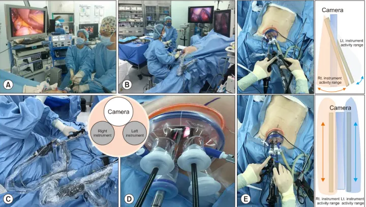
When performing SILC, the operator and camera oper- ator (scopist) share the operative field, which limits the operator’s activity (Fig. 1A). This exacerbates the clashing
Fig. 1. Solo-SILC technical procedure. (A) Single incision laparoscopic cholecystectomy (SILC), composed of operator and cam- era operator (scopist). (B) Solo single incision laparoscopic cholecystectomy (Solo-SILC), surgeon alone manipulates all instru- ments including the camera. (C) Laparoscopic scope holder (LaparostatTM, CIVCO medical Solutions, IA, USA) is located in the level of lowest margin of left rib. (D) Glove port (Nelis, Bucheon-si, Gyeonggi-do, Korea) and instrument location; the body of the camera is kept low and the head is kept high, and each instrument is placed on both side of the camera. (E) Instrument movement; sideways vs. push & pull method.
of instruments, which is the major limitation of SILC procedures.15 These issues result in a steep learning curve for SILC. Our center started performing solo SILC (Solo- SILC) in 2014. Our variation uses the parallel method rather than the crossing method. This widens the activity range of the operator and solves difficulties associated with the crossing method. A solo surgery is defined as a proce- dure in which the surgeon alone manipulates all instru- ments, including the camera, to avoid communication problems and unnecessary camera movements (Fig. 1B).16 Recently, our center reported that Solo-SILC is a feasible technique in a small group of patients.17
With the above in mind, this report describes the tech- nique, short-term outcomes, and learning curve of Solo- SILC with the aim of helping other surgeons overcome the initial learning period smoothly and safely.
MATERIALS AND METHODS
A total of 664 patients underwent Solo-SILC at Seoul
National University Bundang Hospital from July 2014 to December 2016. Of these, we extracted the data of 591 patients who underwent Solo-SILC performed by four hepatobiliary surgeons with ≥2 years of clinical experi- ence in hepatobiliary surgery (surgeons A-D). The pro- spectively recorded data of the patients were collected and retrospectively reviewed. Surgeons A, B, and C had in- dependently performed >50, 100, and 80 consecutive con- ventional LCs. They started to perform Solo-SILC imme- diately despite having no experience of SILC. They train- ed in Solo-SILC and 3-port LC simultaneously. Surgeon D is a highly experienced surgeon who had performed >500 conventional LCs. Moreover, before starting to perform Solo-SILC, surgeon D had experienced 10 cases of SILC before changing the surgical procedure because of diffi- culties with the solo and parallel method.
Solo-SILC is performed for all patients with benign GB diseases at our institution. If the patients had an American Society of Anesthesiologist score (ASA) >3 or were un- able to tolerate general anesthesia due to other reasons,
verity of disease, we categorized the presence of preop- erative percutaneous transhepatic biliary drainage (PTGBD) and emergency surgery. We compared the presence of preoperative fever (℃), white blood cell (WBC) count (103/L), and C-reactive protein (CRP) (mg/dl) levels. The parameters used to assess surgical outcomes included op- erative time (min), estimated blood loss (EBL) (ml), use of additional trocars, the presence of GB stones and their maximal size (mm), insertion of a Jackson-Pratt drain, GB perforation during surgery, length of hospital stay (days), complications, and histologic diagnosis of resected GB.
All patients who underwent Solo-SILC received the same clinical pathway for postoperative management. If there were no problems, patients were permitted sips of water 6 hours after surgery. Laboratory examinations were per- formed on postoperative day 1. If there was no issue with diet or postoperative problems, the patients were dis- charged on postoperative day 1. To clarify the learning curve, we used the simple moving average method with operative time as the measure of improvements in technique. We reviewed the studies through MEDLINE to compare outcomes and learning curves between Solo-SILC and SILC.
Surgical technique Position of the patient
The patient is placed in the supine position with the legs straightened and apart. The operator is positioned be- tween the patient’s legs during the main procedures. The laparoscopic scope holder (LaparostatTM, CIVCO Medical Solutions, IA, USA) is positioned around the level of the lowest margin of the left rib. The monitor is located above the patient’s head. The diathermy pedal is placed near the surgeon’s right or left foot (Fig. 1B, C).
Single port placement and patient position
A longitudinal 2-2.5-cm trans-umbilical incision is made (symptomatic GB stones or chronic cholecystitis suspected: 2 cm; acute cholecystitis suspected: 2.5 cm).
After an incision is made, 0.5-% bupivacaine hydrochloride is injected into the wound to relieve postoperative wound pain. After it has been confirmed that there are no adhe- sions around the umbilicus in the peritoneal cavity, a Glove port (Nelis, Bucheon-si, Gyeonggi-do, Korea) is in- serted through the incision site. Pneumoperitoneum is in-
placed in the reverse-Trendelenburg position with a left- sided tilt.
Manipulating instruments using the parallel method As the camera (10-mm laparoscopic flexible scope [Olym- pus CV-190, Shinjuku-ku, Japan]) is placed at the upper area of the single incision site, two conventional laparo- scopic instruments are inserted through the lower side holes (Fig. 1D). The parallel method is used instead of the crossing method. This establishes a triangular approach between the target organ and the two instruments and al- lows the instruments to perform push/pull movements while minimizing sideways movement. This push/pull movement reduces the overlapping activity range between the two instruments (Fig. 1E). The left instrument grabs the fundus of the GB and pushes it to exposure the hilar area. In the case of adhesions around the liver, a large quadrate lobe hiding the hilar area, or difficulty when ex- posing the hilar area, an Endo-grab or Organ retractor (AESCULAP Inc. U.S.A) can be used to retract the GB.
The operator alters the surgical view during the operation by manipulating the laparoscopic camera holder.
Specimen extraction and closure
After isolating and dividing the cystic duct and cystic artery, the GB is dissected from the liver and extracted through the glove port. If GB perforation occurs during the procedure, we use a laparoscopic vinyl bag to reduce the risk of abdominal cavity infection and bile leakage during GB extraction. The pneumoperitoneum is reduced to 5-6 mmHg to identify the minor bleeding point before the retrieval of instruments.
The umbilical port site is then repaired using inter- rupted sutures in the fascia layer with only 1-2 intra- dermal sutures at both ends of the skin incision site for skin closure and drainage for fluid collection in the um- bilicus.
Statistical analysis
All statistical calculations were performed using the SPSS software package for Windows, version 21 (IBM Corporation, Armonk, NY, USA). The demographic and clinicopathological characteristics were summarized using descriptive analysis, and all qualitative values are pre- sented as means and standard deviations unless expressed otherwise. The statistical analysis was performed with
Table 1. Patients characteristics and pre-operative findings
A (37) B (51) C (33) D (470) p
Age 55±17.02 52.8±15.34 57.6±16.09 53.2±15.52 0.424
Sex (M/F) 18/19 29/22 17/16 207/263 0.74
BMI (kg/m2) 24.77±3.29 25.84±4.14 24.93±3.41 24.72±4.04 0.35
History of abdominal surgery (N/%) 14 (37.83%) 13 (25.49%) 8 (24.24%) 139 (29.6%) 0.361
ASA (N/%) 0.001
1 27 (73.0%) 44 (86.3%) 21 (63.6%) 234 (49.8%)
2 10 (27.0%) 7 (13.7%) 11 (33.3%) 206 (43.8%)
3 0 0 1 (3.10%) 30 (6.4%)
DM (N/%) 0.473
Yes 7 (18.9%) 9 (17.65%) 3 (9.1%) 65 (13.8%)
No 30 (81.1%) 42 (82.35%) 30 (90.9%) 405 (89.6%)
Preoperative biliary drainage
PTGBD or PTBD (N/%) 0.312
Yes 1 (2.7%) 6 (11.76%) 3 (9.1%) 49 (10.4%)
No 36 (97.3%) 45 (88.24%) 30 (90.9%) 421 (86.2%)
Emergency cholecystectomy (N/%) 0.604
Yes 13 (35.1%) 13 (25.49%) 9 (27.3%) 171 (36.6%)
No 24 (64.9%) 38 (74.51%) 24 (72.7%) 298 (63.4%)
Systemic signs of inflammation
Preoperative Fever (N/%) 0.26
Yes 12 (32.4%) 9 (17.6%) 7 (21.2%) 115 (24.5%)
No 25 (67.6%) 42 (82.4%) 26 (78.8%) 355 (75.5%)
Preoperative WBC (103/l) 6614.94±2941.19 8776.23±9078.43 8715.75±4207.18 8034.46±4108.81 0.254
Preoperative CRP (mg/dl) 1.31±4.01 2.66±6.12 3.16±6.05 3.28±6.46 0.34
BMI, body mass index; ASA, American Society of Anesthesiologist score; DM, diabetes mellitus; PTGBD, percutaneous gall- bladder drainage; PTBD, percutaneous biliary drainage
one-way analysis of variance and the Tukey post-hoc hon- est significant difference test. A p-value <0.05 was con- sidered statistically significantly.
RESULTS
Patient characteristics and pre-operative findings During the study period, 121 patients underwent Solo- SILC performed by surgeon A (n=37), B (n=51), and C (n=33). Surgeon D performed 470 cases of Solo-SILC during the same period. The patients’ preoperative base- line demographics and pre-operative findings are shown in Table 1. The mean age was 55.0±17.02, 52.8±15.34, 57.6±16.09, and 53.2±15.52 years for patients treated by surgeon A, B, C, and D (p=0.424). The mean body mass index (kg/m2) was 24.77±3.29, 25.84±4.14, 24.93±3.41, and 24.72±4.04 for patients treated by surgeon A, B, C, and D (p=0.35). The proportion of each gender did not differ significantly among the groups (p=0.74). No sig- nificant differences were found in the history of abdomi-
nal surgery (p=0.361). Although, ASA score was signi- ficantly higher in patients treated by surgeon C and D (p=
0.001), the incidence of diabetes mellitus was not different among the groups (surgeons A:B:C:D=18.9%:17.65%:9.1%:
13.8%, p=0.473). The proportion of patients who under- went insertion of a PTGBD or ERBD before the operation was not different among the groups (p=0.312). Thirteen (35.1%), 13 (25.5%), nine (27.3%), and 171 (36.6%) patients treated by surgeon A, B, C, and D were emergency chol- ecystectomy cases (p=0.604). Local and systemic signs of inflammation, including the presence of fever, preopera- tive WBC counts, and preoperative CRP level, were not significantly different (pre-operative fever, p=0.26; pre- operative WBC counts, p=0.254; pre-operative CRP level, p=0.34).
Surgical outcomes and histologic diagnosis of the resected GB
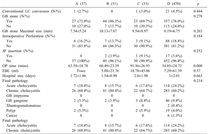
The surgical outcomes and histologic diagnoses of the resected GB are shown in Table 2. The mean operative time
A (37) B (51) C (33) D (470) p
Conventional LC conversion (N/%) 1 (2.7%) 0 1 (3.0%) 21 (4.5%) 0.444
GB stone (N/%) 0.278
Yes 27 (73.0%) 44 (86.3%) 23 (69.7%) 357 (76.0%)
No 10 (27.0%) 7 (13.7%) 10 (30.3%) 113 (24.0%)
GB stone Maximal size (mm) 7.54±5.24 10.13±7.67 9.54±8.97 6.19±6.75 0.263
Intraoperative Perforation (N/%) 0.184
Yes 6 (16.2%) 7 (13.7%) 3 (9.1%) 88 (18.8%)
No 31 (83.8%) 44 (86.3%) 30 (90.9%) 381 (81.2%)
JP insertion (N/%) 0.252
Yes 0 2 (3.9%) 3 (9.1%) 17 (3.6%)
No 37 (100%) 49 (96.1%) 30 (90.9%) 452 (96.4%)
OP time (mins) 63.59±18.70 60.49±23.39 91.36±26.93 54.85±24.72 0.000
EBL (ml) Trace 5.88±23.76 18.78±45.86 7.29±61.59 0.57
Hospital stay (days) 1.72±1.86 1.54±0.90 2.0±1.98 1±2.01 0.663
Final pathology 0.214
Acute cholecystitis 7 (18.4%) 8 (15.7%) 6 (17.6%) 114 (24.2%)
Chronic cholecystitis 26 (68.4%) 41 (80.4%) 22 (64.7%) 283 (60.2%)
GB empyema 0 0 0 0
GB gangrene 2 (5.3%) 2 (3.9%) 3 (8.8%) 46 (9.8%)
Xhantogranulomatous 0 0 0 2 (0.4%)
Polyp 2 (5.3%) 0 2 (5.9%) 19 (4.0%)
Cancer 0 0 0 6 (1.3%)
Final pathology
Acute cholecystitis 7 (18.4%) 8 (15.7%) 6 (17.6%) 114 (24.2%)
Chronic cholecystitis 26 (68.4%) 41 (80.4%) 22 (64.7%) 283 (60.2%) LC, laparoscopic cholecystectomy; GB, gallbladder; JP, Jackson-pratt; OP, operation; EBL, estimated blood loss
(min) was 63.59±18.71, 60.49±23.39, 91.36±26.93, and 54.85±24.72 in patients treated by surgeon A, B, C, and D (p=0.000), respectively. The mean EBL (ml) was <5, 5.88±23.76, 18.78±45.86, and 7.29±61.59 in patients treat- ed by surgeon A, B, C, and D (p=0.57). Additional trocar insertion was required in one (2.7%), zero, one (3.0%), and 21 (4.5%) patients treated by surgeon A, B, C, and D (p=0.444). Conversion to open surgery occurred in one case due to bile duct injury in the patients treated by sur- geon C. A drainage tube was placed in zero, two (3.9%), three (9.1%), and 17 (3.6%) patients (p=0.252). There was no difference in the presence of GB stones or their max- imal size (mm) (GB stones, p=0.263; GB stone maximal size (mm), p=0.184). As mentioned above, all patients who underwent Solo-SILC received the same clinical pathway. The mean hospital stay was 1.72±1.86, 1.54±
0.90, 2.0±1.98, and 1±2.01 days in patients treated by sur- geon A, B, C, and D (p=0.663).
Complications
During the mean follow-up period (24±7.2 months), the incidence of postoperative complications was not signi- ficantly different among the different surgeon groups (p=0.27) (Table 3). Wound infection (total incidence, 10 of 591 [1.69%]) and incisional hernia (total incidence, 5 of 591 [0.85%]), which resolved without special treat- ment, occurred in all study groups. Three postoperative complications that delayed bile leakage occurred in pa- tients treated by surgeon D (0.63%). These complications were resolved by ultrasound-guided puncture and drain- age. One patient underwent endoscopic retrograde chol- angiography due to abnormalities on liver function tests after surgery and was diagnosed with common bile duct stricture.
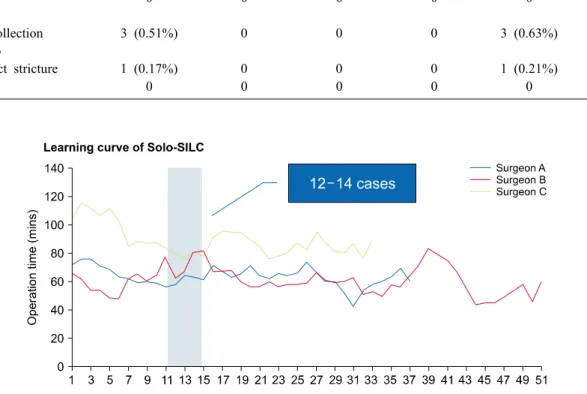
Learning curve of Solo-SILC
Fig. 2 shows the learning curve of each surgeon. The operative time stabilized for surgeon A after 14 cases (learning period: 64.85±6.72 min; experienced period:
Table 3. Post-operative complication
Total (591) A (37) B (51) C (33) D (470) p
0.27 Grade I
wound infection 10 (1.69%) 1 (2.7%) 1 (1.96%) 2 (6.06%) 6 (1.27%) Incisional hernia 5 (0.85%) 1 (2.7%) 1 (1.96%) 1 (3.03%) 2 (0.42%) Grade II
Ileus 0 0 0 0 0
Grade IIIa
Fluid collection 3 (0.51%) 0 0 0 3 (0.63%)
Grade IIIb
Bile duct stricture 1 (0.17%) 0 0 0 1 (0.21%)
Grade IV 0 0 0 0 0
Fig. 2. Learning curve of each surgeon. Learning curve for each surgeon A after 14 cases (learning period: 64.85±6.72 min;
experienced period: 62.73±19.31 min; p=0.04). For the surgeon B, the operative time stabilized after 12 cases (learning period:
62.51±20.86 min; experienced period: 60.89±23.33 min; p=0.04) while surgeon C stabilized after 12 cases of Solo-SILC (learning period: 102.75±29.50 mins; experienced period: 84.38±21.51 mins; p=0.013). Surgeon D did not show a learning curve.
62.73±19.31 min; p=0.04), for surgeon B after 12 cases (learning period: 62.51±20.86 min; experienced period:
60.89±23.33 min; p=0.04), and for surgeon C after 12 cas- es (learning period: 102.75±29.50 mins; experienced peri- od: 84.38±21.51 mins; p=0.013).
DISCUSSION
This study demonstrated that the learning curve of Solo-SILC was approximately 12-14 cases for hepato- biliary surgeons who are experienced in conventional LC.
In addition, the surgeon who was fluent in both LC and SILC had no learning curve when starting to perform Solo-SILC. In the patients treated by surgeons A, B, C, and D, there was one case of bile duct injury (0.17% [1
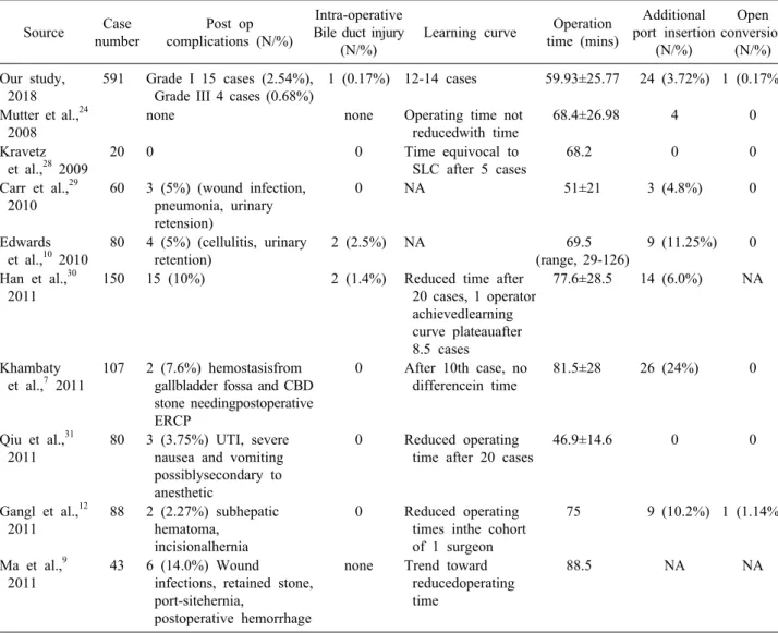
of 591]). This was lower than the overall rate of bile duct injury for SILC (0.39% [9 of 2236]) across all previous studies.3,19,20 Moreover, the mean operative time in this study group (59.93±25.77 min) was shorter than that re- ported in other studies of SILC (median operative time across studies=80.75 [range 40-186] min).21
Hodgett et al. demonstrated a consistent operative time for 29 patients undergoing SILC.22 Pan et al.23 concluded that surgeons who are experienced in conventional LC are likely to pass the learning curve after performing 20 cases of SILC. Mutter et al.24 did not show any significant re- duction in operative time with increasing experience.
Compared to the above results, the present study showed a relatively short learning curve for Solo-SILC, and the mean operative time was shorter than that in other studies.
Table 4. Studies investigating outcomes of SILC Source Case
number
Post op complications (N/%)
Intra-operative Bile duct injury
(N/%)
Learning curve Operation time (mins)
Additional port insertion
(N/%)
Open conversion
(N/%) Our study,
2018
591 Grade I 15 cases (2.54%), Grade III 4 cases (0.68%)
1 (0.17%) 12-14 cases 59.93±25.77 24 (3.72%) 1 (0.17%)
Mutter et al.,24 2008
none none Operating time not
reducedwith time
68.4±26.98 4 0
Kravetz et al.,28 2009
20 0 0 Time equivocal to
SLC after 5 cases
68.2 0 0
Carr et al.,29 2010
60 3 (5%) (wound infection, pneumonia, urinary retension)
0 NA 51±21 3 (4.8%) 0
Edwards et al.,10 2010
80 4 (5%) (cellulitis, urinary retention)
2 (2.5%) NA 69.5
(range, 29-126)
9 (11.25%) 0
Han et al.,30 2011
150 15 (10%) 2 (1.4%) Reduced time after 20 cases, 1 operator achievedlearning curve plateauafter 8.5 cases
77.6±28.5 14 (6.0%) NA
Khambaty et al.,7 2011
107 2 (7.6%) hemostasisfrom gallbladder fossa and CBD stone needingpostoperative ERCP
0 After 10th case, no differencein time
81.5±28 26 (24%) 0
Qiu et al.,31 2011
80 3 (3.75%) UTI, severe nausea and vomiting possiblysecondary to anesthetic
0 Reduced operating time after 20 cases
46.9±14.6 0 0
Gangl et al.,12 2011
88 2 (2.27%) subhepatic hematoma,
incisionalhernia
0 Reduced operating times inthe cohort of 1 surgeon
75 9 (10.2%) 1 (1.14%)
Ma et al.,9 2011
43 6 (14.0%) Wound infections, retained stone, port-sitehernia,
postoperative hemorrhage
none Trend toward reducedoperating time
88.5 NA NA
procedures, the surgical team, and the surgical devices used. When performing SILC, a skilled scopist is required to visualize the surgical field. However, the number of ex- perienced scopists is insufficient and they have a fixed working time, which limits the practice of SILC in Korea.
An unexperienced scopist who is not familiar with the surgical procedures might cause the surgical view to be unstable. However, in the case of Solo-SILC, the proce- dure is performed by one surgeon who understands the entire procedure. This eliminates miscommunication be- tween the operator and scopist. Therefore, the learning curve of SILC is influenced by the scopist, whereas Solo- SILC is influenced only by the skill of the operator. This study revealed that stabilization of surgical skill is ach- ieved more rapidly when Solo-SILC is performed.
Solo-SILC are standardized by the experienced surgeon D. This surgeon used trial and error while implementing SILC, and introduced and standardized the Solo-SILC program to train other hepatobiliary and pancreatic sur- geons. Recently, surgeon D has been performing solo sin- gle incision laparoscopic hepatectomy.25 Surgeons A, B, and C, who have been trained in Solo-SILC with a rela- tively standardized procedure, were able to overcome the learning curve relatively quickly despite having no experi- ence of SILC. The primary concern and source of skepti- cism for many surgeons considering the place of SILC is the frequent deviation from standards.13 However, this study shows that standardization of procedures can ensure stability. Several studies have described SILC using the crossing method to overcome the left-right reversal of in-
struments.14,15 In our center, we use the parallel method, which is similar to the conventional instrumental manipu- lation method. The most important thing when starting a new procedure is patient safety. As shown in Table 3, the total complication rate was 3.21% in the present study.
There were only 24 (3.72%) cases of additional port in- sertion and one case of open conversion (0.17%). As Table 4 shows, when comparing these outcomes with those of SILC, the patient safety provided by our novel approach to Solo-SILC is acceptable.
There are several methods for assessing a surgeon's learning curve. The ideal approach is to use multivariate logistic regression or the cumulative sum method, both of which have been used recently.26,27 These methods usually use outcome-related variables, such as the rate of con- version to open procedures and the occurrence of severe complications. In this study, the rate of open conversion was 0.83%, with a rate of Clavien-Dindo classification
>IIIa complications of 0.83%. Considering these rates, we deemed that it was appropriate to use a moving aver- age method to assess the learning curve. Among the varia- bles for evaluating skill when performing SILC, operative time is the most direct and accurate indicator because it reflects a surgeon’s mastery of the technique.
This study has several limitations that should be noted.
First, this was a retrospective study, which means that the possibility of selection bias cannot be excluded. Second, the follow-up period was short. A longer follow-up will be required to confirm our findings and investigate other issues, such as the rate of incisional hernia. Third, the re- sults of resected GB histology included acute cholecys- titis, GB empyema, GB gangrene, and xanthogranuloma- tous inflammation. In the early period, we performed Solo-SILC for suspected symptomatic GB stones or chron- ic cholecystitis. With the accumulation of cases, the in- dications for Solo-SILC were expanded to include acute cholecystitis.
In conclusion, this study reports that hepatobiliary sur- geons experienced in LC can overcome the learning curve of Solo SILC within 12-14 cases. Moreover, a surgeon with more experience in conventional LC and SILC expe- rienced no learning curve. Solo-SILC is a technique that can overcome the shortcomings of SILC after the learning curve and provide a more stable surgical option. Solo- SILC is a promising treatment for GB disease.
ACKNOWLEDGEMENTS
This research was supported by Seoul National University Bundang Hospital Research Grant (No.
14-2016-025).
REFERENCES
1. Litynski GS. Highlights in the history of laparoscopy: the devel- opment of laparoscopic techniques--a cumulative effort of in- ternists, gynecologists, and surgeons. Frankfurt: Barbara Bernert Verlag, 1996:165-168.
2. Navarra G, Pozza E, Occhionorelli S, Carcoforo P, Donini I.
One-wound laparoscopic cholecystectomy. Br J Surg 1997;84:695.
3. Aprea G, Coppola Bottazzi E, Guida F, Masone S, Persico G.
Laparoendoscopic single site (LESS) versus classic video-laparo- scopic cholecystectomy: a randomized prospective study. J Surg Res 2011;166:e109-e112.
4. Lai EC, Yang GP, Tang CN, Yih PC, Chan OC, Li MK. Pro- spective randomized comparative study of single incision laparo- scopic cholecystectomy versus conventional four-port laparo- scopic cholecystectomy. Am J Surg 2011;202:254-258.
5. Lee SK, You YK, Park JH, Kim HJ, Lee KK, Kim DG. Single- port transumbilical laparoscopic cholecystectomy: a preliminary study in 37 patients with gallbladder disease. J Laparoendosc Adv Surg Tech A 2009;19:495-499.
6. Asakuma M, Hayashi M, Komeda K, Shimizu T, Hirokawa F, Miyamoto Y, et al. Impact of single-port cholecystectomy on postoperative pain. Br J Surg 2011;98:991-995.
7. Khambaty F, Brody F, Vaziri K, Edwards C. Laparoscopic ver- sus single-incision cholecystectomy. World J Surg 2011;35:967- 972.
8. Tsimoyiannis EC, Tsimogiannis KE, Pappas-Gogos G, Farantos C, Benetatos N, Mavridou P, et al. Different pain scores in single transumbilical incision laparoscopic cholecystectomy versus clas- sic laparoscopic cholecystectomy: a randomized controlled trial.
Surg Endosc 2010;24:1842-1848.
9. Ma J, Cassera MA, Spaun GO, Hammill CW, Hansen PD, Aliabadi-Wahle S. Randomized controlled trial comparing sin- gle-port laparoscopic cholecystectomy and four-port laparoscopic cholecystectomy. Ann Surg 2011;254:22-27.
10. Edwards C, Bradshaw A, Ahearne P, Dematos P, Humble T, Johnson R, et al. Single-incision laparoscopic cholecystectomy is feasible: initial experience with 80 cases. Surg Endosc 2010;
24:2241-2247.
11. Fronza JS, Linn JG, Nagle AP, Soper NJ. A single institution's experience with single incision cholecystectomy compared to standard laparoscopic cholecystectomy. Surgery 2010;148:731-734;
discussion 734-736.
12. Gangl O, Hofer W, Tomaselli F, Sautner T, Függer R. Single incision laparoscopic cholecystectomy (SILC) versus laparoscopic cholecystectomy (LC)-a matched pair analysis. Langenbecks Arch Surg 2011;396:819-824.
13. Greaves N, Nicholson J. Single incision laparoscopic surgery in general surgery: a review. Ann R Coll Surg Engl 2011;93:437- 440.
14. Ishikawa N, Arano Y, Shimizu S, Morishita M, Kawaguchi M, Matsunoki A, et al. Single incision laparoscopic surgery (SILS) using cross hand technique. Minim Invasive Ther Allied Technol 2009;18:322-324.
15. Jaspers JE, Breedveld P, Herder JL, Grimbergen CA. Camera
vasive surgery. Surg Laparosc Endosc Percutan Tech 2004;14:
145-152.
16. Singh M, Mehta KS, Yasir M, Kaur A, Aiman A, Sharma A, et al. Single-incision laparoscopic cholecystectomy using con- ventional laparoscopic instruments and comparison with three- port cholecystectomy. Indian J Surg 2015;77(Suppl 2):546-550.
17. Choi Y, Han HS, Yoon YS, Cho JY, Jang JY, Choi HL, et al.
Single incision laparoscopic cholecystectomy without a camera operator. J Minim Invasive Surg 2017;20:63-68.
18. Daabiss M. American Society of Anaesthesiologists physical sta- tus classification. Indian J Anaesth 2011;55:111-115.
19. Philipp SR, Miedema BW, Thaler K. Single-incision laparo- scopic cholecystectomy using conventional instruments: early ex- perience in comparison with the gold standard. J Am Coll Surg 2009;209:632-637.
20. Podolsky ER, Rottman SJ, Curcillo PG 2nd. Single port access (SPA) cholecystectomy: two year follow-up. JSLS 2009;13:528- 535.
21. Hall TC, Dennison AR, Bilku DK, Metcalfe MS, Garcea G.
Single-incision laparoscopic cholecystectomy: a systematic re- view. Arch Surg 2012;147:657-666.
22. Hodgett SE, Hernandez JM, Morton CA, Ross SB, Albrink M, Rosemurgy AS. Laparoendoscopic single site (LESS) cholecys- tectomy. J Gastrointest Surg 2009;13:188-192.
23. Pan MX, Liang ZW, Cheng Y, Jiang ZS, Xu XP, Wang KH, et al. Learning curve of transumbilical suture-suspension sin- gle-incision laparoscopic cholecystectomy. World J Gastroenterol
24. Mutter D, Leroy J, Cahill R, Marescaux J. A simple technical option for single-port cholecystectomy. Surg Innv 2008;15:332- 333.
25. Choi YR, Han HS, Yoon YS, Cho JY, Jang JS, Kwon SU, et al. Solo single incision laparoscopic left hemihepatectomy using a laparoscopic scope holder. Videoscopy 2018;28. doi: 10.1089/
vor.2017.0473.
26. Jin SH, Kim DY, Kim H, Jeong IH, Kim MW, Cho YK, et al.
Multidimensional learning curve in laparoscopy-assisted gas- trectomy for early gastric cancer. Surg Endosc 2007;21:28-33.
27. Ramsay CR, Grant AM, Wallace SA, Garthwaite PH, Monk AF, Russell IT. Statistical assessment of the learning curves of health technologies. Health Technol Assess 2001;5:1-79.
28. Kravetz AJ, Iddings D, Basson MD, Kia MA. The learning curve with single-port cholecystectomy. JSLS 2009;13:332-336.
29. Carr A, Bhavaraju A, Goza J, Wilson R. Initial experience with single-incision laparoscopic cholecystectomy. Am Surg 2010;76:
703-707.
30. Han HJ, Choi SB, Park MS, Lee JS, Kim WB, Song TJ, et al.
Learning curve of single port laparoscopic cholecystectomy de- termined using the non-linear ordinary least squares method based on a non-linear regression model: an analysis of 150 con- secutive patients. J Hepatobiliary Pancreat Sci 2011;18:510-515.
31. Qiu Z, Sun J, Pu Y, Jiang T, Cao J, Wu W. Learning curve of transumbilical single incision laparoscopic cholecystectomy (SILS): a preliminary study of 80 selected patients with benign gallbladder diseases. World J Surg 2011;35:2092-2101.