J Korean Soc Surg Hand 2013;18(3):124-131.
http://dx.doi.org/10.12790/jkssh.2013.18.3.124
THE HAND
INTRODUCTION
The hand fracture is one of the most common traumas, and it accounts for up to 10% of all the human body fractures1,2. Its etiologic factors include sports activities, traffic accidents and industrial work activities. There is a variability in the management of hand fracture. This poses challenging prob- lems for surgeons. The treatment goal is to obtain good out- comes, for which surgeons should consider 1) restoration of
the normal alignment, 2) achievement of the appropriate union, 3) recovery of the early range of movement and earlier return to full activities, and 4) absence of residual disabilities or deformities
It is difficult to maintain reduction without causing unde- sirable side effects. The complications of hand fracture include infection, non-union, malunion, tendon adhesion and joint stiffness3. Of these, the most serious potential prob- lem is an inability to attain a full range of movement. There
Percutaneous Multiple Kirschner Wire Fixation in the Treatment of Hand Fractures
Seong Jae Hong1, Hyeung Gyo Seo2,
Jong Ick Whang2, Sanghun Cho1
Department of 1Plastic and Reconstructive Surgery, Dongguk University Ilsan Hospital, Goyang; 2Department of Plastic and Reconstructive Surgery, Duson Hospital, Ansan, Korea
Received:June 5, 2013 Revised:September 3, 2013 Accepted:September 7, 2013 Correspondence to:Hyeung Gyo Seo Department of Plastic and Reconstructive Surgery, Duson Hospital, 114
Seonbugwangjang 1-ro,
Danwon-gu, Ansan 425-140, Korea TEL:+82-31-402-0114-201 FAX:+82-31-402-1805
E-mail:[email protected]
Purpose:We reported results of percutaneous multiple K-wire fixation tech- nique without passing through the joint in patients with a hand fracture.
Methods:We evaluated a total of 116 cases in 94 patients who underwent per- cutaneous multiple K-wire fixation on dorsal cortex over a 10-year period between 2001 and 2010. The treatment outcomes were evaluated based on total active motion (TAM), as proposed by Widegrow.
Results:Our clinical series of patients achieved good functional outcomes. Of total patients, 89% (84/94) had excellent TAM, 2% (2/94) did good TAM and 9%
(8/94) did poor TAM. Postoperatively, our clinical series of patients had such a good compliance as to achieve a TAM of >181 when performing the early active movement. There were no notable postoperative complications during the fol- low-up period.
Conclusion:Our results indicate that percutaneous multiple K-wire fixation technique without passing through the joint from normal bone density patients is effective in providing the rigid fixation. Thus, our patients could perform the early movement as promptly as possible and maintaining the full mobility of the rest of the hand.
Keywords:Hand, Fractures, bone, Fracture fixation, Bone wires, Motion
This is an Open Access article distributed under the terms of the Creative Commons Attribution Non-Commercial License (http://creativecommons.org/ licenses/by- nc/3.0/) which permits unrestricted noncommercial use, distribution, and reproduction in any medium, provided the original work is properly cited.
are multiple factors which limit the range of motion, and these include structural problems such as concurrent injuries to the joint or tendon, i.e., the overlying structures that per- form a gliding function, and soft tissue injuries including neurovascular injuries. Moreover, the limited range of motion is correlated with infection, swelling, pain and non- rigid, unstable fixation4. All of these factors cause tendon adhesion or joint stiffness by maintaining the immobilized hand.
For the effective treatment of hand fracture, the early active motion as well as the rigid fixation should be achieved. In addition, the postoperative complications should be prevent- ed and the normal functions of the hand based on the maxi- mum range of motion should be restored5.
We performed the percutaneous multiple K-wire tech- nique on dorsal cortex without passing through the joint in patients with hand fracture and obtained good treatment outcomes. Here, we present our surgical methods and their outcomes based on our 10-year single institution experience.
MATERIALS AND METHODS
1. Patients
A total of 94 patients (116 cases) underwent percutaneous multiple K-wire fixation over a 10-year period between 2001 and 2010. We performed a retrospective analysis of such vari- ables as location, direction of the fracture line, amount of dis- placement, degree of malalignment, angulation, subluxation, dislocation, rotational deformity (malrotation), presence or absence of comminution and articular involvement.
Inclusion/exclusion criteria for the current study are as fol- lows: 1) Inclusion criteria, patients with phalangeal or metacarpal fractures of the hand with normal bone density.
2) Exclusion criteria, patients with low bone density; patients with concurrent carpal fractures; patients with fractures with large bone defects; patients with fractures associated with replantation; patients with severely multilated injuries requiring primary amputation; patients who required skin grafting.
We finally enrolled a total of 94 patients (116 cases) in the current study. The current study was approved by the Institutional Review Board of our medical institution.
Because of its retrospective nature, the requirement for obtaining a written informed consent was waived.
2. Surgical techniques
The basic principle of the surgical treatment for hand frac- tures is to realign the mobile segment to the less mobile one.
Under local anesthesia, axillary block anesthesia in some cases, with or without standard C-arm fluoroscopic guidance, the fractures were properly treated with closed or open reduction when there was a concurrent presence of injury.
Reduction of the fracture was done by pulling or traction of the finger with an even distribution of the sufficient pressure.
After the fractures were well aligned and good reduction was obtained by closed or open manipulation, multiple K-wires were placed using a power driver percutaneously starting from the dorsal cortex to the other side dorsal cortex above the mid-lateral line for maximizing rigidity. In metacarpal fractures, multiple K-wires were percutaneously inserted in a transverse direction and then horizontally placed in the sta- ble unit of the hand. The non-fractured adjacent bone was served as the stable unit that is expected to plays a role as the cornerstone of the bridge (Fig. 1). In comminuted fractures, we passed multiple K-wires through the largest segment into the smaller one and then fixed to each other, thus attempting to increase the rigidity, and thereby stabilized the fracture without passing K-wires through the joint (Figs. 2, 3). Then, we encouraged the patients to perform the early movement.
We applied the sufficient compression by manual forces or encouraged the patients to move the hand for daily activities.
Thus, we attempted to evaluate whether the range of motion and the stability of fixation are so sufficient as to maintain the reduction (Figs. 3, 4). Unless the fixation had been sufficient or bone fractures had been immobilized, we would have per- formed the longitudinal percutaneous K-wire fixation. But there were no such cases. The pins were withdrawn within a mean period of four weeks postoperatively (range, 3-6 weeks).
3. Postoperative management
After the internal fixation, we initiated the early mobilization without delay while maintaining the adequate alignment of the fragments with rigid fixation. When there were no con-
current injuries in such body areas as the tendon, collateral ligament and neurovascular structures, the patients were allowed to perform early mobilization within 7 to 14 days postoperatively. We encouraged the patients to perform active motion. In addition, we also added the passive range of
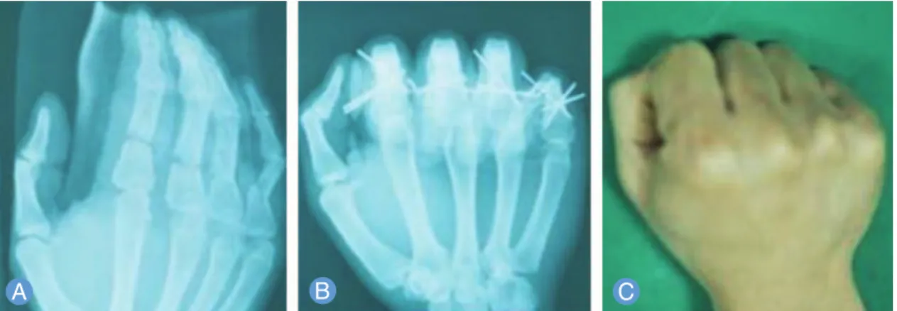
motion when there was a limitation in the movement because of swelling or pain. Postoperatively, there were no patients who needed splint protection because there was a sufficient stability due to a rigid fixation with multiple K-wires although the patients with concurrent tendon injuries were Fig. 1.Spiral metacarpal fracture with rotational malalignment.(A)Spiral fractures with rotational deformity of the index
metacarpal shaft and long metacarpal base.(B)Closed reduction and internal fixation of each spiral fracture with percu- taneous multiple K-wires stabilized the fracture yet allow movement. The third and fourth metacarpal function as a foun- dation stone. No wire was transfixed through the metacarpophalangeal joint surface.(C) On postoperative year 1, no rotational malalignment was observed and patient restored alignment with a full range of motion.
Fig. 2.Rigid fixation of comminuted diaphyseal fracture.(A)Diaphyseal fracture of the index proximal phalanx with com- minution.(B)Immediately after surgery, with the fixation by percutaneous multiple K-wires without passing through the joint, the rigid fixation was maintained during the active flexion.
in need of dynamic splinting. In patients who had injuries to neurovascular structures, however, the early mobilization was permitted after 14 days postoperatively.
4. Evaluation of outcomes
The treatment outcomes are based on such variables as the range of joint motion, the degree of deformity, grasping func- tion, pain and sensory recovery. In an actual clinical setting, however, it is difficult to objectively evaluate all the above variables. Moreover, there was a variability in the period of
long-term follow-up. We therefore evaluated the total active movement (TAM), as proposed by Widgerow (Table 1). The TAM refers to an additive sum of three movements: the flex- ion at the metacarpophalangeal, the proximal interpha- langeal, and the distal interphalangeal joints minus the extension deficit at the same joints6. Thus, we classified the treatment outcomes based on the TAM into three grades:
‘excellent (TAM >250。)’, ‘good (181。<TAM<249。)’, and
‘poor (TAM <180。)’.
Fig. 3.Early mobilization after percutaneous multiple K-wire fixation.(A) Proximal phalangeal shaft, base fracture with comminution of the index finger.(B)Closed reduction and percutaneous multiple K-wires.(C)With the rigid fixation with- out passing through the joint, the early active movement was promoted. The degree of motion was satisfactory on post- operative day 3.
Fig. 4.Multiple transverse fractures with concurrent injures.(A)Multiple transverse fractures of the shaft with the tendon injures in the proximal phalanges accompanied by the dorsal, concave angulation.(B)Immediately after surgery, the rigid fixation and the early active motion were achieved using the percutaneous multiple K-wire technique.(C)On postopera- tive year 1, the full range of motion was restored and there were no residual disabilities or deformities.
RESULTS
1. Characteristics of the patients
The mean age of the patients was 53-year-old (range, 23-82 year-old), and they consisted of 54 men and 40 women. The most common fracture was metacarpal fracture, accounting for 48% (56/116) of total cases (Table 2). Our clinical series of patients include six cases of intra-articular fracture, 18 cases of spiral fracture and 12 cases of comminuted fracture.
2. Clinical outcomes
As shown in Table 1, our clinical series of patients achieved good functional outcomes. Of total patients, 89% (84/94) had excellent TAM, 2% (2/94) did good TAM and 9% (8/94) did poor TAM. Postoperatively, our clinical series of patients had such a good compliance as to achieve a TAM of >181。when performing the early active movement. All of our clinical series of patients were followed up postoperatively, and underwent uneventful course and achieved a recovery of hand motion. Furthermore, there were no patients who had malunion or nonunion of the fracture as well as residual deformities (Figs. 1, 4).
DISCUSSION
The hand is one the most essential organs of the human
body, and it is characterized by the sophisticated functions and complicated architecture. In addition, it is vulnerable to trauma with the industrial development and the increased use of transportation. The hand fracture accounts for 30% of all industrial fractures, and it is the most frequently seen7.
The management of hand fracture is a Cinderella subject; it is subject to the extent and mechanism of injury, patient’s age, concurrent injuries and surgeons’technical expertise.
To put this in another way, there is no established treatment modality for hand fracture. The optimal treatment modality should be chosen on a case-by-case basis. According to Swanson, patients with hand fracture may be complicated; it may lead to the deformity when left untreated, stiffness when overtreated and both deformity and stiffness when incorrect- ly treated8. This suggests that it would be mandatory to select the optimal treatment modality for each case although many cases of hand fracture can be treated by conservative man- agement at the present9. In an actual clinical setting, there are some patients who achieve a recovery even when almost or completely left untreated. If overtreated or incorrectly treat- ed, however, patients will present with deformity or other severe complications than the deformity. According to Curry10, the basic principle of the surgical treatment for hand fractures is to realign the mobile distal segment to the less mobile proximal one.
Active measures can be taken if a satisfactory reduction cannot be achieved or if there are concurrent injuries in such areas the vessels, nerves, tendons or other soft tissues.
Surgical treatments of hand fracture can be classified into the internal fixation and the external one. In the internal fixation, the reduction is achieved by percutaneous K-wire pinning, interosseous wiring and the fixation with a plate and a screw.
Percutaneous K-wire pinning is a common surgical tech- nique that is both simple and cost-effective. In addition, it is advantageous in that it can be versatile used for skeletal stabi- Table 1.Functional outcomes based on the total active movement
Excellent >249 84 (89)
Good 181 -249 2 (2)
Poor <180 8 (9)
The total active motion (TAM) refers to an additive sum of three movements: the flexion at the metacarpophalangeal, the proximal interphalangeal and the distal interphalangeal joints minus the exten- sion deficit at the same joints.
Outcomes TAM No. of patients (%)
Table 2.Fracture site and accompanying injuries
Distal phalanx 2 0 0 2
Middle phalanx 4 4 2 10 (S: 2, C: 2)
Proximal phalanx 32 10 6 48 (I: 6, S: 2, C: 6)
Metacarpal 38 12 6 56 (S: 14, C: 4)
I, intra-articular fracture; S, spiral fracture; C, comminuted fracture.
Fracture site No concurrent injuries Tendon injuries Neurovascular structure injuries Total no. of cases
lization and it causes less complications11. Black et al.12ana- lyzed the rigidity of three methods, 1) the dorsal plating with or without lag screws, 2) the intraosseous wiring, and 3) the K-wire pinning, for the treatment of metacarpal fracture. This showed that the dorsal plate fixation and the wired tech- niques provided the most and the least rigid fixation, respec- tively. With the most stable fixation, patients can initiate the early active movement as promptly as possible. Despite the stable fixation, the plate system reveals several disadvantages due to a plate and a screw. That is, the plate is somewhat huge and then cannot be easily used for the fixation of hand phalanges. In addition, it needs an wide exposure for the extensive periosteal stripping. With the inappropriate loca- tion of the plate, the normal gliding of the tendon would be compromised13. Moreover, it would be mandatory to remove both a screw and a plate at the second operation. To date, however, no attempts have been made to stabilize the rigidity of K-wire fixation because it has the great advantages. By con- trast, the K-wire fixation minimally needs the periosteal ele- vation, but it is also known to provide the unstable fixation that limits the early active movement in patients with hand fracture12.
Rush and Rush14 attempted the use of immediate intramedullary wiring in the treatment of patients with metacarpal fracture. But these authors did not use splints but recommended that patients perform the early movement of the hand and finger.
To date, many authors have emphasized the importance of the early mobilization in patients with hand fracture. In asso- ciation with this, Dobyns et al.15reported that final treatment outcomes would be improved if at least a partial arc of motion is restored as promptly as possible. The treatment outcomes would vary depending on the type of treatment methods and the duration of immobilization. It is well known that the small joints of the hand are prone to stiffen with immobilization. In the rehabilitation program, the early active mobilization based on the flexion and extension of the hand and finger should be considered of primary concern and it can attained when patients maintain the rigid fixation of reduction. Other digits and joints of the extremities should be mobilized as promptly as possible. Thus, attempts can be made to prevent the sympathetic dystrophy as well as total
loss of hand function8. Our clinical series of patients had no immobilized joints and they were permitted to perform the early mobilization.
According to Clifford16, the treatment goal of hand fracture is to reduce the fractures and to immobilize them. But we assume that the treatment goal of hand fracture is to reduce the fractures with stable fixation and to provide patients with the early active movement without immobilization.
We treated patients with hand fracture simply with percu- taneous multiple K-wire fixation without passing through the joint and thereby obtained such satisfactory outcomes that it achieved a sufficiently rigid fixation in the reduced position without joint immobilization. Then, we could shorten the usual postoperative course by subsequently performing the early active movement. In our series, the early movement including both the active and passive motion was effective in minimizing the occurrence of soft tissue swelling, achieving a recovery of the gliding function of tendon around the joints, thus preventing the occurrence of posttraumatic tendon adhesions, and allowing unrestricted motion of the joint, thus preventing the occurrence of joint stiffness and enabling patients to return to the daily lives. After achieving a rigid fix- ation, patients appeared to recover joint stability and could initiate the early mobilization without delay.
Our surgical methods were also effective in correcting rota- tional deformities in patients with spiral fracture or commin- uted one because the percutaneous multiple K-wire fixation provided such a sufficient stability as to maintain the reduc- tion. In particular, the percutaneous multiple K-wire fixation technique is appropriate for patients with metacarpal frac- ture. Therefore, our clinical series of patients achieved an adequate fixation even when they had a concurrent presence of rotational malalignment. Unlike the plate and screw sys- tem, the percutaneous multiple K-wire fixation is a cost- effective procedure that it does not need a second operation.
Moreover, there is no need to pass the K-wire through the joint. Furthermore, the periosteal elevation for the direct visualization of the fracture is needed only at a minimal level in patients who are indicated in open reduction.
Our results cannot be applied to patients with vascular dis- eases, those with low bone density and pediatric ones who had epiphyseal growth plate. In addition, surgeons cannot
use the K-wire fixation in patients with hemodynamic derangement13.
With the percutaneous insertion of multiple K-wires into the soft tissues, the range of early mobilization can be limited because of the restricted tendon gliding or intrinsic muscle movement. In our series, however, the above findings promptly disappeared immediately after the removal of pins.
In addition, there were also a small number of patients where the final range of active movement was left intact. These patients restored the functions of the hand.
To summarize, for the past ten years, we have used the per- cutaneous multiple K-wire fixation technique in 94 patients with hand fracture and thereby obtained satisfactory treat- ment outcomes. Our clinical series of patients showed no postoperative complications and a good prognosis. Our sur- gical technique is straightforward and fairly simple but the effect of surgeon experience is required for overcoming the learning curve.
CONCLUSION
In conclusion, our results indicate that the percutaneous multiple K-wire fixation technique without passing through the joint is effective in providing the rigid fixation. Thus, our patients could perform the early movement as promptly as possible and maintaining the full mobility of the rest of the hand.
REFERENCES
1. Green DP, Rowland SP. Fractures and dislocations in the hand. In: Rockwood CA Jr, Green DP, editors.
Fractures in adults. 3rd ed. Philadelphia, PA: Lippincott Williams & Wilkins; 1975. 265-344.
2. Emmett JE, Breck LW. A review and analysis of 11,000 fractures seen in a private practice of orthopaedic
surgery, 1937-1956. J Bone Joint Surg Am. 1958;40:1169- 75.
3. Brown PW. The management of phalangeal and metacarpal fractures. Surg Clin North Am.
1973;53:1393-437.
4. Jupiter JB, Koniuch MP, Smith RJ. The management of delayed union and nonunion of the metacarpals and phalanges. J Hand Surg Am. 1985;10:457-66.
5. Wright TA. Early mobilization in fractures of the metacarpals and phalanges. Can J Surg. 1968;11:491-8.
6. Widgerow AD, Edinburg M, Biddulph SL. An analysis of proximal phalangeal fractures. J Hand Surg Am.
1987;12:134-9.
7. Watson-Jones R. Fractures and joint injuries. 4th ed.
London: E.&S. Livingstone; 1955.
8. Swanson AB. Fractures involving the digits of the hand.
Orthop Clin North Am. 1970;1:261-74.
9. Stern PJ. Management of fractures of the hand over the last 25 years. J Hand Surg Am. 2000;25:817-23.
10. Curry GJ. Treatment of finger fractures, simple and compound. Am J Surg. 1946;71:80-3.
11. Hsu LP, Schwartz EG, Kalainov DM, Chen F, Makowiec RL. Complications of K-wire fixation in procedures involving the hand and wrist. J Hand Surg Am.
2011;36:610-6.
12. Black D, Mann RJ, Constine R, Daniels AU. Comparison of internal fixation techniques in metacarpal fractures. J Hand Surg Am. 1985;10:466-72.
13. Lamphier TA. Improper reduction of fractures of the proximal phalanges of fingers. Am J Surg. 1957;94:926- 30.
14. Rush LV, Rush HL. Evolution of medullary fixation of fractures by the longitudinal pin. Am J Surg. 1949;78:
324-33.
15. Dobyns JH, Linscheid RL, Cooney WP 3rd. Fractures and dislocations of the wrist and hand, then and now. J Hand Surg Am. 1983;8:687-90.
16. Clifford RH. Intramedullary wire fixation of hand frac- tures. Plast Reconstr Surg (1946). 1953;11:366-71.
다발성 K-강선을 이용한 수부 골절의 치료
홍성재
1∙서형교
2∙황종익
2∙조상헌
11동국대학교 일산병원 성형외과, 2두손병원 성형외과
목적: 정상 골밀도 소견을 보이는 환자군의 수부 골절 치료에 있어 다발성 K-강선을 이용하여 관절면의 통과 없이 고
정한 결과를 보고하고자 한다.
대상 및 방법:2001년부터 2010년까지 94명 116예의 수부골절 환자들을 대상으로 다발성 K-강선을 이용하여 배측 피 질골에 고정을 시행하였으며 Widegrow가 제안한 총능동운동(total active motion, TAM)으로 수술 후 결과를 측정, 평가하였다.
결과:대부분의 환자에서 만족할 만한 결과를 보였다. 89% (84/94)에서 TAM ≥250。를 보였으며 2% (2/94)에서 181。
<TAM<250。, 그리고 9% (8/94)에서 TAM <180。의 결과를 보였다. 견고한 고정 후 조기운동을 실시한 환자군에서 TAM >181。의 좋은 결과를 얻었다. 추적기간 동안 합병증은 발생하지 않았다.
결론:정상 골밀도 소견을 보이는 환자의 수부 골절 치료에 있어 다발성 K-강선을 이용하여 관절면의 관통없이 골절편
을 견고하게 고정한다면 조기에 능동 및 수동운동을 시행할 수 있으며 이것은 수부의 빠른 기능 회복에 도움을 줄 것으 로 생각된다.
색인단어:수부, 골절, 골절고정, 금속강선, 운동
접수일2013년 6월 5일수정일2013년 9월 3일 게재확정일2013년 9월 7일
교신저자서형교
경기도 안산시 단원구 선부광장 1로 114 두손병원 성형외과
TEL031-402-0114-201 FAX031-402-1805 [email protected]