Biomedical Science Letters 2016, 22(4): 127~139 http://dx.doi.org/10.15616/BSL.2016.22.4.127 eISSN : 2288-7415
Antiplatelet Effects of Cordycepin-Enriched WIB-801CE from Cordyceps militaris: Involvement of Thromboxane A2, Serotonin, Cyclooxygenase-1, Thromboxane A2 Synthase,
Cytosolic Phospholipase A2
Woo Jeong Ok1,§, Gi Suk Nam1,§, Min Ji Kim1,§, Hyuk-Woo Kwon1, Hyun-Hong Kim1, Jung-Hae Shin1, Deok Hwi Lim1, Ho-Kyun Kwon2, Chang-Hwan Lee2,
Soo-Hak Chung2, Jong-Lae Kim2,† and Hwa-Jin Park1,†
1Department of Biomedical Laboratory Science, College of Biomedical Science and Engineering, Inje University, Gimhae, Gyungnam 50834, Korea
2Central Research Center, Whanin Pharm. Co., Ltd., Suwon, Gyeonggi-do 16229, Korea
A species of the fungal genus Cordyceps has been used as an ingredient of traditional Chinese medicine. In this study, we prepared cordycepin-enriched WIB-801CE, an ethanol extract from culture solution of Cordyceps militaris-hypha, and evaluated its antiplatelet effects on human platelet aggregation. WIB-801CE dose-dependently inhibited ADP-, collagen-, and thrombin-induced platelet aggregation. These antiplatelet effects by WIB-801CE were associated with the attenuation of thromboxane A2 (TXA2) production and serotonin release by ADP, collagen, and thrombin. The inhibition of TXA2 production by WIB-801CE was due to the inhibition of cyclooxygenase-1, TXA2 synthase, and cytosolic phospholipase A2 activity. Therefore, these data suggest that WIB-801CE may be a beneficial component against protection from platelet aggregation-mediated thrombotic disease.
Key Words: Cordycepin, WIB-801CE, TXA2, Serotonin, TXAS, COX-1, cPLA2
INTRODUCTION
Platelet aggregation by various agonists (i.e., collagen, thrombin, ADP, adrenaline) is absolutely required for the formation of a hemostatic plug when normal blood vessels are injured. However, this physiological reaction can also cause cardiovascular diseases such as thrombosis, atheroscle-
rosis and myocardial infarction (Schwartz et al., 1990). When various platelet agonists bind to their receptors (i.e., P2Y, glycoprotein VI, protease-activated receptors) and activate platelets, membrane phosphatidylinositol 4,5-bisphosphate (PIP2) is hydrolyzed by phospholipase Cγ2 and Cβ to pro- duce inositol 1,4,5-trisphosphate (IP3), and diacylglycerol (DG) (Samuelsson et al., 1978; Berridge et al., 1984; Guidetti et al., 2008; Jennings, 2009). IP3 binds to its receptor type I
Original Article
*Received: September 4, 2016 / Revised: October 18, 2016 / Accepted: November 14, 2016
§These authors contributed equally to this work.
†Corresponding author: Hwa-Jin Park. Department of Biomedical Laboratory Science College of Biomedical Science and Engineering Inje University Gimhae, Gyungnam 50834, Korea.
Tel: +82-55-320-3538, Fax: +82-55-334-3426, e-mail: [email protected]
†Corresponding author: Jong-Lae Kim. Central Research Center, Whanin Pharm. Co., Ltd. Suwon, Gyeonggi-do 16229, Korea.
Tel: +82-31-259-6837, Fax: +82-31-259-6825, e-mail: [email protected]
○CThe Korean Society for Biomedical Laboratory Sciences. All rights reserved.
○CCThis is an Open Access article distributed under the terms of the Creative Commons Attribution Non-Commercial License (http://creativecommons.org/licenses/by-nc/3.0/) which permits unrestricted non-commercial use, distribution, and reproduction in any medium, provided the original work is properly cited.
(IP3RI) which then mobilizes cytosolic free Ca2+ ([Ca2+]i) from the dense tubular systems (DTS). Increased [Ca2+]i
activates both the Ca2+/calmodulin-dependent phosphoryla- tion of myosin light chain (20 kDa) and the DG-dependent phosphorylation of pleckstrin (47 kDa) to induce granule secretion (i.e., serotonin, ATP, ADP) and platelet aggre- gation (Nishikawa et al., 1980; Kaibuchi et al., 1982). DG is hydrolyzed by DG- and monoacylglycerol-lipase to produce arachidonic acid (AA), which is metabolized to TXA2 via cyclooxygenase-1 (COX-1) and thromboxane A2 synthase (TXAS). TXA2 is known to intensify platelet aggregation and the formation of thrombus by binding to its receptors in resting platelets, and induces vasoconstriction as an autacoidal action (Hamberg et al., 1975; Samuelsson et al., 1978; Gresele et al., 1991; He and Yang, 1999). An important role in the agonist-induced platelet aggregation is played by aggregation-inducing molecules TXA2 and granule secretion (i.e., serotonin, ATP) (Malmsten et al., 1975; Lewis and Watts, 1982; Li et al., 2010). Therefore, inhibition of TXA2
production and granule secretion is very useful to evaluate an antiplatelet effect of any substance or compound. For instance, COX-1 inhibitor aspirin that inhibits TXA2 pro- duction has used as antiplatelet drug (Patrono, 2001), which has a characteristics that secondarily prevents cardiovascular events, such as myocardial infarction, stroke and cardio- vascular death (Tendera and Wojakowski, 2003).
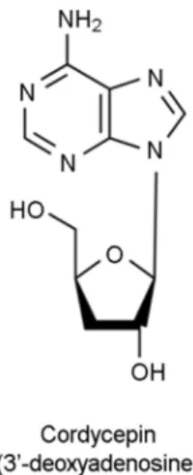
A species of the fungal genus Cordyceps is an ingredient in the traditional Chinese medicine and is known for its anti- inflammatory and anti-cancerous properties (Cunningham et al., 1951; Ng and Wang, 2005). It is well established that cordycepin (3'-deoxyadenosine), a major component of Cordyceps militaris, attenuates [Ca2+]i and TXA2 production in collagen-induced human platelet aggregation (Cho et al., 2007) (Fig. 1). Recently, we reported that cordycepin- enriched n-butanol extract (WIB801C) from Cordyceps militaris has antiplatelet effects by inhibiting TXA2 pro- duction and [Ca2+]i mobilization in collagen-, and ADP- induced human platelet aggregation (Lee et al., 2014; Lee et al., 2015).
In this study, we prepared WIB-801CE (Compound from 2008 First Project of Biotechnology, Whanin Pharm. Co., Ltd., Suwon, Korea), an ethanol extract from culture solution
of Cordyceps militaris-hypha, and analyzed the composition of cordycepin with high performance liquid chromatography (HPLC). In present study, we observed that WIB-801CE contains enough cordycepin, and investigated whether WIB- 801CE involves in attenuation of TXA2 production and its associated COX-1 and TXAS activities, Ca2+-dependent cytosolic phospholipase A2 (cPLA2) activity, and serotonin release.
MATERIALS AND METHODS Materials
WIB-801CE was provided from Whanin Pharmaceutical Corporation (Suwon, Korea). Thrombin, collagen and ADP were obtained from Chrono-Log Corporation (Havertown, PA., USA). Serotonin ELISA kit was purchased from Labor Diagnostika Nord GmbH & Corporation (Nordhorn, Germany). Cordycepin, protease inhibitor cocktail, and 9,11- dideoxy-9α,11α-methanoepoxyprostaglandin F2α (U46619) were obtained from Sigma Chemical Corporation (St. Louis, MO., USA). TXB2 enzyme immunoassay (EIA) kit, COX-1 fluorescent activity assay kit, and prostaglandin H2 for TXAS assay were purchased from Cayman Chemical (Ann Arbor, MI., USA). Anti-phosphor-cPLA2 and anti-rabbit IgG- horseradish peroxidase conjugate (HRP), and lysis buffer were obtained from Cell Signaling (Beverly, MA., USA).
Polyvinylidene difluoride (PVDF) membrane was from GE Healthcare (Piseataway, NJ., USA). Enhanced chemilumine- sence solution (ECL) was from GE Healthcare (Chalfont St., Giles, Buckinghamshire, UK).
Preparation of WIB-801CE
Cordyceps militaris was cultivated in Whanin Pharma- ceutical Corporation (Suwon, Korea) and culture-solution of Cordyceps militaris-hypha was concentrated up to 50°
Brix with a rotary vacuum evaporator (Eyela N3000, Rikakikai Co., Ltd., Tokyo, Japan) at 60℃. The Brix was measured with refractometer (Atago Co., Ltd., Tokyo, Japan).
The concentrate was extracted by extraction-shaker (Cosmos 660, Kyungseo Co., Ltd., Seoul, Korea) for 4 h at 40℃ one time with distilled water/95% ethanol (1 : 3.5, v/v), which was filtered one time using a filter paper (Advantec No.2).
The filtrate was completely concentrated by an evaporator (Eyela N3000, Rikakikai Co., Ltd., Tokyo, Japan) under reduced pressure (60℃), and was lyophilized and stored at -20℃ until used. This was named as cordycepin-enriched WIB-801CE (Compound from 2008 First Project of Bio- technology, Whanin Pharm. Co., Ltd., Suwon, Korea).
Analysis of cordycepin in WIB-801CE with HPLC WIB-801CE was dissolved with 75% methanol, then analyzed by high performance liquid chromatography (HPLC). An Alliance 2695 liquid chromatography system (Milford, MA, USA), equipped with vacuum degasser, qua- ternary gradient pump, autosampler and photodiode array detector, was connected to Empower software. A hydrosphere C18 column (250 mm×4.6 mm id, 5 μm, YMC) was used at a column temperature of 30℃. The applied-mobile phase gradient program was 0.01 M KH2PO4/methanol (95 : 5, v/v) at 0 min and held for 5 min; 0.01 M KH2PO4/methanol (70 : 30, v/v) at 20 min and held for 6 min; 0.01M KH2PO4
/methanol (95 : 5, v/v) at 27 min and held 6 min for chro- matographic balance. In this step, 99.8% of methanol was used. The flow rate was at 1.0 mL/min and sample injec- tion volume was 10 μL. The UV detection was operated at 254 nm.
Preparation of human platelets
Human platelet-rich plasma (PRP) with acid-citrate- dextrose solution (0.8% citric acid, 2.2% sodium citrate, 2.45% glucose) was supplied from Korean Red Cross Blood Center (Changwon, Korea). PRP was centrifuged for 10 min at 125 ×g to remove red blood cells and white cells, and was centrifuged for 10 min at 1,300 ×g to obtain platelet pellets. The platelets were washed twice with washing buffer (138 mM NaCl, 2.7 mM KCl, 12 mM NaHCO3, 0.36 mM NaH2PO4, 5.5 mM glucose, and 1 mM Na2EDTA, pH 6.5).
The washed platelets were then resuspended in suspension buffer (138 mM NaCl, 2.7 mM KCl, 12 mM NaHCO3, 0.36 mM NaH2PO4, 0.49 mM MgCl2, 5.5 mM glucose, 0.25% gelatin, pH 6.9) to a final concentration of 5×10⁸/mL.
Washed platelets were used to investigate the platelet aggre- gation by collagen and thrombin, PRP (5×10⁸/mL) was used to investigate the platelet aggregation by ADP and
U46619. All of the above procedures were carried out at 25℃ to avoid platelet aggregation from any effect of low temperature. The Korea National Institute for Bioethics Policy Public Institutional Review Board (Seoul, Korea/
PIRB12-072-01) approved these experiments.
Measurement of platelet aggregation
To investigate the effect on human platelet aggregation, human washed platelets (108/mL) were pre-incubated for 3 min at 37℃ with or without substances in the presence of 2 mM CaCl2, then stimulated with thrombin (0.025 U/mL) and collagen (5 μg/mL). ADP (20 μM)- and U46619 (10 μM)-induced platelet aggregation was measured with PRP, removed red blood cells and white cells, in the absence of 2 mM CaCl2. After adding agonists, each aggregation was performed for 5 min using an aggregometer (Chrono-Log Corporation, Havertown, PA, USA) at a constant stirring speed of 1,000 rpm. Each aggregation rate was determined as an increase in light transmission. The suspension buffer and PPP were used as the reference (transmission 0). WIB- 801CE was dissolved in platelet suspension buffer (pH 6.9).
U46619 was dissolved in 0.01% dimethyl sulfoxide (DMSO).
0.01% DMSO did not affect the platelet aggregation (0.3
± 0.6%).
Measurement of TXB2
To investigate the effect of WIB-801CE on autacoidal aggregation-inducing molecule, TXA2 production, the aggre- gation was terminated by adding ice-cold 5 mM EDTA and 0.2 mM indomethacin to inhibit subsequent conversion of AA to TXA2. The amounts of TXB2, a stable metabolite of TXA2, were determined using a TXB2 EIA kit according to the procedure described by the manufacturer.
Determination of serotonin release
To investigate the effect of WIB-801CE on autacoidal aggregation-inducing molecule, serotonin release, the aggre- gation was centrifuged at 4℃ for 10 min at 200 ×g. The supernatants were used for the assay of serotonin release.
Serotonin release was measured with a Synergy HT multi- Model Microplate Reader (BioTek Instruments, Winoosku, VT, USA) using serotonin ELISA kit.
Preparation of platelet lysates
We prepared platelet lysates (homogenates) for measure- ment of COX-1 and TXAS. Non-stimulated platelets (108 /mL), and ADP-, collagen-, and thrombin-stimulated platelets (108/mL) in presence of 1% protease inhibitor cocktail (Sigma Chemical Co., St. Louis, MO., USA) were sonicated 10 times at sensitivity 100% for 20 sec on ice with a model HD 2070 sonicator (Bandelin Electronic, Bandelin, Germany) to obtain platelet lysates. Next, the platelet lysates were centrifuged at 12,000 ×g for 15 min at 4℃ to remove cell debris. The supernatant was used to measure COX-1 and TXAS activity.
Measurement of COX-1 activity
For measure COX-1 activity, platelet lysates containing 10 μg of protein were used. COX-1 activities were measured with Synergy HT multi-model microplate reader (BioTek Instruments, Winooski, VT., USA) using COX fluorescent activity assay kit according to the procedure described by manufacturer.
Measurement of TXAS activity
For measure TXAS activity, platelet lysates containing 20 μg of protein were used. The reaction for assay of TXAS activity was initiated by the addition of TXAS substrate prostaglandin H2 (PGH2) and allowed to proceed for 1 min at 37℃. The reaction was terminated by the addition of 1 M citric acid, then was neutralized with 1 N NaOH. The con- centration of TXA2 was determined as thromboxane B2
(TXB2), a stable metabolite of TXA2, which was measured with Synergy HT multi-model microplate reader (BioTek Instruments, Winooski, VT., USA) using TXB2 EIA kit.
Western blot for analysis of cPLA2 phosphorylation
The aggregation was terminated by adding an equal volume (250 μL) of lysis buffer (20 mM Tris-HCl, 150 mM NaCl, 1 mM Na2EDTA, 1 mM EGTA, 1% Triton X-100, 2.5 mM sodium pyrophosphate, 1 mM serine/threonine phos- phatase inhibitor β-glycerophosphate, 1 mM ATPase, alkaline and acid phosphatase, and protein phosphotyrosine phospha- tase inhibitor Na3VO4, 1 μg/mL serine and cysteine protease
inhibitor leupeptin, and 1 mM serine protease and acetyl- cholinesterase inhibitor phenylmethanesulfonyl fluoride, pH 7.5). Platelet lysates were suspended in their equal volume of sodium dodecyl sulfate-polyacrylamide gel electrophoresis (SDS-PAGE) buffer (62.5 mM Tris-HCl, 10% glycerol, 1%
SDS, 1% β-mercaptoethanol, 0.01% bromphenol blue, pH 6.8), then were boiled to completely denature the proteins for 5 min. Aliquots containing 15 μg of protein from each sample tube were subjected to SDS-PAGE (8%, 1.5 mm gel).
Proteins in the gel were transferred to PVDF membrane in the presence of transfer buffer (25 mM Tris-HCl, 192 mM glycine, 20% methanol, pH 8.3). PVDF membrane was washed one time for 5 min with Tris-buffered saline with tween 20 (25 mM Tris-HCl, 140 mM NaCl, 2.7 mM KCl, 0.1% tween 20, pH 7.4), then was blocked with blocking buffer (25 mM Tris-HCl, 140 mM NaCl, 2.7 mM KCl, 0.1%
tween 20, 5% skim milk, pH 7.4) for 1 h at room tempera- ture, and subsequently was washed three times for 5 min.
The protein phosphorylation was observed by using Western blotting. The dilutions for anti-phosphor-cPLA2 and anti- rabbit IgG-HRP were 1:1,000 and 1:10,000, respectively. The membranes were visualized using ECL. Blots were analyzed by using the Quantity One, Ver. 4.5 (BioRad, Hercules, CA, USA).
Fig. 1. Chemical structure of cordycepin (3'-deoxyadenosine).
Statistical analyses
The experimental results are indicated as the mean ± standard deviation accompanied by the number of obser- vations. Data were determined by analysis of variance (ANOVA). If this analysis showed significant differences among the group means, then each group was compared by the Newman-Keuls method. Statistical analysis was carried out according to the SPSS 21.0.0.0 (SPSS, Chicago, IL, USA). P<0.05 was considered to be statistically significant.
RESULTS
Composition of cordycepin in WIB-801CE
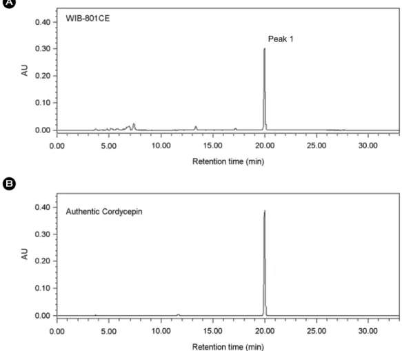
Cordyceps militaris, source of WIB-801CE, contains
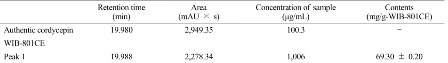
cordycepin as detected by HPLC in our experiment (Fig. 1) (Huang et al., 2003). As shown in Fig. 2, peak 1 was ob- served at 19.988 min of the retention time (Fig. 2A), which was almost in accord with the retention time (19.980 min) of authentic cordycepin (Fig. 2B). The concentration of peak 1 in WIB-801CE corresponding to cordycepin was 69.30
± 0.2 mg/g-WIB-801CE (approximately 6.93 ± 0.02%, Table 1). Whole fruiting body myelia of Cordyceps militaris is known to contain 0.16% of cordycepin, and whole fruiting body, stroma, and larva of Cordyceps sinensis does not con- tain cordycepin (Yue et al., 2008). Therefore, the cordycepin content in WIB-801CE that we used in this study was much higher than those in whole fruiting body myelia of Cordyceps militaris, and in whole fruiting body, stroma, and larva of Cordyceps sinensis.
Fig. 2. HPLC chromatograms of WIB-801CE and authentic cordycepin. (A) The chromatogram of WIB-801CE. (B) The chromatogram of authentic cordycepin. HPLC was performed as described in "Materials and Methods".
Peak 1
Effects of WIB-801CE on ADP-, collagen-, and thrombin- induced human platelet aggregation
The concentration of ADP which induced maximal human platelet aggregation was approximately 10 μM (Fig. 3A, inset). But, as cordycepin and WIB801C, an analogue sub- stance of WIB-801CE, inhibited 20 μM of ADP-induced
platelet aggregation (Lee et al., 2015) in this study 20 μM of ADP was used as a platelet agonist. When PRP (108/mL) were activated with ADP (20 μM), the aggregation rate was increased to 67.7 ± 1.5%. However, various concentrations of WIB-801CE (50 to 400 μg/mL) significantly suppressed ADP-induced human platelet aggregation in a dose-dependent manner (Fig. 3A).
Table 1. Content of cordycepin in WIB-801CE Retention time
(min)
Area (mAU × s)
Concentration of sample (μg/mL)
Contents (mg/g-WIB-801CE)
Authentic cordycepin 19.980 2,949.35 100.3 -
WIB-801CE
Peak 1 19.988 2,278.34 1,006 69.30 ± 0.20
The content of cordycepin in mg/g-WIB-801CE was expressed using the following equation : mg/g-WIB-801CE = (area of peak 1 / area of authentic cordycepin) × (concentration of authentic cordycepin / concentration of WIB-801CE) × (% of purity of authentic cordycepin / 100%) × (1,000 mg / 1 g). Purity of authentic cordycepin was 98% by manufactured. The data are given as the mean ± standard deviation (n=3).
Fig. 3. Effects of WIB-801CE on various agonists-induced platelet aggregation. (A) The concentration threshold of ADP on human platelet aggregation (inset). Effects of WIB-801CE on ADP- induced human platelet aggregation. (B) The concentration threshold of collagen on human platelet aggregation (inset). Effects of WIB- 801CE on collagen-induced human platelet aggregation. (C) The concentration threshold of thrombin on human platelet aggregation (inset). Effects of WIB-801CE on thrombin-induced human platelet aggregation. The data are expressed as the mean ± standard devi- ation (n=4). *P<0.05, **P<0.001 versus the each agonist-stimulated platelets. IC50, half maximal inhibitory concentration.
The concentration of collagen-induced maximal human platelet aggregation was approximately 5 μg/mL, and its degree was 83.3 ± 3.1% (Fig. 3B, inset). Therefore, 5 μg/
mL of collagen was used as a platelet agonist in this study.
As shown in Fig. 3B, when washed human platelets (108/ mL) were activated with collagen (5 μg/mL) in the presence of 2 mM CaCl2, the aggregation rate was increased to 83.0
± 1.0%. However, WIB-801CE (50 to 300 μg/mL)-dose dependently inhibited collagen-induced human platelet aggre- gation (Fig. 3B).
The concentration of thrombin-induced maximal human platelet aggregation was approximately 0.025 U/mL, and its degree was 91.7 ± 3.2% (Fig. 3C, inset). Therefore, 0.025 U/mL of thrombin was used as a platelet agonist in this study.
As shown in Fig. 3C, when washed human platelets (108 /mL) were stimulated with thrombin (0.025 U/mL) in the presence of 2 mM CaCl2, the aggregation rate was increased to 92.3 ± 1.0%. However, WIB-801CE (50 to 400 μg/mL)- dose dependently attenuated thrombin-induced human plate- let aggregation (Fig. 3C).
Effects of WIB-801CE on TXA2 production, and its analogue U46619-induced platelet aggregation
We, next, investigated whether the inhibition of ADP-, collagen-, and thrombin-induced human platelet aggregation by WIB-801CE resulted from the reduction of TXA2 pro- duction. The TXA2 level (determined as TXB2) in resting platelets was 1.4 ± 0.2 ng/108 platelets, and ADP (20 μM) Fig. 4. Effects of WIB-801CE on TXB2 production and U46619-induced platelet aggregation. (A) Effects of WIB-801CE on ADP- induced TXB2 production. (B) Effects of WIB-801CE on collagen-induced TXB2 production. (C) Effects of WIB-801CE on thrombin- induced TXB2 production. (D) Effects of WIB-801CE on U46619-induced platelet aggregation. Measurement of TXB2 production and platelet aggregation were carried out as described in "Materials and Methods". The data are expressed as the mean ± standard deviation (n=4).
aP<0.05 versus the unstimulated platelets, *P<0.05, **P<0.001 versus the each agonist-stimulated platelets. NS, not significant versus the unstimulated platelets. Δ (%) = [(WIB-801CE + agonist) - agonist] / agonist × 100.
potently increased TXA2 level to 24.5 ± 1.6 ng/108 platelets (Fig. 4A). This suggests that ADP increased TXA2 produc- tion to 1,650% as compared with that (1.4 ± 0.2 ng/108 platelets) by resting platelets. But, WIB-801CE alone (300, 400 μg/mL) did not affect the TXA2 production (Fig. 4A) in resting platelets. WIB-801CE dose (100 to 400 μg/mL)- dependently reduced TXA2 production in ADP (20 μM)- activated platelets (Fig. 4A). 150 μg/mL of WIB-801CE, IC50 to ADP-induced platelet aggregation (Fig. 3A), attenu- ated ADP-elevated TXA2 level (24.5 ± 1.6 ng/108 platelets) to 14.9 ± 1.2 ng/108 platelets (39.2%) (Fig. 4A).
Collagen (5 μg/mL) strongly increased TXA2 level to 120.9 ± 7.1 ng/108 platelets (Fig. 4B). This suggest that collagen increased TXA2 production to 8,537.7% as com- pared with that (1.4 ± 0.2 ng/108 platelets) by resting plate- lets. WIB-801CE alone (100, 150 μg/mL) did not affect the TXA2 production in resting platelets, but WIB-801CE dose (50 to 150 μg/mL)-dependently inhibited TXA2 production in collagen (5 μg/mL)-activated platelets (Fig. 4B). 100 μg/
mL of WIB-801CE, IC50 to collagen-induced platelet aggre- gation (Fig. 3B), reduced collagen-elevated TXA2 level (120.9 ± 7.1 ng/108 platelets) to 63.2 ± 3.4 ng/108 plate- lets (47.7%) (Fig. 4B).
Thrombin (0.025 U/mL) potently elevated TXA2 level to 47.5 ± 0.8 ng/108 platelets (Fig. 4C). This suggest that thrombin increased TXA2 production to 3,292.8% as com- pared with that (1.4 ± 0.2 ng/108 platelets) by resting plate- lets. WIB-801CE alone (300, 400 μg/mL) did not affect the TXA2 production in resting platelets, however, WIB-801CE attenuated TXA2 level in thrombin (0.025 U/mL)-activated platelets in a dose (100 to 400 μg/mL)-dependent manner (Fig. 4C). 200 μg/mL of WIB-801CE, IC50 to thrombin- induced platelet aggregation (Fig. 3C), decreased thrombin- increased TXA2 level (47.5 ± 0.8 ng/108 platelets) to 41.1
± 1.7 ng/108 platelets (13.5%) (Fig. 4C).
TXA2 is produced by agonist-activated platelets and sub- sequently acts as a positive feedback promotor on resting platelets, which is connected to the intensification of throm- bus (Halushka, 1995). This means that TXA2 is a strong agonist against resting platelets and TXA2 antagonistic sub- stance or compound may be used beneficially as an anti- thrombotic agent. Therefore, we investigated whether WIB-
801CE, inhibiting TXA2 production, inhibits TXA2 mimetic compound U46619-induced platelet aggregation. As shown in Fig. 4D, the concentration of U46619-induced maximal Fig. 5. Effects of WIB-801CE on COX-1 activity. (A) Effects of WIB-801CE on ADP-induced COX-1 activity. (B) Effects of WIB-801CE on collagen-induced COX-1 activity. (C) Effects of WIB-801CE on thrombin-induced COX-1 activity. Measurements of COX-1 activity was carried out as described in "Materials and Methods". The data are expressed as the mean ± standard deviation (n=4). aP<0.05 versus the basal. NS, not significant versus each agonist-stimulated human platelets, *P<0.05 versus each agonist- stimulated human platelets. Δ (%) = [(ADP-WIB-801CE) / (ADP- basal)] × 100.
platelet aggregation was 10 μM (Fig. 4D, inset). WIB-801CE dose (12.5 to 300 μg/mL)-dependently inhibited U46619 (10 μM)-induced human platelet aggregation (Fig. 4D), and its inhibitory dose is very low as compared with doses (50 to 400 μg/mL) that attenuated ADP-, collagen-, and thrombin- induced human platelet aggregation (Fig. 3A-C).
Effects of WIB-801CE on COX-1 activity
We investigated whether the inhibition of TXA2 produc- tion by WIB-801CE resulted from the inhibition of COX-1 activity. ADP (20 μM) increased COX-1 activity from basal activity (0.24 ± 0.02 nmoL/protein-mg/min) to 0.36 ± 0.04 nmoL/protein-mg/min (Fig. 5A). In contrast, WIB- 801CE dose (100 to 400 μg/mL)-dependently inhibited ADP-induced COX-1 activity (Fig. 5A). 150 μg/mL of WIB-801CE, IC50 to ADP-induced platelet aggregation (Fig.
3A), attenuated ADP-induced COX-1 activity (100%) to 33.3% (Fig. 5A). 400 μg/mL of WIB-801CE decreased ADP-induced COX-1 activity (0.36 ± 0.04 nmoL/protein- mg/min) to 0.24 ± 0.04 nmoL/protein-mg/min (100%) (Fig. 5A).
Collagen (5 μg/mL) and thrombin (0.025 U/mL) increased COX-1 activity from basal activity (0.24 ± 0.02 nmoL/
protein-mg/min) to 0.71 ± 0.02 nmoL/protein-mg/min, and 0.80 ± 0.06 nmoL/protein-mg/min, respectively (Fig. 5B, C). However, WIB-801CE did not inhibit collagen-, and thrombin-induced COX-1 activity (Fig. 5B, C). These results suggest that WIB-801CE inhibited ADP-induced COX-1 activity to reduce TXA2 production but did not attenuate collagen- and thrombin-induced COX-1 activity.
Effects of WIB-801CE on TXAS activity
We investigated whether the inhibition of TXA2 produc- tion by WIB-801CE was due to the attenuation of TXAS activity. Collagen (5 μg/mL) elevated TXAS activity from basal activity (264.90 ± 1.88 ng/protein-mg/min) to 309.80
± 5.01 ng/protein-mg/min (Fig. 6A). However, WIB-801CE dose (50 to 150 μg/mL)-dependently attenuated collagen- induced TXAS activity (Fig. 6A). 100 μg/mL of WIB- 801CE, IC50 to collagen-induced platelet aggregation (Fig.
3B), decreased collagen-elevated TXAS activity (309.80 ± 5.01 ng/protein-mg/min) to 278.3 ± 5.5 ng/protein-mg/
min (70.2%) (Fig. 6A).
ADP (20 μM) increased TXAS activity from basal activity Fig. 6. Effects of WIB-801CE on TXAS activity. (A) Effects of WIB-801CE on collagen-induced TXAS activity. (B) Effects of WIB-801CE on ADP-induced TXAS activity. (C) Effects of WIB- 801CE on thrombin-induced TXAS activity. Measurements of TXAS activity was carried out as described in "Materials and Methods". The data are expressed as the mean ± standard deviation (n=4). aP<0.05 versus the basal. NS, not significant versus each agonist-stimulated human platelets, *P<0.05 versus each agonist- stimulated human platelets. Δ (%) = [(collagen-WIB-801CE) / (collagen-basal)] × 100.
(264.90 ± 1.88 ng/protein-mg/min) to 297.40 ± 2.54 ng/
protein-mg/min (Fig. 6B), which suggests that ADP (20 μM)
elevated TXAS activity to 12.3%. But WIB-801CE did not inhibit ADP-induced TXAS activity (Fig. 6B). In addition, thrombin (0.025 U/mL) increased very weakly TXAS acti- vity from basal (264.90 ± 1.88 ng/protein-mg/min) to 275.30 ± 0.30 ng/protein-mg/min, which was not inhibited by WIB-801CE (Fig. 6C). The above results suggest that WIB-801CE attenuated collagen-induced TXAS activity to inhibit TXA2 production, but did not inhibit ADP- and thrombin-induced TXAS activity.
Effects of WIB-801CE on serotonin release
Platelets do not synthesize neurotransmitter serotonin, which is incorporated by platelets and stored in dense body of platelets (Fraer and Kilic, 2015). Serotonin is released out of dense body when platelets are aggregated by various agonists (i.e., ADP, collagen, thrombin), and subsequently intensify platelet aggregation to generate the thrombosis (Moerland et al., 2011; Fraer and Kilic, 2015). Therefore, we investigate the effect of WIB-801CE on agonist-released serotonin. WIB-801CE alone (100 to 400 μg/mL) did not release serotonin out of resting platelets (Fig. 7A-C). This means that WIB-801CE alone did not affect serotonin release, a marker of platelet activation, out of resting platelets. ADP (20 μM), collagen (5 μg/mL), and thrombin (0.025 U/mL) released serotonin 207.6 ± 8.1 ng/108 platelets (Fig. 7A), 154.9 ± 9.5 ng/108 platelets (Fig. 7B), and 384.4 ± 4.9 ng/108 platelets (Fig. 7C), respectively. However, 150 μg/
mL of WIB-801CE, IC50 to ADP-induced platelet aggre- gation (Fig. 3A), attenuated ADP-induced serotonin release (207.6 ± 8.1 ng/108 platelets) to 100.9 ± 1.4 ng/108 plate- lets (51.4%) (Fig. 7A). 100 μg/mL of WIB-801CE, IC50 to collagen-induced platelet aggregation (Fig. 3B), reduced collagen-elevated serotonin release (154.9 ± 9.5 ng/108 platelets) to 63.6 ± 9.5 ng/108 platelets (58.9%) (Fig. 7B).
200 μg/mL of WIB-801CE, IC50 to thrombin-induced plate- let aggregation (Fig. 3C), diminished thrombin-elevated serotonin release (384.4 ± 4.9 ng/108 platelets) to 275.0
± 1.3 ng/108 platelets (28.5%) (Fig. 7C).
Effects of WIB-801CE on cPLA2 activity
cPLA2 is Ca2+-dependently activated, and subsequently hydrolyzed membrane phospholipids (i.e., phosphatidyl- Fig. 7. Effects of WIB-801CE on serotonin release. (A) Effects
of WIB-801CE on ADP-induced serotonin release. (B) Effects of WIB-801CE on collagen-induced serotonin release. (C) Effects of WIB-801CE on thrombin-induced serotonin release. The data are expressed as the mean ± standard deviation (n=4). aP<0.05 versus the unstimulated platelets, NS, not significant versus the unsti- mulated platelets. *P<0.05, **P<0.001 versus the each agonist- stimulated platelets. Δ (%) = [Agonist- (agonist + WIB-801CE)] / agonist × 100.
choline) to release AA, precursor of TXA2. Therefore, we investigated whether WIB-801CE inhibits cPLA2 activity to attenuate the production of TXA2 (Fig. 4). cPLA2 activity is determined by its phosphorylation status. ADP (20 μM), collagen (5 μg/mL), and thrombin (0.025 U/mL) potently stimulated cPLA2 activity, respectively (Fig. 8). WIB-801CE, however, inhibited these agonist-induced cPLA2 activities in a dose-dependent manner (Fig. 8). Especially, WIB-801CE without having inhibitory effect on COX-1 (Fig. 5C) and TXAS (Fig. 6C) activities attenuated cPLA2 activity in thrombin-induced platelet aggregation (Fig. 8C).
DISCUSSION
Platelet aggregation is a marker of platelet activation which is controlled by various agonists (i.e., ADP, collagen, thrombin)-produced TXA2. Therefore, to observe the inhib- ition of agonist-induced TXA2 production it is important to evaluate the antiplatelet effect of any substance or compound (Schwartz et al., 1990; Duguid, 1946; Cahill and Newland, 1993; Grau et al., 1998). WIB-801CE potently attenuated TXA2 production to inhibit ADP-, collagen- and thrombin- induced platelet aggregation. Especially, ADP is an autac- oidal platelet agonist that is released by thrombin- and collagen-stimulation. Since WIB-801CE inhibits ADP- induced platelet aggregation and TXA2 production it may be considered as a beneficial antiplatelet agent. TXA2 precursor AA is produced by phospholipase C (PLC) or cPLA2, and subsequently metabolized to TXA2 by COX-1 and TXAS (Hamberg et al., 1975; Samuelsson et al., 1978; Gresele et al., 1991). COX-1 produces PGG2 from AA, which is oxidized to PGH2 by endoperoxidase. TXAS produces finally TXA2
from PGH2. Accordingly, it is considered that WIB-801CE inhibited TXA2 production by protecting the use of AA by COX-1 in ADP-activated platelets, and PGH2 by TXAS in collagen-activated platelets. In addition, there is the possibility that WIB-801CE diminishes Ca2+-dependent cPLA2 activity to reduce AA supply in ADP-, collagen- and thrombin- activated platelets. Accordingly, WIB-801CE seems to in- hibit the activities of COX-1, TXAS, and cPLA2 to attenuate TXA2 production in ADP- and collagen-activated platelets.
Although WIB-801CE did not inhibit COX-1 and TXAS Fig. 8. Effects of WIB-801CE on cPLA2 phosphorylation. (A)
Effects of WIB-801CE on ADP-induced cPLA2 phosphorylation.
(B) Effects of WIB-801CE on collagen-induced cPLA2 phosphory- lation. (C) Effects of WIB-801CE on thrombin-induced cPLA2
phosphorylation. cPLA2 phosphorylation was determined as de- scribed in "Materials and Methods" section. The data are expressed as the mean ± standard deviation (n=4). *P<0.05, **P<0.001 versus the each agonist-stimulated platelets.
activities, it attenuated TXA2 production and cPLA2 activity in thrombin-activated platelets. These results mean that WIB-801CE attenuated the production of TXA2 by inhi- biting the supply of AA via inhibition of cPLA2 activity in thrombin-activated platelets.
With regard to PLC activity, it has been shown by our previous report that cordycepin does not inhibit PLCγ2
activity, and IP3 production by PLCγ2 in collagen-induced human platelet aggregation (Cho et al., 2007). In this study, because we did not measure PLC activity, it is unknown whether WIB-801CE inhibits PLC activity to attenuate the supply of AA. This should be investigated in the future.
Various agonist-elevated [Ca2+]i also involves in activation of Ca2+/calmodulin-dependent myosin light chain kinase (MLCK) and integrin glycoprotein IIb/IIIa (αIIb/β3) to acti- vate platelets. The release of granule compounds is an index of platelet activation, and is generated by MLCK- phosphorylated MLC (20 kDa) (Nishikawa et al., 1980;
Kaibuchi et al., 1982). It is well established that cordycepin in WIB-801CE attenuates [Ca2+]i, and subsequently inhibits the phosphorylation of 20 kDa by Ca2+-dependent MLCK in collagen-, and U46619-activated human platelets (Cho et al., 2006; Cho et al., 2007). WIB-801CE also inhibited ADP-, collagen- and thrombin-elevated [Ca2+]i (data not shown).
Therefore, the decrease of [Ca2+]i by WIB-801CE and cor- dycepin is resulted in the inhibition of ADP-, collagen-, and thrombin-induced serotonin release.
Because agonist-produced TXA2 itself binds to its receptor as autacoidal agonist, and subsequently elevates [Ca2+]i to stimulate the granule secretion (Halushka et al, 1995), the inhibition of U46619-induced human platelet aggregation by WIB-801CE might be also involved in attenuation of sero- tonin and [Ca2+]i as evidenced that cordycepin in WIB-801CE attenuated U46619-elevated [Ca2+]i and Ca2+-dependent 20 kDa phosphorylation (Cho et al., 2006). Because WIB- 801CE contains cordycepin as well as other unknown com- pounds, it is unknown whether cordycepin is the key ingre- dient in WIB-801CE that contributes to the inhibition of platelet aggregation. Recently, we showed that cordycepin purified from butanol extracts (BE) of Cordyceps militaris has antiplatelet effect (Lee et al., 2015a; Lee et al., 2015b), and it has a synergistic inhibitory effect with BE. As shown
in Table 1, because WIB-801CE contains cordycepin, it is thought cordycepin in WIB-801CE might contribute to the antiplatelet effect of WIB-801CE.
In conclusion, cordycepin-enriched WIB-801CE, an etha- nol extract from Cordyceps militaris, inhibited thrombosis- generation molecules (i.e. TXA2, serotonin) increased by platelet aggregation. Therefore, we suggest that WIB-801CE and cordycepin might be considered a beneficial and effec- tive agent for the treatment or protection from thrombosis, atherosclerosis, and myocardial infarction via inhibition of platelet aggregation.
Acknowledgements
This study was supported by a grant (20150361, to Hwa- Jin Park) from Whanin Pharm. Co., Ltd. (Suwon, Korea).
Conflict of interest
The authors declare no conflict of interest.
REFERENCES
Berridge MJ, Irvine RF. Inositol trisphosphate, a novel second messenger in cellular signal transduction. Nature. 1984. 312:
315-321.
Cahill MR, Newland AC. Platelet activation in coronary artery disease. Br J Biomed Sci. 1993. 50: 221-234.
Cho HJ, Cho JY, Rhee MH, Lim CR, Park HJ. Cordycepin (3'- deoxyadenosine) inhibits human platelet aggregation induced by U46619, a TXA2 analogue. J Pharm Pharmacol. 2006. 58:
1677-1682.
Cho HJ, Cho JY, Rhee MH, Park HJ. Cordycepin (3'-deoxy- adenosine) inhibits human platelet aggregation in a cyclic AMP- and cyclic GMP-dependent manner. Eur J Pharmacol.
2007. 558: 43-51.
Cunningham KG, Hutchinson SA, Manson W, Spring FS. Cordy- cepin, a metabolic product from cultures of Cordyceps militaris (Linn.) Link. Part I. Isolation and Characterization. Journal of Chemical Society. 1951. 2: 2299-2300.
Duguid JB. Thrombosis as a factor in the pathogenesis of coronary atherosclerosis. J Pathol Bacteriol. 1946. 58: 207-212.
Fraer M, Kilic F. Serotonin: a different player in hypertension- associated thrombosis. Hypertension. 2015. 65: 942-948.
Grau AJ, Ruf A, Vogt A, Lichy C, Buggle F, Patscheke H, Hacke W.
Increased fraction of circulating activated platelets in acute and previous cerebrovascular ischemia. Thromb Haemost.
1998. 80: 298-301.
Gresele P, Deckmyn H, Nenci GG, Vermylen J. Thromboxane synthase inhibitors, thromboxane receptor antagonists and dual blockers in thrombotic disorders. Trends Pharmacol Sci.
1991. 12: 158-163.
Guidetti GF, Lova P, Bernardi B, Campus F, Baldanzi G, Graziani A, Balduini C, Torti M. The Gi-coupled P2Y12 receptor regulates diacylglycerol-mediated signaling in human platelets. J Biol Chem. 2008. 283: 28795-28805.
Halushka PV, Allan CJ, Davis-Bruno KL. Thromboxane A2 rece- ptors. J Lipid Mediat Cell Signal. 1995. 12: 361-378.
Hamberg M, Svensson J, Samuelsson B. Thromboxanes: a new group of biologically active compounds derived from pro- staglandin endoperoxides. Proc Natl Acad Sci USA. 1975. 72:
2994-2998.
He GW, Yang CQ. Inhibition of vasoconstriction by the throm- boxane A2 antagonist GR32191B in the human radial artery.
Br J Clin Pharmacol. 1999. 48: 207-215.
Huang LF, Liang YZ, Guo FQ, Zhou ZF, Cheng BM. Simultaneous separation and determination of active components in Cordy- ceps sinensis and Cordyceps militaris by LC/ESI-MS. J Pharm Biomed Anal. 2003. 33: 1155-1162.
Jennings LK. Role of platelets in atherothrombosis. Am J Cardiol.
2009. 103: 4A-10A.
Kaibuchi K, Sano K, Hoshijima M, Takai Y, Nishizuka Y. Phos- phatidylinositol turnover in platelet activation; calcium mobi- lization and protein phosphorylation. Cell Calcium. 1982. 3:
323-335.
Lee DH, Kim HH, Cho HJ, Yu YB, Kang HC, Kim JL, Lee JJ, Park HJ. Cordycepin-Enriched WIB801C from Cordyceps militaris Inhibits Collagen-Induced [Ca2+]i Mobilization via Camp-Dependent Phosphorylation of Inositol 1, 4, 5-Tris- phosphate Receptor in Human Platelets. Biomol Ther. 2014.
22: 223 -231.
Lee DH, Kim HH, Lim DH, Kim JL, Park HJ. Effect of Cordycepin-enriched WIB801C from Cordyceps militaris sup- pressing fibrinogen binding to glycoprotein IIb/IIIa. Biomol Ther. 2015. 23: 60-70
Lee DH, Kwon HW, Kim HH, Lim DH, Nam GS, Shin JH, Kim
YY, Kim JL, Lee JJ, Kwon HK, Park HJ. Cordycepin- enriched WIB801C from Cordyceps militaris inhibits ADP- induced [Ca2+]i mobilization and fibrinogen binding via phos- phorylation of IP3R and VASP. Arch Pharm Res. 2015. 38:
81-97.
Lewis GP, Watts IS. Prostaglandin endoperoxides, thromboxane A2 and adenosine diphosphate in collagen-induced aggregation of rabbit platelets. Br J Pharmacol. 1982. 75: 623-631.
Li Z, Delaney MK, O'Brien KA, Du X. Signaling during platelet adhesion and activation. Arterioscler Thromb Vasc Biol. 2010.
30: 2341-2349.
Malmsten C, Hamberg M, Svensson J, Samuelsson B. Physiological role of an endoperoxide in human platelets: hemostatic defect due to platelet cyclo-oxygenase deficiency. Proc Natl Acad Sci USA. 1975. 72: 1446-1450.
Moerland M, Kemme M, Dijkmans A, Bergougnan L, Burggraaf J. Modulation of vasoactivity and platelet aggregation by selective 5-HT receptor antagonism in humans. J Cardiovasc Pharmacol. 2011. 58: 575-580.
Ng TB, Wang HX. Pharmacological actions of Cordyceps, a prized folk medicine. J Pharm Pharmacol. 2005. 57: 1509 -1519.
Nishikawa M, Tanaka T, Hidaka H. Ca2+-calmodulin-dependent phosphorylation and platelet secretion. Nature. 1980. 287: 863 -865.
Patrono C. Aspirin: new cardiovascular uses for an old drug. Am J Med. 2001. 110: 62S-65S.
Samuelsson B, Goldyne M, Granström E, Hamberg M, Hammarström S, Malmsten C. Prostaglandins and throm- boxanes. Annu Rev Biochem. 1978. 47: 997-1029.
Schwartz SM, Heinmark RL, Majesky MW. Developmental mech- anisms underlying pathology of arteries. Physiol Rev. 1990.
70: 1177-1209.
Tendera M, Wojakowski W. Role of antiplatelet drugs in the pre- vention of cardiovascular events. Thromb Res. 2003. 110: 355 -359.
Yue GG, Lau CB, Fung KP, Leung PC, Ko WH. Effects of Cordyceps sinensis, Cordyceps militaris and their isolated compounds on ion transport in Calu-3 human airway epithelial cells. J Ethnopharmacol. 2008. 117: 92-101.