교신저자 : 신승우. 서울특별시 서초구 잠원동
38-25 기린한방병원
Tel : 02-515-7300, Fax : (02) 516-9393, E-mail : [email protected]
E ffec ts o f P e ro xiso m e P rolife ra to r-A c tiva te d R ec e ptor-γ2 P ro 12 A la P o lym orph ism on B o d y F a t D istrib u tio n in Fe m ale K orea n S ub je c ts
Kil-Soo Kim*, Sun-Mi Choi, Seung-Uoo Shin*, Hyun-Sung Yang, and Yoo-Sik Yoon
*Kirin Oriental Hospital, Korea Institute of Oriental Medicine
Objectives : The effects of peroxisome proliferator-activated receptor γ2 (PPARγ2) Pro12Ala (P12A) polymorphism on body mass index (BMI) and type 2 diabetes are well documented; however, until now, only a few studies have evaluated the effects of this polymorphism on body fat distribution. This study was conducted to elucidate the effects of this polymorphism on computed tomography (CT)-measured body fat distribution and other obesity-related parameters in Korean female subjects.
Methods & Results : The frequencies of PPARγ2 genotypes were: PP type, 93.0%; PA type, 6.8%; and AA type, 0.2%. The frequency of the A allele was 0.035. Body weight (P .012), BMI (P .012), and waist-to-hip ratio (WHR) (P .001) were significantly higher in subjects with PA/AA compared with subjects with PP. When body composition was analyzed by bioimpedance analysis, lean body mass and body water content were similar between the 2 groups. However, body fat mass (P .003) and body fat percent (P .025) were significantly higher in subjects with PA/AA compared with subjects with PP.
Among overweight subjects with BMI of greater than 25, PA/AA was associated with significantly higher abdominal subcutaneous fat (P .000), abdominal visceral fat (P .031), and subcutaneous upper and lower thigh adipose tissue (P .010 and .013). However, among lean subjects with BMI of less than 25, no significant differences associated with PPARγ2 genotype were found, suggesting that the fat-accumulating effects of the PA/AA genotype were evident only among overweight subjects, but not among lean subjects. When serum lipid profiles, glucose, and liver function indicators were compared among overweight subjects, no significant difference associated with PPARγ2 genotype was found. Changes in body weight, BMI, WHR, and body fat mass were measured among overweight subjects who finished a 1-month weight lose program of a hypocaloric diet and exercise; no significant differences associated with PPARγ2 genotype were found.
Conclusions : The results of this study suggest that the PPARγ2 PA/AA genotype is associated with increased subcutaneous and visceral fat areas in overweight Korean female subjects, but does not significantly affect serum biochemical parameters and outcomes of weight loss programs.1)
ꠏꠏꠏꠏꠏꠏꠏꠏꠏꠏꠏꠏꠏꠏꠏꠏꠏꠏꠏꠏꠏꠏꠏꠏꠏꠏꠏꠏ
Key words : Peroxisome proliferator-activated receptor γ2(PPARγ2), Polymorphism, Body fat distribution
Peroxisome Proliferator-Activated Receptor-γ 2 (PPARγ2) Pro12Ala (P12A) 유전자 다형성이 한국여성의
체지방분포에 미치는 영향
김길수*․최선미․신승우*․양현성․윤유식
*기린한방병원, 한국한의학연구원
■ 교신저자 : 신승우, 서울특별시 서초구 잠원동 38-25 기린한방병원 (02) 515-7300, [email protected]
Ⅰ.
서 론Proliferator-Activated Receptor-γ(PPARγ)는 지 방세포 분화에 중요한 역할을 하는 전사인자1)로, 프로모터의 선택적 이용과 splicing에 의해 PPAR γ1와 PPARγ2의 두 가지 isoform을 가지게 된다.
PPArγ2는 아미노 말단에 28개의 추가 아미노산을 가진다고 알려져 있다2). PPARγ1 와 PPARγ2은 모두 지방조직에서 발현되지만 PPARγ2가 인슐린 이 매개된 전사활성화와 지방세포분화에 더 민감 하다. 이것은 비만과 인슐린저항성에 PPARγ2의 역할이 더 분명한 것을 제시한다3). 아주 드물게 발 견되는 PPARγ2의 우성돌연변이는 심한 인슐린 저항성과 당뇨를 가진 가계에서 나타나며4), 또한 극도로 비만한 개개인에서 드물게 PPARγ2의 기 능적 돌연변이가 발견되기도 한다5).
PPARγ2 유전자의 12번째 아미노산의 proline이 alanine으로 대체된 다형성이 Yen 등에 의해 발견 되었다6). 많은 연구에서 PPARγ2의 이 부분이 인 슐린 매개된 전사 활성화와 관련되어 있고, pro- line이 alanine으로 치환되면 단백질의 구조가 확 실히 바뀌기 때문에 Pro12Ala 다형성과 대사 증후 군사이의 관계가 추정되어 왔다. 최근에 보고된 3000명이 넘는 데이터에 근거한 분석은 Pro12Ala 유전자 다형성이 제2형 당뇨병의 위험도에 영향을 준다는 것을 제시하고 있고, Pro allele 는 Ala allele에 비해 당뇨 위험을 1.25배 높다는 것을 규 명하였다7). 또한 최근 Masud와 Ye8)는 PPARγ2 유전자 다형성과 비만도와의 meta 분석에서 19,136명의 데이터를 종합한 분석을 실시하여 비 만도의 지표인 BMI가 Pro allele 보다 Ala allele
에서 통계적으로 의미 있게 더 높은 것을 보고하 였다.
그러나 CT-측정 체지방 면적에 대한 PPARγ2 유전자 다형성의 영향에 대하여서는 소수의 연구 결과만이 보고되고 있을 뿐이다. 본 연구에서는 한 국여성에 있어서 비만과 관련된 지표로서 CT로 측정된 복부와 허벅지의 지방면적에 대한 PPARγ2 유전자 다형성의 영향을 평가하였다.
Ⅱ.
연구대상 및 방법1. 연구대상
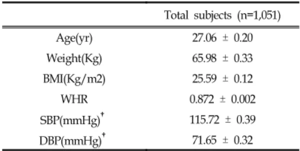
기린한방병원에서 진료를 받은 1,051명의 한국여 성을 대상으로 하였다. 일반적인 피험자의 특징은 Table I에 기재되어 있다. 남성도 있었으나 통계학 적 분석에 충분한 수가 되지 않아 분석에서 제외 하였다. 체성분 구성은 시중에서 판매되고 있는 bio-impedance analysis 측정기기 (Inbody 2.0, Biospace)를 통해 측정되었다. 471명을 대상으로 복부피하지방, 복부 내장지방, 허벅지 피하지방 면 적이 CT(Hispeed CT/e, GE, USA)를 이용하여 측정되었다. 198명의 과체중 그룹을 대상으로 1달 동안 500 kcal/day의 저열량 식이와 유산소 운동 으로 구성된 체중감량 프로그램을 수행하여 프로 그램이 진행되는 동안의 체중, BMI, WHR, 체지 방변화를 측정하였다.
Table I. General Characteristics of Study Subjects Total subjects (n=1,051)
Age(yr) 27.06 ± 0.20
Weight(Kg) 65.98 ± 0.33
BMI(Kg/m2) 25.59 ± 0.12
WHR 0.872 ± 0.002
SBP(mmHg)† 115.72 ± 0.39
DBP(mmHg)† 71.65 ± 0.32
Data are mean ± SE
†SBP : Systolic blood pressure, DBP : diastolic blood pressure.
2. 연구방법
1) PPARγ2 유전자형의 결정
Gemomic DNA는 Qiagen kit를 사용하여 분리 하였다. PCR반응을 통하여 PPARγ2 유전자의 Pro12Ala 위치를 포함한 부분을 증폭시켰다. Up- stream primer, downstream primer, 3㎕ dNTP mix (1 mM), 0.2㎕ Taq DNA polymerase (1 unit), 3㎕ PCR buffer (10x)를 섞고 증류수로 30
㎕의 전체 부피를 맞추었다. 증폭 방법은 94도에서 30초 변성, 52도에서 30분 접합, 72도에서 30초 신 장단계로 이루어진 35 cycle로 구성되었다.
증폭된 PCR 생성물은 3% agarose gel로 154bp 의 크기가 맞는지 확인한 다음 제한효소 HhaI로 2 시간동안 37도에서 절단하고 3% agarose gel에서 전기영동하였다. 그 결과 밴드는 PP type (단일 밴드154 bp), PA type (3개의 밴드, 154, 132, 22 bp), AA type(2개의 밴드, 132, 22 bp)의 패턴이 나타난다.
2) 혈액 생화학적 분석
혈액 샘플은 12시간이상 동안 절식한 후에 정맥 에서 채혈하여 30분동안 2000rpm으로 원심분리하 였다. 혈청에서의 글루코스, cholesterol, HDL cholesterol, triglyceride, GOT, GPT, 전체 bil- irubin의 수준을 자동화된 혈액생화학분석기를 통 해 측정하였다. LDL cholesterol은 Friedewald equation [LDL cholesterol = TC - HDL cho- lesterol - TG/5]을 이용하여 계산하였다.
3) 통계분석
모든 값은 평균 ± 표준오차로 나타내었다. Gen- eral Linear Model에 의한 univariate 분석으로 연령을 보정한 PPARγ2 유전자형의 독립적 효과
를 알아보았다. Chi-square test은 고지방그룹과 정상그룹사이의 PPARγ2 유전자형의 빈도를 비교 하는데 사용하였다. PPARγ2 유전자형, 나이, lean body mass, 혈중triglyceride를 포함한 multivar- iate analysis은 General Linear Model의 type III sum of square를 이용해 측정되었다. 이 방법은 Robitaille 등9) 의 연구에 따르면 연구대상에서 모 든 다른 변수를 조정한 후에 독립적인 변수의 영 향을 측정할수 있다고 보고되었다. 통계적인 유의 성은 p<0.05으로 하였고 모든 분석들은 SPSS ver.
10.0를 사용하여 실행하였다.
Ⅲ.
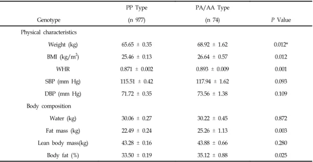
결 과PPARγ2 유전자의 Pro12Ala (P12A) 다형성의 빈도를 1,051명의 한국여성을 대상으로 측정한 결 과, PP type은 93%(978명), PA type은 6.8%(71 명), AA type은 0.2%(2명)으로 나타났으며 이 빈 도는 Hardy-Weinberg equilibrium과 일치하였 다. Ala(A) allele의 빈도인 0.035는 중국 0.03910), 대만 0.04011), 일본 0.04112)의 다른 동아시아에서 보고된 빈도와 유사하였다. 그러나 0.1113), 0.1214), 0.1315)의 백인에게서 보고된 빈도보다는 상당히 작 게 나타났다. <Table II>는 PPARγ2 유전자형에 따른 신체지수와 체성분의 자료를 보여준다. AA type은 단지 2명뿐으로 통계적으로 원활한 분석을 위하여 PA type과 합쳐서 PA/AA type을 구성 하였다. 체중, BMI, WHR은 PP type에 비해 PA/
AA type에서 뚜렷하게 높게 나타났다. 체성분을 bio-impedance analysis 방법으로 측정했을 때 lean body mass와 water content는 두 그룹에서 비슷하였으나 PP type과 비교하여 PA/AA type 에서 체지방량과 체지방률이 뚜렷하게 높았다. 이 것은 PA/AA type의 체중증가가 lean body
mass보다는 체지방의 증가의 결과임을 의미한다.
또한PP type에서보다 PA/AA type에서 체중과 BMI가 각각 5%와 4.6% 높은 반면에 체지방량은 12.3% 높아서 PPARγ2 유전자형이 체중보다 체 지방량에서 더 확실한 영향이 있다는 것을 제시 하였다.
PPARγ2 유전자형이 체지방 과축적 위험에 영향 을 주는 것으로 예측되었으므로 여성에게 제시되
는 국제체지방 기준에 의해 정상지방 그룹과 고지 방 그룹으로 나누고16) PPARγ2 유전자형의 분포를 두 그룹 사이에서 비교하였다(Table III). 분석결과 는 PA/AA type이 PP type보다 지방 과축적에 대하여 1.7배 더 높은 위험성 (95% 신뢰구간, 1.017~2.885) 을 가진다는 것을 보여주었다.
Table II. Comparisons of Physical Characteristics and Body Compositions by Genotypes of PPARγ2
PP Type PA/AA Type
Genotype (n 977) (n 74) P Value
Physical characteristics
Weight (kg) 65.65 ± 0.35 68.92 ± 1.62 0.012*
BMI (kg/m2) 25.46 ± 0.13 26.64 ± 0.57 0.012
WHR 0.871 ± 0.002 0.893 ± 0.009 0.001
SBP (mm Hg) 115.51 ± 0.42 117.94 ± 1.62 0.093
DBP (mm Hg) 71.72 ± 0.35 73.56 ± 1.38 0.109
Body composition
Water (kg) 30.06 ± 0.27 30.22 ± 0.45 0.872
Fat mass (kg) 22.49 ± 0.24 25.26 ± 1.13 0.003
Lean body mass(kg) 43.28 ± 0.16 43.88 ± 0.66 0.280
Body fat (%) 33.50 ± 0.19 35.12 ± 0.88 0.025
NOTE. Data are mean SE.
* P values were obtained by general linear model (covariance) analysis adjusted for age.
Table III. Distribution of PPARγ2 Genotypes in Subjects With Normal and Unhealthy Body Fat Levels Normal Range
(fat < 32%)
Unhealthy Range
(fat > 32%) Total P value*
PP type 395 (40.4%)† 582 (59.6%) 977 (100.0%)
PA/AA type 21 (28.4%) 53 (71.6%) 74 (100.0%) 0.048
Total 416 (39.6%) 565 (60.4%) 1051 (100.0%)
* P value and odd ratio were obtained by chi-square test.
†Number of subjects (%).
Table IV. Comparison of CT-Measured Fat Areas by PPARγ2 Genotype
Genotype PP type
(n=434)
PA/AA type
(n=37) p-value
Abdominal Subcutaneous Fat (mm2) 25,478 ± 490 32,641 ± 2,687 0.000*
Abdominal Visceral Fat (mm2) 5,574 ± 135 6,477 ± 509 0.117
Total Abdominal Fat†(mm2) 30,510 ± 555 39,118 ± 3,083 0.000
V/S ratio‡ (mm2) 0.234 ± 0.007 0.206 ± 0.011 0.376
Upper Thigh Subcutaneous Fat (mm2) 14,715 ± 166 16,325 ± 898 0.009 Lower Thigh Subcutaneous Fat (mm2) 9,807 ± 151 11,593 ± 863 0.002 Data are mean SE.
* P values were obtained by general linear model (covariance) analysis adjusted for age.
†Total abdominal fat is the sum of abdominal subcutaneous fat and abdominal visceral fat.
‡V/S ratio is the ratio of abdominal visceral fat to abdominal subcutaneous fat.
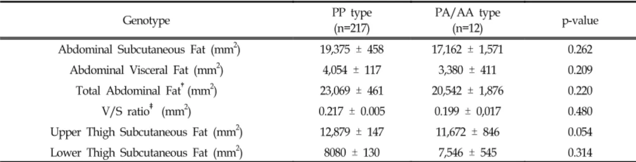
Table V. Comparison of CT-Measured Fat Areas by PPARγ2 Genotype in Lean Subjects With BMI of less than 25
Genotype PP type
(n=217)
PA/AA type
(n=12) p-value
Abdominal Subcutaneous Fat (mm2) 19,375 ± 458 17,162 ± 1,571 0.262
Abdominal Visceral Fat (mm2) 4,054 ± 117 3,380 ± 411 0.209
Total Abdominal Fat†(mm2) 23,069 ± 461 20,542 ± 1,876 0.220
V/S ratio‡ (mm2) 0.217 ± 0.005 0.199 ± 0,017 0.480
Upper Thigh Subcutaneous Fat (mm2) 12,879 ± 147 11,672 ± 846 0.054 Lower Thigh Subcutaneous Fat (mm2) 8080 ± 130 7,546 ± 545 0.314 Data are mean SE.
* P values were obtained by general linear model (covariance) analysis adjusted for age.
†Total abdominal fat is the sum of abdominal subcutaneous fat and abdominal visceral fat.
‡V/S ratio is the ratio of abdominal visceral fat to abdominal subcutaneous fat.
Table VI. Comparison of CT-Measured Fat Areas by PPARγ2 Genotype in Overweight Subjects With BMI of Greater Than 25
Genotype PP type
(n=225)
PA/AA type
(n=25) p-value
Abdominal Subcutaneous Fat (mm2) 31,363 ± 645 40,071 ± 2,898 0.000
Abdominal Visceral Fat (mm2) 7,064 ± 193 7,963 ± 503 0.031
Total Abdominal Fat†(mm2) 37,816 ± 717 48,034 ± 3,179 0.000
V/S ratio‡ (mm2) 0.250 ± 0.014 0.210 ± 0.014 0.426
Upper Thigh Subcutaneous Fat (mm2) 16,518 ± 241 18,558 ± 996 0.013 Lower Thigh Subcutaneous Fat (mm2) 11,517 ± 216 13,534 ± 1,050 0.010 Data are mean SE.
* P values were obtained by general linear model (covariance) analysis adjusted for age.
†Total abdominal fat is the sum of abdominal subcutaneous fat and abdominal visceral fat.
‡V/S ratio is the ratio of abdominal visceral fat to abdominal subcutaneous fat.
인체내의 지방축적에 대한 PPARγ2 유전자형의 효과에 대한 정밀한 평가를 위하여 471명을 대상 으로 CT를 이용하여 신체 각부위의 지방조직 단 면적을 측정하였다(Table IV). 복부피하지방면적은 PP type과 비교하여 PA/AA type에서 28% 더 크게 나타났다. 복부 내장지방면적은 통계상 확실 하게 의미 있는 차이가 아닐지라도 PA/AA type 에서 16% 높게 관찰되었다. 전체 복부지방면적(피 하지방과 내장지방의 합)는 PA/AA형에서 의미있 게 높았다. 피하지방에 대한 내장지방의 비율은 크 게 다르지 않았다. 위 및 아래 허벅지에서 측정한 피하지방면적은 PP type과 비교했을 때 PP/AA type에서 윗부분 허벅지에서 11% 넓고 아래쪽 허 벅지에서 18% 넓었다.
CT 측정 지방면적에 대한 PPARγ2 유전자형의 효과를 BMI를 기준으로 25이하의 마른 그룹과 25 이상의 과체중 그룹으로 나누어 관찰하였다. PA/
AA형은 마른 그룹에서보다 과체중에서 더 빈도가 높았으나, chi-square test에 의한 통계학적 유의성 은 발견되지 않았다. BMI가 25이하인 마른 그룹에 서는 피하지방과 내장지방면적에서 PPARγ2 유전 자형에 따라 차이가 관찰되지 아니하였다(Table V). 그러나 BMI 25가 넘는 과체중 그룹에서는 PA /AA형에서 복부피하지방, 복부내장지방, 허벅지 피하지방면적에서 의미있게 높은 결과가 나타났다 (Table VI). 이러한 결과는 PPARγ2 유전자형의 지방축적에 대한 효과가 마른 그룹에서는 뚜렷하
지 않은 반면 과체중 그룹에서는 분명하다는 것을 보여주고 있다.
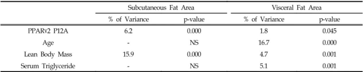
피하지방과 내장지방면적에 대한 PPARγ2 유전 자형의 영향을 다변량 분석을 이용하여 평가하였 다(Table VII). 나이, lean body mass, serum tri- glyceride가 모델에 포함되었다. PPARγ2 유전자 형은 피하지방면적변화의 6.25%를 내장지방면적 변화의 1.8%를 설명하였다. 이러한 결과는 PPARγ 2 유전자형이 내장지방보다 피하지방에서 더 큰 영향을 가진다는 것을 명백히 보여준다. 나이와 혈 청 triglyceride는 내장지방면적의 변화를 부분적 으로 설명하였고 피하지방면적의 변화는 lean body mass로 부분적으로 설명되었다.
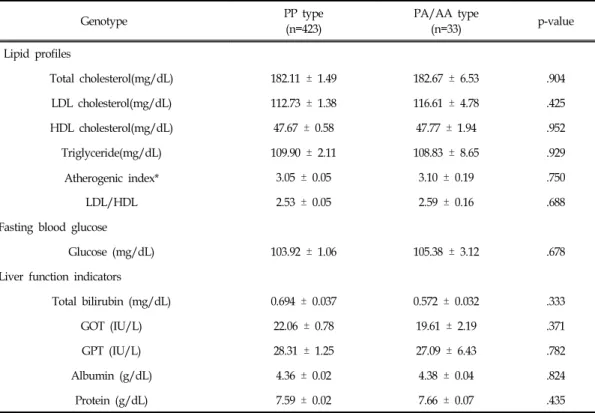
혈청지질, glucose, 간기능 지표들은 PPARγ2 유 전자형에 관련하여 명백한 차이를 보이지 않았다 (Table VIII). 과체중집단 198명을 대상으로 한달 동안의 저열량식이와 운동으로 구성된 체중감량 프로그램을 하는 동안 PPARγ2 유전자형에 따른 체중, BMI, WHR, 체지방량의 변화를 비교하였으 나 의미 있는 차이를 나타내지는 아니하였다 (Table IX).
전체적으로 본 연구의 결과는 PPARγ2 유전자 형이 과체중 여성에서 복부와 허벅지 지방조직의 지방축적에 영향을 주는 것을 증명하였다. 또한 PPARγ2 유전자형은 내장지방조직보다 피하지방 의 축적에 더 큰 영향을 주는 것이 관찰되었다.
Table VII. Source of Variation in Subcutaneous and Visceral Fat Areas in Overweight Subjects With BMI of Greater Than 25
Subcutaneous Fat Area Visceral Fat Area
% of Variance p-value % of Variance p-value
PPARγ2 P12A 6.2 0.000 1.8 0.045
Age - NS 16.7 0.000
Lean Body Mass 15.9 0.000 4.7 0.001
Serum Triglyceride - NS 5.1 0.001
NS : not significant.
Table VIII. Comparison of Serum Biochemical Parameters by PPARγ2 Genotype in Overweight Subjects With BMI of Greater Than 25
Genotype PP type
(n=423)
PA/AA type
(n=33) p-value
Lipid profiles
Total cholesterol(mg/dL) 182.11 ± 1.49 182.67 ± 6.53 .904
LDL cholesterol(mg/dL) 112.73 ± 1.38 116.61 ± 4.78 .425
HDL cholesterol(mg/dL) 47.67 ± 0.58 47.77 ± 1.94 .952
Triglyceride(mg/dL) 109.90 ± 2.11 108.83 ± 8.65 .929
Atherogenic index* 3.05 ± 0.05 3.10 ± 0.19 .750
LDL/HDL 2.53 ± 0.05 2.59 ± 0.16 .688
Fasting blood glucose
Glucose (mg/dL) 103.92 ± 1.06 105.38 ± 3.12 .678
Liver function indicators
Total bilirubin (mg/dL) 0.694 ± 0.037 0.572 ± 0.032 .333
GOT (IU/L) 22.06 ± 0.78 19.61 ± 2.19 .371
GPT (IU/L) 28.31 ± 1.25 27.09 ± 6.43 .782
Albumin (g/dL) 4.36 ± 0.02 4.38 ± 0.04 .824
Protein (g/dL) 7.59 ± 0.02 7.66 ± 0.07 .435
Data are mean ± SE.
* Atherogenic index (AI) = (total cholesterol - HDL cholesterol) / HDL cholesterol.
†LDL cholesterol to HDL cholesterol ratio.
‡P values were obtained by general linear model (covariance) analysis adjusted for age.
Table IX. Changes in Physical Characteristics and Body Fat Mass During a 1-Month Weight Loss Program among Overweight Subjects With BMI of Greater Than 25
Genotype PP type
(n=181)
PA/AA type
(n=17) p-value
Weight(Kg) - 7.06 ± 0.19 - 7.23 ± 0.79 0.799*
BMI(Kg/m2) - 2.92 ± 0.12 - 2.81 ± 0.33 0.755
WHR - 0.035 ± 0.005 -0.054 ± 0.010 0.243
Fat mass(Kg) - 5.05 ± 0.23 - 5.98 ± 0.91 0.249
Data are mean SE.
* P values were obtained by general linear model (covariance) analysis adjusted for age.
Ⅳ.
고 찰최근 Masud and Ye8) 은 30개의 독립적인 연구 로부터 얻은 총인원 19,136명의 데이터를 사용하 여 meta분석을 수행한 결과 PPARγ2 유전자의 Ala allele이 명백히 더 높은 BMI와 연관되어 있 다고 보고하였다. 본 연구의 결과에서도 체중, BMI, WHR은 Ala allele에 의해 확실히 더 높고 이것은 meta-analysis 결과와 일치한다(Table II).
이러한 meta-analysis 결과와의 일관성은 본 연구 의 피험자 집단이 일반적인 집단을 잘 대표하고 있으며 본 연구의 다른 데이터도 신뢰성이 있음을 제시한다.
지금까지 CT로 측정된 체지방 분포에서의 PP ARγ2 유전자 다형성의 영향은 연구된 바가 극히 적다. Mori 등17)은 215명의 남자 일본인에서 BMI, 피하지방면적, 내장지방면적에 있어서 PPARγ2 유 전자형의 영향이 없다고 보고하였다. 그러나 이 보 고의 경우 연구대상의 수가 작았으며(203 Pro homozygotes와 12 Ala allele carriers) BMI에 미 치는 영향이 meta분석결과와 일치하지 않아 일반 집단을 잘 반영하고 있다고 보기 어렵다. 반면에 Robitaille 등9)은 720명의 French Canadians을 대 상으로 한 Quebec Family Study에서 Ala allele 이 피하지방, 내장지방, BMI, 허리둘레, 체지방량 을 증가시키는 효과가 있음을 보고하였다. 그들은 Ala allele이 내장지방과 피하지방의 면적을 14%
와 27% 증가시킨다고 보고하였다. 한국여성을 대 상으로 한 본 연구에서 Ala allele의 효과는 내장 지방면적과 피하지방면적이 16%와 28% 높아져서 French Canadian 집단에서의 효과와 거의 동일하 다(Table IV). 한국인과 Quebec Family Study 의 Caucasian 집단 사이에는 유전적 배경과 식이패 턴이 분명히 다를 것이 예상되지만 <Table II>와
<Table IV>의 결과에서 나타난 Ala allele의 BMI, 체지방량, CT로 측정된 복부지방면적에 대한 영향 은 유사하다. 본 연구에서는 허벅지의 지방면적이 추가적으로 측정되었으며 Ala allele은 복부와 허 벅지의 지방조직에 비슷한 영향이 있었다(Table IV).
<Table V>와 <Table VI>에서 보여주는 자료는 피험자의 비만도 상태에 따른 체지방면적에 대한 Ala allele의 영향을 보여준다. 마른 피험자에서는 Ala allele은 지방면적에 뚜렷한 영향이 없는 반면 과체중집단에서는 통계적으로 뚜렷한 영향이 있었 다. 이 결과는 Ala allele 이 BMI가 27보다 큰 집 단에서 비만도와 확실히 관련되어있지만 BMI가 27보다 작은 집단내에서는 명백한 관련이 발견되 지 않는다는 Masud and Ye8) 의 meta분석과 일 치한다. 마른 피험자와 과체중 피험자에서 Ala allele이 다른 효과를 나타내는 것은 PPARγ2 유전 자형의 영향이 다른 요소에 의해 변형될 수 있다는 것을 알려준다. 기존 연구에 의하면 마른 피험자에 서 보다 과체중 피험자의 지방조직에서 PPARγ2 mRNA의 발현 level이 더 높으며 그 level은 저칼 로리 식이조절에 의해 감소된다18). 비만 그룹에서 PPARγ2 mRNA의 높은 발현 level은 Pro12Ala 유전자 다형성에 의한 PPARγ2 활성의 작은 차이 를 크게 증폭시킬 수 있다.이러한 증폭은 마른 피 험자에서는 충분하지 못하여 유의적인 영향에 도 달하지 못할 것이다. 더불어 PPARγ2 유전자형의 영향은 식이중의 포화지방과 불포화지방의 비율에 의하여 조절될 수 있다고 보고되어 있는데19) 이러 한 Gene- nutrient 상호작용이 지방축적에 대한 Ala allele의 영향을 조절할 것이다. <Table IV>는 전체 피험자에서 PPARγ2 유전자형에 따라 피하 지방은 유의적인 차이가 있으나 내장지방에는 뚜 렷한 차이가 없음을 보여주고 있다. 과체중그룹의 경우에도 Ala allele에 의하여 복부피하지방과 내
장지방이 각각 28%와 13%로 높게 나타나 PPARγ 2 유전자형이 내장지방에서보다 피하지방에서 더 큰 효과가 있다는 것을 알려준다(Table VI). Table VII은 내장지방보다 피하지방에서 PPARγ2의 더 큰 영향을 더 분명히 보여준다. Lefebvre 등20) 은 PP ARγ2가 내장지방조직보다 피하지방에서 더 높은 level로 발현된다고 보고하였다. 피하지방조 직의 PPARγ2의 더 높은 발현수준은 Pro12Ala 유 전자 다형성의 의한 활성의 미묘하게 차이를 충분 히 증폭시킬 수 있을 것이지만 내장지방에서는 증 폭이 더 낮을 것이다21).
내장지방축적이 대사증후군과 연관되어 있다는 것은 잘 알려져 있으나 피하지방과의 연관은 알려 져 있지 않다. PPARγ activator인 Troglitazone은 대사증후군을 개선시키는 동시에 피하지방면적을 증가시킨다고 보고되어있어 피하지방축적과 대사 증후군과의 관련성을 부정하는 결과를 제시한다22). 또한Matsuzawa 등23) 은 피하지방에 대한 복부지 방의 비율이 대사증후군을 예측하는 대단히 우수 한 지표라고 제시하고 있으며 본 연구에서 그 비 율은 Ala allele에 의하여 차이를 보이고 있지 않 다(Table IV, Table VI). 위의 사실은 <Table VIII>
에서 보이는 PPARγ2 유전자형에 의한 대사지표 에의 뚜렷하지 못한 영향을 설명하고 있다.
지방형성과정에 대한 PPARγ2 유전자 다형성의 분자적 메카니즘은 현재까지 설명되고 있지 않다.
Deeb 등24) 은 12번째 코돈의 Pro에 대한 Ala의 대체는 PPARγ2의 promotor에 대한 binding affinity의 감소를 가져오고 시험관 실험에서 전사 활성을 줄인다고 보고하였다. 반대로 이 연구의 결 과는 Robitaille 등9) 의 연구와 함께 Ala allele이 지방형성을 증가시키는 것을 제시한다. Ala allele 의 효과는 시험관과 생체내의 조건에서 서로 다를 것으로 생각된다. 시험관 시험에 사용된 세포주는 종종 몇몇 생체기작이 없음이 알려져 있고 인체
내에서의 상황과는 다를 수 있다. Soukas 등25) 은 시험관내와 생체내의 지방세포에서 유전자 발현의 중요한 차이점을 발견하였다. 또한 시험관 실험에 서는 고효율 벡터의 도입에 의해 PPARγ2 단백질 이 비생리적으로 많은 양 발현되어 생체내와 다른 반응을 나타낼 수 있다. 너 나아가 시험관 실험은 매우 짧은 기간에 행해지지만 인체내의 지방축적 은 태아시기에 시작되어 일생동안 진행된다. 긴 기 간의 효과는 짧은 시간의 효과와는 다를 것이다.
앞으로의 연구는 PPARγ2 유전자 다형성의 생체 내에서의 메카니즘을 밝히는 것이 필요하다.
참고문헌
1. Spiegelman BM. PPARγ: adipogenic regu- lator and thiazolidinedione receptor. Diabe- tes Care. 1998;47:507-14
2. Elbrecht A, Chen Y, Cullinan CA, Hayes N, Leibowitz MD, Moller DE, Berger J. Moleular cloning, expression and characterization of human peroxisome proliferators activated receptors γ1 and γ2. Biochem Biophys Res Commun. 1996;224:431-7
3. Werman A, Hollenberg A, Solanes G, Bjorbaek C, Vidal-Puig AJ, Flier JS. Lig- and-independent activation domain in the N-terminus of peroxisome proliferators acti- vated receptor γ : differential activity of PPARγ-1 and γ-2 isoforms and influence of insulin. J Biol Chem. 1997;272:20230-5 4. Barroso I, Gurnell M, Growley VE, Agostini
M, Schwabe JW, Soos MA et al. Dominant negative mutations in human PPARγ asso- ciated with severe insulin resistance, diabetes
mellitus and hypertension. Nature. 1999; 402:
880-3
5. Ristow M, Muller-Wieland D, Pfeiffer A, Krone W, Kahn CR. Obesity associated with a mutation in a genetic regulator of adi- pocyte differentiation. N Engl J Med. 1998;
339:953-9
6. Yen C-J, Beamer BA, Negri C, Silver K, Brown KA, Yarnall DP et al. Molecular scanning of the human peroxisome prolif- erators activated receptor γ(hPPARγ) gene in diabetic Caucasians: identification of a Pro 12Ala PPARγ-2 missense mutation. Biochem Biophys Res Commun. 1997;241:270-4 7. Altshuler D, Hirschhorn JN, Klannemark M,
Lindgren CM, Vohl MC, Nemesh J, et al. The Common PPARγ Pro12Ala polymorphism is associated with decreased risk of type 2 diabetes. Nat Genet. 2000;26:76-80
8. Masud S, Ye S. Effect of the peroxisome proliferators activated receptor-γ gene Pro 12Ala variant on body mass index: a meta analysis. J Med Genet. 2003;40:773-80 9. Robitaille J, Despres JP, Perusse L, Vohl MC.
The PPAR-γ P12A polymorphism modulates the relationship between dietary fat intake and components of the metabolic syndrome:
results from the Quebec Family Study. Clin Genet. 2003;63:109-16
10. Fu M, Chen H, Li X, Li J, Wu B, Cheng L, et al. Association of Pro12Ala variant in peroxisome proliferator-activated receptor-γ 2 gene with type 2 diabetes mellitus.
Zhonghua Yi Xue Yi Chuan Xue Za Zhi.
2002;19:234-8
11. Lei HH, Chen MH, Yang WS, Chiu MC, Chen MC, Tai TY, et al. Peroxisome pro- liferator-activated receptor γ 2 Pro12Ala gene variant is strongly associated with larger body mass in the Taiwanese. Me- tabolism. 2000;49:1267-70
12. Mori H, Ikegami H, Kawaguchi Y, Seino S, et al. The Pro12Ala substitution in PPARγ is associated with resistance to development of Diabetes in the General Population.
Diabetes. 2001; 50:891-894
13. Beamer BA, Yen CJ, Andersen RE, Muller D, Elahi D, Cheskin LJ, et al. Association of the Pro12Ala variant in the peroxisome proliferator-activated receptor-γ2 gene with obesity in two Caucasian populations. Dia- betes. 1998;47:1806-8
14. Schaffler A, Barth N, Schmitz G, Zietz B, Palitzsch KD, Scholmerich J. Frequency and significance of Pro12Ala and Pro115Gln polymorphism in gene for peroxisome pro- liferation-activated receptor-γ regarding met- abolic parameters in a Caucasian cohort.
Endocrine. 2001;14:369-73
15. Kolehmainen M, Uusitupa MI, Alhava E, Laakso M, Vidal H: Effect of the Pro12Ala polymorphism in the peroxisome prolif- erator-activated receptor (PPAR) γ2 gene on the expression of PPARγ target genes in adipose tissue of massively obese subjects. J Clin Endocrinol Metab. 2003;88:1717-22 16. Nieman DC. Exercise testing and prescrip-
tion: A health related approach, 4th ed.
Mayfield, CA, Mountain View. 1999 17. Mori Y, Kim-Motoyama H, Katakura T,
Yasuda K, Kadowaki H, Beamer BA, et al.
Effect of the Pro12Ala variant of the human peroxisome proliferators-activated receptor γ2 gene on adiposity, fat distribution, and insulin sensitivity in Japanese men. Bio- chem Biophys Res Commun. 1998;251:
195-198
18. Vidal-PuigAN, Considine RV, Jimenez- Linan M, Werman A, Pories WJ, Caro JF, et al. Peroxisome proliferator-activated recep- tor gene expression in human tissues.
Effects of obesity,weight loss, and regula- tion by insulin and glucocorticoids. J Clin Invest. 1997;99:2416-22
19. Luan J, Browne PO, Harding AH, Halsall DJ, O'Rahilly S, Chatterjee VK, et al. Evi- dence for gene-nutrient interaction at the PPARγ locus. Diabetes. 2001;50:686-9 20. Lefebvre A, Laville M, Vaga N, Riou J, Van
Gall L, Auwerx J, et al. Depot-specific dif- ferences in adipose tissue gene expression in lean and obese subjects. Diabetes. 1998;
47:98-103
21. Bjorntorp P: Metabolic implication of body fat distribution. Diabetes Care. 1991;14:1132- 43
22. Akazawa S, Kawasaki E, Sun F, Eguchi K, Ito M. Efficacy of troglitazone on body fat distribution in type 2 diabetes. Diabetes Care. 2000;23:1067-71
23. Matsuzawa Y, Nakamura T, Shimomura I, Kotani K. Visceral fat accumulation and cardiovascular disease. Obes Res. 1995;
Suppl 5:645-7
24. Deeb SS, Fajas L, Nemoto M, Pihlajamaki J, Mykkanen L, Kuusisto J, et al. A Pro12Ala substitution in PPARγ2 associated with decreased receptor activity, lower body mass index and improved insulin sensi- tivity. Nat Genet. 1998;20:284-7
25. Soukas A, Socci ND, Saatkamp BD, Novelli S, Friedman JM. Distinct transcriptional profiles of adipogenesis in vivo and in vitro. J Biol Chem. 2001;36:34167-74