Received August 20, 2018; revised October 12, 2018; accepted October 23, 2018.
Corresponding author: Seung-Woon Rha, Cardiovascular Center, Korea University Guro Hospital, 148 Gurodong-ro, Guro-gu, Seoul 08308, Korea.
E-mail: [email protected]
Copyright Ⓒ 2018 The Korean Academy of Clinical Geriatrics
This is an open access article distributed under the term s of the Creative Com m ons Attribution Non-Com m ercial License (http://creativecom m ons.org/licenses/by-nc/4.0) which perm its unrestricted non-com m ercial use, distribution, and reproduction in any m edium , provided the original work is properly cited.
Popliteal Pseudoaneurysm Complicated by Massive Hematoma following Catheter-Directed Thrombolysis for a May-Thurner Syndrome Patient with Deep Vein Thrombosis
Eun-Jin Park, Seung-Woon Rha
Cardiovascular Center, Korea University Guro Hospital, Seoul, Korea
We present a patient with massive deep vein thrombosis (DVT) resulting from May-Thurner syndrome as known as the iliac vein compression syndrome. He underwent an angioplasty procedure in which stents were placed at ileofemoral vein and cathe- ter-directed thrombolysis was performed. This angioplasty procedure was complicated by blood vessel wall injuries such as pseu- doaneurysm at his right popliteal blood vessel. Manual compression, thrombin injection to pseudoaneurysm and hematoma aspira- tion could not resolve the complications. Finally, surgical drainage could completely resolve the hemorrhagic complications.
Therefore, catheter-directed thrombolysis requires particular attention to post-procedure close observation, especially, within one week of onset of DVT as well as within 48 hours of procedure.
Key Words: Fibrinolytic agent, May-thuner syndrome, Peripheral Catheterization, Venous thrombosis
INTRODUCTION
Conventional anticoagulant treatment for acute deep vein thrombosis (DVT) effectively prevents thrombus extension and recurrence. It’s not through the way of dissolving the clot directly, rather, they inhibit new clot forming, activate the body’s own fibrinolytic system and restore venous patency and function [1]. When anticoagulation is insufficieint to cure the DVT, there remains post thrombotic syndrome char- acterised by pain, swelling, a sensation of heaviness, edema, pigmentation, and deterioration of the skin, including venous ulcers in severe cases [2]. Therefore, in many cases, cathe- ter-directed thrombolysis which actively dissolves clot rather than inhibits clot formation, is necessary to get optimal ther-
aputic result of DVT despite of its hemorrhaging complica- tions [3,4]. The present case describes a pateint who experi- enced massive DVT resulting from May-Thurner syndrome.
The patient had undergone catheter-directed thrombolysis be- cause he was not completely healed despite of balloon angio- plasty, stenting and systemic anticoagulation, resulting in a popliteal pseudoaneurysm and massive hematoma, which was completely resolved by surgical drainage.
CASE REPORT
A 66-year old man, who was injured his left tibia by acci- dent a month ago and undergone tibia fixation surgery, was transferred to our center due to sustained pain and edema in
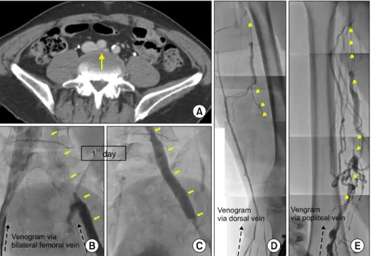
Figure 1. Stent implantation and follow-up venography after cath- eter-directed thrombolysis. (A), Axial plane of computed tomography venography (iliac bifurcation level) of the patient when he was transferred in. Long arrow indi- cates compressed left common iliac vein (CIV). (B), Invasive femoral venography of both side via femoral vein. (C), Stent implan- tation in left femoral vein. Short arrows indicate filling defect of left CIV (B) and well contrasted left CIV after stenting (C). (D), (E), Invasive femoral venography via left dorsal vein and left popliteal vein after 4 days. There was still a considerable amount of thrombus (Arrow heads).
his left leg. He was on anticoagulation therapy for more than 20 days with diagnosis of ileofemoral DVT. He had no past clinical history and on admission, his laboratory exams were within normal range but computed tomography (CT) venog- raphy of lower extremity showed thrombotic filling defect from left external iliac vein to popliteal vein. His left com- mon iliac vein was being compressed between right common iliac artery and vertebral body, indicating May-Thurner syn- drome (Figure 1A). We decided to do a mechanical re- vascularization by percutaneous transluminal angioplasty for his left common iliac vein because of his refractory symptoms and poor response to anticoagulation. Furthermore, May-thurner syndrome may cause ileofemoral DVT recurrently without mechanical intervention.
At first day, we tried to access left popliteal vein in prone position under fluoroscopic guidance using popliteal arterial pulse and vascular calcification marking. However, we punc- tured popliteal artery several times rather than vein, so we could identify filling defect from left common femoral vein to iliac vein bifurcation site only after bilateral femoral vein ap- proaching (Figure 1B). After quick placement of intravenous vena caval filter and balloon angioplasty, we implanted a stent in left iliac vein (Smart control 10.0×80 mm, Cordis, USA, Figure 1C) and started systemic anticoagulation with rivarox-
aban, aspirin and intravenous heparin. Unfortunately, 4 days later, in follow up venography via dorsal vein of foot, pop- liteal vein was not visible due to thrombus and venous flow was being drained via saphenous vein to femoral vein (Figure 1D). To evaluate the status of the iliac vein, we punctured his left popliteal vein again as we tried the previous day, and finally, confirmed large amount of remnant thrombi in pop- liteal and femoral vein (Figure 1E). After balloon angioplasty using Armada 5.0×250 mm (Abbott vascular, USA) at his left femoral vein, we started intravenous urokinase infusion through multi-side-ported infusion catheter (Cook, USA) through left popliteal vein to femoral vein. After maintaining for 3 days of urokinase infusion, we confirmed decreased thrombotic burden at follow-up venography but still remained. But because the patient had to be transferred out to the hospital where he had taken tibial surgery to get a treatment for his wound, we transferred out the patient ceas- ing infusion of urokinase while maintaining of rivaroxaban and aspirin.
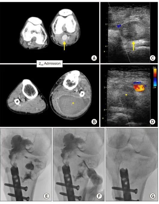
The patient was re-transferred to our center 10 days later because of persistent painful swelling in his left leg. In follow up CT venography, the thrombosis was resolved except small amount of thrombus in femoral vein. But there were pseu- doaneurysm with massive intramuscular hematoma (fluid col-
Figure 2. Popliteal pseudoan- eurysm and massive hematoma as complications of catheter-dir- ected thrombolysis. (A, B) Axial plane of computed tomography venography of the patient (pop- liteal level) when he was re-tran- ferred in. (C, D) Doppler guided thrombin injection to pseudoaneu- rysm. Arrows in (A) and (C) indi- cate popliteal artery and sur- rounding hematoma. Asterisks in (B) and (D) indicate huge fluid collection. (E, F, G), Manual aspir- ation of hematoma and pseudoan- eurysm under the guidance of fluoroscopy.
lection) which were expected to be originated from popliteal artery (arrow in Figure 2A, asterisk in 2B). The lesion might be complications of trans-catheter intravenous thrombolysis combined with injury of popliteal artery during multiple at- tempts to puncture popliteal vein under fluoroscopic guidance. Without mechanical method they would not resolve naturally, we injected thrombin into pseudoaneurysm under Doppler guidance (Figure 2C, D), and aspirated hematoma under fluoroscopic guidance (Figure 2E, F, G). Despite such procedure and conservative management for 1 week, pseudo- aneurysm accompanied by massive hematoma did not resolve.
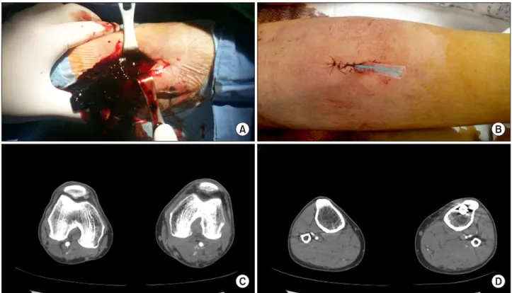
Thereafter we opened the calf and directly removed the hem-
atoma by incision and drainage (Figure 3A, B). Finally, after the surgical manipulation, hematoma was completely resolved (Figure 3C, D) and patient’s symptoms were improved.
DISCUSSION
DVT has a higher prevalence in elderly than younger pop- ulation, because the elderly has less mobility. When the eld- erly person gets hurt and becomes bed-ridden or if there is a congenital vascular problem such as may-thurner syndrome, the incidence of DVT is further increased. May–Thurner syndrome is an anatomically variable condition of venous out-
Figure 3. Resolution of pseudoaneurysm and hematoma by surgical drainage. (A, B) Incision and drainage of pseudoaneurysm and hematoma. (C, D) Completely resolved aneurysm hematoma in follow up computed tomography.
flow obstruction caused by extrinsic venous compression by the arterial system against bony structures. Especially a con- dition that left common iliac vein is compressed by the over- lying right common iliac artery so that leads stasis of venous return. The exact incidence and prevalence of May-Thurner syndrome is still unknown [5].
Previous studies are showing that the catheter-directed thrombolysis especialy in ileofemoral DVT is more effective than conventional systemic anticoagulation to prevent post thrombotic syndrome [3,4,6]. Eden et al. showed significant reduction of post thrombotic syndrome after addiitonal cathe- ter-directed thrombolysis compared with conventional treat- ment in their randomised trial [3]. Bashir et al. showed that the mortality between two groups, catheter-directed thrombol- ysis plus anticoagulation group and anticoagulation alone group, was not significantly different in there nation wide ob- servatoinal study. The adverse events were higher in cathe- ter-directed thrombolysis group, but they decreased over study period reflecting learning curve in terms of dose and duration [4].
In our case, because DVT which was developed secondary
to May-Thurner syndrome so that had little response to an- gioplasty combined with stenting, we conducted cathe- ter-directed thrombolysis and resulted in serious complications at puncture site. However, there are several reasons why such complications may have occurred, and we should check the point. First, since the patient was tranfered in 20 days after DVT was diagnosed, the thrombus would not have been fresh. Moreover, since sysemic anticoagulation including in- travenous heparin and non-vitamine K oral anticoagulant did not dissolve the thrombus, we started catheter-directed throm- bolysis 4 days after he arrived at our hospital. Catheter-di- rected thrombolysis is known to be effective in clots whithin a week of its occurenc [1]. Therefore, the timing of our treatment was not appropriate. Second, when we puncture the popliteal vein, ultrasound guidance would have been safer than fluoroscopic guidance. The popliteal artery was uninten- tionally damaged because we tried to puncture the vein rely- ing only on popliteal arterial pulse and calcific vascular mark- ing under fluoroscopic guidnce. Third, we maintained the in- fusion of urokinase more than 48 hours. Hofmann et al. re- command that catheter-directed thrombolysis should be com-
pleted within 1∼1.5 days and not exceed 2 days [7]. If the clot does not dissolve within this time, it should be consid- ered chronic in nature. Given these points, we would not have caused the such severe hemorrhagic complications of this patient if we used safer devices and tools to punture, start catheter-directed thrombolysis earlier, and shorten its maintenance period. Fortunately, the patient’s pseudoaneurysm and hematoma was successfully resolved through surgical drainage. If we follow the ideal treatment time point and du- ration of maintenance, and can handle the complications as well, catheter-directed thrombolysis will still be better option than anticoagulation alone in ileofemoral DVT patinets.
CONFLICTS OF INTEREST
No potential conflict of interest relevant to this article was reported.
REFERENCES
1. Shuman LS. Catheter-directed thrombolysis of extensive deep venus thrombosis. J Lanc Gen Hosp 2008;3:61.
2. Kahn SR. The post‐thrombotic syndrome: progress and pitfalls.
Br J Haematol 2006;134:357-65.
3. Enden T, Haig Y, Klow NE, Slagsvold CE, Sandvik L, Ghanima W, et al. Long-term outcome after additional cathe- ter-directed thrombolysis versus standard treatment for acute iliofemoral deep vein thrombosis (the CaVenT study): a rando- mised controlled trial. Lancet 2012;379:31-8.
4. Bashir R, Zack CJ, Zhao H, Comerota AJ, Bove AA.
Comparative outcomes of catheter-directed thrombolysis plus anticoagulation vs anticoagulation alone to treat low- er-extremity proximal deep vein thrombosis. JAMA Intern Med 2014;174:1494-501.
5. Mousa AY, AbuRahma AF. May-Thurner syndrome: update and review. Ann Vasc Surg 2013;27:984-95.
6. Liew A, Douketis J. Catheter-directed thrombolysis for ex- tensive iliofemoral deep vein thrombosis: review of literature and ongoing trials. Expert Rev Cardiovasc Ther 2016;14:
189-200.
7. Hofmann LV, Kuo WT. Catheter-directed thrombolysis for acute DVT. Lancet 2012;379:3-4.