Background and Purpose Fibroblast growth factor 23 (FGF23) is associated with athero- sclerosis via nitric-oxide-associated endothelial dysfunction and calcium-phosphate-related bone mineralization. This study aimed to determine the association of the plasma FGF23 concentration with intracranial cerebral atherosclerosis (ICAS) and extracranial cerebral ath- erosclerosis (ECAS).
Methods We prospectively enrolled 262 first-ever ischemic stroke patients in whom brain magnetic resonance was performed and a blood sample acquired within 24 h after admis- sion. Plasma FGF23 concentrations were measured using an enzyme-linked immunosorbent assay. The presence of ICAS or ECAS was defined as a ≥50% decrease in arterial diameter in magnetic resonance angiography. The burden of cerebral atherosclerosis was calculated by adding the total number of vessels defined as ICAS or ECAS.
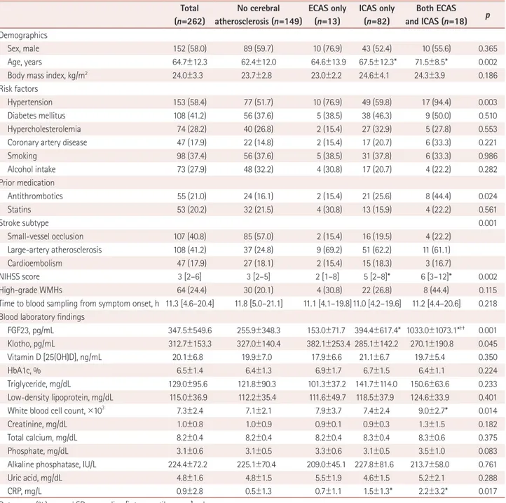
Results Our study population included 152 (58.0%) males. The mean age was 64.7 years, and the plasma FGF23 concentration was 347.5±549.6 pg/mL (mean±SD). ICAS only, ECAS only, and both ICAS and ECAS were present in 31.2% (n=82), 4.9% (n=13), and 6.8% (n=18) of the subjects, respectively. In multivariate binary and ordinal logistic analyses, after adjust- ing for sex, age, and variables for which p<0.1 in the univariate analysis, the plasma FGF23 concentration (per 100 pg/mL) was positively correlated with the presence of ICAS [odds ra- tio (OR)=1.07, 95% CI=1.00–1.15, p=0.039], burden of ICAS (OR=1.09, 95% CI=1.04–1.15, p=0.001), and burden of ECAS (OR=1.06, 95% CI=1.00–1.12, p=0.038), but it was not signifi- cantly related to the presence of ECAS (OR=1.05, 95% CI=0.99–1.12, p=0.073).
Conclusions The plasma FGF23 may be a potential biomarker for cerebral atherosclerosis, particularly the presence and burden of ICAS in stroke patients.
Key Words fibroblast growth factor 23, Klotho, cerebral atherosclerosis, intracranial atherosclerosis.
Plasma Fibroblast Growth Factor 23 Concentration Is Associated with Intracranial Cerebral Atherosclerosis in Acute Ischemic Stroke Patients
INTRODUCTION
Intracranial cerebral atherosclerosis (ICAS) is a major risk factor for cerebrovascular dis- ease, particularly in Asians. ICAS is also associated with recurrent stroke, stroke-related morbidity, and future mortality.1 The importance of ICAS for stroke patients and the as- sociated factors and risk factors remain unclear, although they have been suggested to be aging, racial differences (mainly Asian), hypertension, diabetes mellitus, smoking, obstruc- tive sleep apnea, and obesity.2-4 Thus, further evaluations to find the factors that explain ICAS are necessary.
Fibroblast growth factor 23 (FGF23) is an endocrine FGF and phosphaturic hormone Yoonkyung Changa*
Jinkwon Kimb* Ho Geol Wooc* Dong-Ryeol Ryud Hyung Jung Ohe Tae-Jin Songa
a Department of Neurology, Ewha Womans University Mokdong Hospital, Ewha Womans University College of Medicine, Seoul, Korea
b Department of Neurology, Gangnam Severance Hospital, Yonsei University College of Medicine, Seoul, Korea
c Department of Neurology, Ewha Womans University Seoul Hospital, Ewha Womans University College of Medicine, Seoul, Korea
d Department of Internal Medicine, Ewha Womans University College of Medicine, Seoul, Korea
e Ewha Institute of Convergence Medicine, Ewha Womans University
Mokdong Hospital, Seoul, Korea
pISSN 1738-6586 / eISSN 2005-5013 / J Clin Neurol 2020;16(1):29-36 / https://doi.org/10.3988/jcn.2020.16.1.29
Received April 17, 2019 Revised August 21, 2019 Accepted August 21, 2019 Correspondence Tae-Jin Song, MD, PhD Department of Neurology, Ewha Womans University Mokdong Hospital, Ewha Womans University College of Medicine,
1071 Anyangcheon-ro, Yangcheon-gu, Seoul 07985, Korea
Tel +82-2-2650-2677 Fax +82-2-2650-5958 E-mail [email protected]
*These authors contributed equally to this work.
cc This is an Open Access article distributed under the terms of the Creative Commons Attribution Non-Com- mercial License (https://creativecommons.org/licenses/by-nc/4.0) which permits unrestricted non-commercial use, distribution, and reproduction in any medium, provided the original work is properly cited.
JCN
Open Access ORIGINAL ARTICLEFGF23 and Intracranial Cerebral Atherosclerosis
JCN
mainly formed by osteoblasts and osteocytes.5 In harmony with Klotho (a single-pass transmembrane protein expressed in renal tubules), as an obligate coreceptor to bind and acti- vate FGF receptors, FGF23 is activated during bone miner- alization and turnover defects.6 In addition, kidney dysfunc- tion, adipose tissue inflammation, and vitamin D dysregulation are involved in systemic FGF23 regulation.7 FGF23 is also in- volved in systemic atherosclerosis via nitric-oxide-associated endothelial dysfunction and calcium-phosphate-related bone mineralization.8 Previous studies have found increased FGF23 to be associated with subclinical atherosclerosis and an in- creased left ventricular mass.9-11 Furthermore, FGF23 is an independent predictor of cardiovascular events in the general population.12
Major features of ICAS include atherosis caused by choles- terol deposition and inflammation and sclerosis secondary to endothelial dysfunction, leading to arterial stiffness.1 Be- cause FGF23 is also involved in the development of athero- sclerosis and endothelial dysfunction,9-11,13 this growth factor might also be associated with the atherosclerosis of intracra- nial vessels. We hypothesized that a higher circulating FGF23 concentration is associated with the presence and burden of cerebral atherosclerosis in patients with acute ischemic stroke.
METHODS
Subjects
We prospectively registered 262 patients with first-ever isch- emic stroke who were admitted to our institution between June 2014 and May 2016 within 7 days after symptom onset and had a stroke subtype classified as large-artery atheroscle- rosis, cardioembolism, or small-vessel occlusion. Standard protocols for the stroke registry were applied to all patients, which included a chest X-ray; 12-lead electrocardiography;
routine blood tests at admission (white blood cell count, WBC) and creatinine) and after a 12-h fast {vitamin D [25(OH)D], fasting glucose, HbA1c, triglyceride, total cholesterol, low- density lipoprotein, total calcium, phosphate, albumin, alka- line phosphatase, uric acid, and CRP}; brain imaging with CT and/or MRI; and vascular imaging with CT angiography, magnetic resonance angiography (MRA), or digital subtrac- tion angiography.14,15 Patients who did not agree to participate in the study, refused to provide blood samples, or who had a history of cancer, autoimmune disease, or bone fractures dur- ing the previous 2 months were excluded.
Risk factors were defined as in a previous study and the Supplementary Materials (in the online-only Data Supple- ment).16 The stroke subtype was classified using the Trial of Org 10172 in Acute Stroke Treatment (TOAST) classifica- tion system.17 Briefly, patients with potential cardiac emboli
were classified as the cardioembolic subtype. If a patient ex- hibited substantial stenosis of the relevant artery, they were classified as the large-artery atherosclerosis subtype. The small-vessel occlusion subtype was defined if all of the follow- ing criteria were met: clinical manifestation of classical lacu- nar syndrome, relevant subcortical or brainstem lesion, infarc- tion smaller than 15 mm, no significant stenosis in the relevant artery, and no potential cardiac embolism.18 The Fazekas scor- ing system was used to assess white-matter hyperintensities (WMHs) in MRI FLAIR.19 A Fazekas score of ≥2 in the peri- ventricular or deep white matter was defined as high-grade WMH. The renal function was investigated using the estimat- ed glomerular filtration rate according to the Modification of Diet in Renal Disease study equation.20 This study was ap- proved by our Institutional Review Board (approval no. ECT 2014-04-023), and we obtained informed consent from all participants and their next of kin.
MRI protocol and definition of vascular stenosis Every patient enrolled in this study underwent intracranial and extracranial MRA. All MR images were obtained using a 3.0-T imaging system (Magnetom Verio, Siemens Health- care, Erlangen, Germany). Three-dimensional time-of-flight sequences were used to evaluate the intracranial arteries (in- tracranial internal carotid artery, middle cerebral artery, an- terior cerebral artery, posterior cerebral artery, distal vertebral artery, and basilar artery) and the extracranial arteries (extra- cranial internal carotid artery and common carotid artery).
Extracranial cerebral atherosclerosis (ECAS) was not assessed in the extracranial vertebral arteries because it is difficult to distinguish hypoplasia of the vertebral artery from significant stenosis.3
The method used in this study to evaluate stenosis has been described previously,21 and >50% stenosis was defined as ICAS.22 ECAS was assessed using the criteria from the North American Symptomatic Carotid Endarterectomy Tri- al, with its presence defined as >50% stenosis at the bifurca- tion of the bilateral carotid artery.23 The most serious lesion was chosen when multiple stenotic lesions were present.22, 24 The total number of vessels with ICAS was considered as the ICAS burden (ranging from 0 to 11), while the total number of vessels with ECAS was considered the ECAS burden (rang- ing from 0 to 4).24 Two neurologists blinded to clinical infor- mation (Y.C. and T.J.S.) independently assessed the pres- ence of ICAS. The kappa value for interobserver agreement was 0.936 for the presence of ICAS. Any initial disagreements in the neurologist assessments of ICAS were resolved by discussion.
Chang Y et al.
JCN
Measurement of plasma FGF23 concentrations To measure the plasma FGF23 concentration, a blood sample were collected after a 12-h fast in EDTA tubes. The blood was centrifuged at 1,900×g and 4°C for 15 min to obtain plasma, and then kept at -80°C until being analyzed. The plasma FGF23 concentrations were measured using an ELISA with a commercial reagent (Kainos Laboratories, Tokyo, Japan).25 The concentration detection range was 3–800 pg/mL, and so the assay was repeated using a 1/10 dilution for concentrations
>800 pg/mL.25 The plasma FGF23 concentration was mea- sured twice by two investigators blinded to the medical data (Y.C. and D.R.R.) and then averaged. Intra- and interassay co- efficients of variability were 4.2% and 6.5%, respectively.
Statistical analysis
Statistical analyses were conducted using the SPSS software package for Windows (version 21.0, IBM Corp., Armonk, NY, USA). Continuous variables were analyzed using the indepen- dent t-test, Mann-Whitney test, one-way ANOVA with Bon- ferroni’s post-hoc analysis, and Kruskal-Wallis test. Categor- ical variables were analyzed using the chi-square or Fisher’s exact test. Univariate and multivariate binary logistic regres- sion analyses were performed to investigate the association be- tween the plasma FGF23 concentration and cerebral athero- sclerosis. To investigate the associations of age, body mass in- dex, stroke severity, time to blood sampling from symptom onset, and blood laboratory findings with FGF23, Spearman correlation analysis was performed for the correlations be- tween the National Institutes of Health Stroke Scale (NIHSS) score, blood laboratory findings, and FGF23 concentration.
The association between the plasma FGF23 concentra- tion and the burden of cerebral atherosclerosis was investi- gated by performing a Spearman correlation analysis and univariate and multivariate ordinal logistic regression anal- yses. Ordinal logistic regression was used to analyze the as- sociation between the FGF23 concentration and the number of arteries with cerebral atherosclerosis. The odds ratio (OR) in ordinal regression expresses the common odds for the in- crease in the number of arteries with cerebral atherosclero- sis at each count. Sex, age, and variables for which p<0.1 in the univariate analysis were included in the multivariate lo- gistic regression. In the multivariate analysis, sex, age, body mass index, coronary artery disease, prestroke antithrombot- ics, stroke subtype, NIHSS score, high-grade WMHs, Klotho, triglyceride, WBC count, total calcium, and C-reactive pro- tein were adjusted for the presence and burden of ICAS; sex, age, hypertension, stroke subtype, NIHSS score, high-grade WMHs, WBC count, phosphate, and uric acid were adjusted for the presence and burden of ECAS; and sex, age, hyper- tension, prestroke antithrombotics, NIHSS score, high-grade
WMHs, Klotho, triglyceride, WBC count, and C-reactive pro- tein were adjusted for the presence and burden of both ICAS and ECAS. For sensitivity analysis, we investigated the pres- ence of statistical interactions according to underlying renal dysfunction (<60 mL/min/1.73 m2), which was closely relat- ed to the plasma FGF23 concentration.11 A two-tailed proba- bility value of p<0.05 was considered statistically significant.
RESULTS
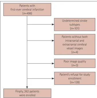
Demographic data and comparative analysis according to presence of cerebral atherosclerosis Fig. 1 shows a flowchart of participants according to the in- clusion and exclusion criteria applied in this study. The de- mographics and accompanying risk factors and prior stroke medications did not differ significantly between the includ- ed and excluded patients (Supplementary Table 1 in the on- line-only Data Supplement). Table 1 lists the demographics of the patients in our study. The 262 patients included 58.0%
(n=152) males. The mean age was 64.7 years, and the plasma FGF23 concentration was 347.5±549.6 pg/mL (mean±SD).
The most common stroke subtype was large-artery athero- sclerosis (41.2%, n=108), followed by small-vessel occlusion (40.8%, n=107) and cardioembolism (17.9%, n=47). The plas- ma FGF23 concentration did not differ among stroke sub- types (p=0.174). ICAS only, ECAS only, and both ICAS and ECAS were present in 31.2% (n=82), 4.9% (n=13), and 6.8%
(n=18) of the subjects, respectively (Table 1). The patients with ICAS and both ICAS and ECAS were older (p=0.002)
Patients with first-ever cerebral infarction
(n=498)
Undetermined stroke subtypes
(n=101)
Patient’s refusal for study enrollment
(n=128) Poor image quality
(n=3) Patients without both
intracranial and extracranial cerebral
vessel images (n=4)
Finally, 262 patients were enrolled
Fig. 1. Flowchart of participants according to inclusion and exclu- sion criteria.
FGF23 and Intracranial Cerebral Atherosclerosis
JCN
and had higher NIHSS scores (p=0.002) than those without cerebral atherosclerosis. Moreover, patients with ICAS only, ECAS only, or both ICAS and ECAS more often had a histo- ry of hypertension (p=0.003) and large-artery atherosclerosis stroke subtype (p=0.001).
The blood laboratory findings indicated that the plasma FGF23 concentration (p=0.001), WBC count (p=0.014), and
C-reactive protein concentration (p=0.017) were higher in patients with both ICAS and ECAS than in patients without cerebral atherosclerosis. Patients with ICAS had higher plasma concentrations of FGF23 (p=0.037) and C-reactive protein (p=0.043) compared with patients without cerebral athero- sclerosis. Patients with both ICAS and ECAS had a higher plas- ma FGF23 concentration compared with patients with only
Table 1. Comparison of clinical characteristics and blood laboratory findings according the presence of cerebral atherosclerosis Total
(n=262)
No cerebral atherosclerosis (n=149)
ECAS only (n=13)
ICAS only (n=82)
Both ECAS and ICAS (n=18) p Demographics
Sex, male 152 (58.0) 89 (59.7) 10 (76.9) 43 (52.4) 10 (55.6) 0.365
Age, years 64.7±12.3 62.4±12.0 64.6±13.9 67.5±12.3* 71.5±8.5* 0.002
Body mass index, kg/m2 24.0±3.3 23.7±2.8 23.0±2.2 24.6±4.1 24.3±3.9 0.186
Risk factors
Hypertension 153 (58.4) 77 (51.7) 10 (76.9) 49 (59.8) 17 (94.4) 0.003
Diabetes mellitus 108 (41.2) 56 (37.6) 5 (38.5) 38 (46.3) 9 (50.0) 0.510
Hypercholesterolemia 74 (28.2) 40 (26.8) 2 (15.4) 27 (32.9) 5 (27.8) 0.553
Coronary artery disease 47 (17.9) 22 (14.8) 2 (15.4) 17 (20.7) 6 (33.3) 0.221
Smoking 98 (37.4) 56 (37.6) 5 (38.5) 31 (37.8) 6 (33.3) 0.986
Alcohol intake 73 (27.9) 48 (32.2) 4 (30.8) 17 (20.7) 4 (22.2) 0.282
Prior medication
Antithrombotics 55 (21.0) 24 (16.1) 2 (15.4) 21 (25.6) 8 (44.4) 0.024
Statins 53 (20.2) 32 (21.5) 4 (30.8) 13 (15.9) 4 (22.2) 0.561
Stroke subtype 0.001
Small-vessel occlusion 107 (40.8) 85 (57.0) 2 (15.4) 16 (19.5) 4 (22.2)
Large-artery atherosclerosis 108 (41.2) 37 (24.8) 9 (69.2) 51 (62.2) 11 (61.1)
Cardioembolism 47 (17.9) 27 (18.1) 2 (15.4) 15 (18.3) 3 (16.7)
NIHSS score 3 [2–6] 3 [2–5] 2 [1–8] 5 [2–8]* 6 [3–12]* 0.002
High-grade WMHs 64 (24.4) 30 (20.1) 4 (30.8) 22 (26.8) 8 (44.4) 0.115
Time to blood sampling from symptom onset, h 11.3 [4.6–20.4] 11.8 [5.0–21.1] 11.1 [4.1–19.8]11.0 [4.2–19.6] 11.2 [4.4–20.6] 0.218 Blood laboratory findings
FGF23, pg/mL 347.5±549.6 255.9±348.3 153.0±71.7 394.4±617.4* 1033.0±1073.1*†‡ 0.001
Klotho, pg/mL 312.7±153.3 327.0±140.4 382.1±253.4 285.1±142.2 270.1±190.8 0.045
Vitamin D [25(OH)D], ng/mL 20.1±6.8 19.9±7.0 17.9±6.6 21.1±6.7 19.7±5.4 0.350
HbA1c, % 6.5±1.4 6.4±1.3 6.9±1.7 6.7±1.5 6.4±1.1 0.224
Triglyceride, mg/dL 129.0±95.6 121.8±90.3 101.3±37.2 141.7±114.0 150.6±63.6 0.233
Low-density lipoprotein, mg/dL 115.0±36.9 112.2±35.4 111.6±49.7 118.5±37.9 124.6±33.9 0.401
White blood cell count, ×103 7.3±2.4 7.1±2.1 7.9±3.7 7.4±2.4 9.0±2.7* 0.014
Creatinine, mg/dL 1.0±0.8 1.0±0.9 0.9±0.1 0.9±0.3 1.3±1.5 0.182
Total calcium, mg/dL 8.2±0.4 8.2±0.4 8.2±0.4 8.3±0.4 8.3±0.6 0.375
Phosphate, mg/dL 3.1±0.6 3.1±0.5 3.3±0.6 3.1±0.5 3.5±1.0 0.083
Alkaline phosphatase, IU/L 224.4±72.2 225.1±70.4 209.0±45.1 227.8±81.6 213.7±58.0 0.761
Uric acid, mg/dL 4.8±1.6 4.8±1.5 5.5±1.9 4.6±1.5 5.2±2.1 0.288
CRP, mg/L 0.9±2.8 0.5±1.3 0.7±1.1 1.5±1.3* 2.2±3.2* 0.017
Data are n (%), mean±SD, or median [interquartile range] values.
*p<0.05 compared with no cerebral atherosclerosis in a Bonferroni post-hoc analysis, †p<0.05 compared with ECAS only in a Bonferroni post-hoc analysis, ‡p<0.05 compared with ICAS only in a Bonferroni post-hoc analysis.
ECAS: extracranial cerebral atherosclerosis, FGF23: fibroblast growth factor 23, ICAS: intracranial cerebral atherosclerosis, NIHSS: National Institute of Health Stroke Scale, WMHs: white-matter hyperintensities.
Chang Y et al.
JCN
ICAS (p=0.001) or only ECAS (p=0.001) (Table 1).
The plasma FGF23 concentration was positively correlated with age (ρ=0.150, p=0.015), NIHSS score (ρ=0.198, p=0.001), vitamin D [25(OH)D] concentration (ρ=0.143, p=0.020), HbA1c (ρ=0.162, p=0.008), triglyceride concentration (ρ=
0.319, p<0.001), WBC count (ρ=0.130, p=0.035), and creat- inine (ρ=0.178, p=0.004), total calcium (ρ=0.153, p=0.043), phosphate (ρ=0.148, p=0.048), and C-reactive protein (ρ=
0.134, p=0.030) concentrations. Moreover, the plasma FGF23 concentration was negatively correlated with the Klotho con- centration (ρ=-0.325, p<0.001).
Association of FGF23 with presence of cerebral atherosclerosis
In a multivariate binary logistic analysis, the plasma FGF23 concentration was positively correlated with the presence of ICAS even after adjustment as a continuous variable (per 100 pg/mL) (OR=1.07, 95% CI=1.00–1.15, p=0.039) or di- chotomizing based on the median value (cutoff value=182.0 pg/mL; OR=2.52, 95% CI=1.36–4.68, p=0.003) and tertiles [comparing the upper tertile (≥235.16 pg/mL) with the lower tertile (0–146.79 pg/mL): OR=3.28, 95% CI=1.53–7.05] (Ta- ble 2, Supplementary Table 2 in the online-only Data Supple- ment). The plasma FGF23 concentration was marginally re- lated to the presence of ECAS after adjustment as a continuous variable (per 100 pg/mL; OR=1.05, 95% CI=0.99–1.12, p=
0.073), but it was not associated with the presence of ECAS after adjustment using the median cutoff value or tertiles (Table 2, Supplementary Table 2 in the online-only Data Sup- plement). The plasma FGF23 concentration was significantly and positively correlated with the presence of both ICAS and ECAS after adjustment as a continuous variable (per 100 pg/
mL; OR=1.10, 95% CI=1.02–1.19, p=0.013) (Table 2, Sup- plementary Table 2 in the online-only Data Supplement).
There was no statistical interaction between the presence of ICAS (p for interaction=0.872) and ECAS (p for interac- tion=0.764) with the presence of renal dysfunction (<60 mL/min/1.73 m2).
Association of FGF23 with the burden of cerebral atherosclerosis
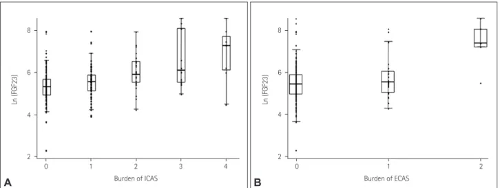
The plasma FGF23 concentration was associated with the ICAS burden (ρ=0.317, p=0.001) and the ECAS burden (ρ=
0.145, p=0.019). The association of FGF23 with the burden of cerebral atherosclerosis is presented as Fig. 2. In a multi- variate ordinal logistic analysis, the plasma FGF23 concentra- tion was positively correlated with the burden of ICAS even after adjustment as a continuous variable (per 100 pg/mL;
OR=1.09, 95% CI=1.04–1.15, p=0.001) or dichotomizing based on the median value (cutoff value=182.0 pg/mL; OR=
1.10, 95% CI=1.58–5.06, p=0.001) and tertiles [comparing the upper tertile (≥235.16 pg/mL) with the lower tertile (0–
146.79 pg/mL): OR=3.30, 95% CI=1.60–6.75] (Table 2, Sup- plementary Table 3 in the online-only Data Supplement).
The plasma FGF23 concentration was related to the burden of ECAS after adjustment as a continuous variable (per 100 pg/mL; OR=1.06, 95% CI=1.00–1.12, p=0.038) but not after adjustment using the median cutoff value or tertiles (Table 2, Supplementary Table 3 in the online-only Data Supplement).
The plasma FGF23 concentration was significantly and pos- itively correlated with the burden of both ICAS and ECAS after adjustment as a continuous variable (per 100 pg/mL;
OR=1.10, 95% CI=1.02–1.19, p=0.013) (Table 2, Supplemen- tary Table 3 in the online-only Data Supplement).
Fig. 2. Association of FGF23 with the burden of ICAS (A) and the burden of ECAS (B). X-axis indicates the burden of each type of cerebral athero- sclerosis, and Y-axis indicates the value of natural logarithm of FGF23. ECAS: extracranial cerebral atherosclerosis, FGF23: fibroblast growth factor 23, ICAS: intracranial cerebral atherosclerosis.
8
6
4
2
Ln (FGF23)
0 1 2 3 4 Burden of ICAS
A
8
6
4
2
Ln (FGF23)
0 1 2 Burden of ECAS
B
FGF23 and Intracranial Cerebral Atherosclerosis
JCN
DISCUSSION
The key finding of our study is that a higher plasma FGF23 concentration was associated with the presence and burden of cerebral atherosclerosis, particularly for ICAS. An associ- ation of FGF23 with cerebral atherosclerosis has rarely been reported. A community-based study found that a higher cir- culating FGF23 concentration was associated with systemic atherosclerosis.26 Moreover, a higher FGF23 concentration was found to be a risk factor for chronic kidney diseases, es- pecially in older, disabled, community-dwelling females.27 In patients with chronic kidney diseases, an elevated FGF23 con- centration reportedly contributes directly to a higher rate of left ventricular hypertrophy28 and a higher coronary artery disease burden.29 Combined with previous research, the pres- ent study is significant because it confirms the association between the plasma FGF23 concentration and the presence and burden of cerebral atherosclerosis, specifically in stroke patients.
While our study cannot suggest the exact mechanism link- ing plasma FGF23 and cerebral atherosclerosis, there are plausible hypotheses. First, vascular inflammation of cere- bral arteries may explain the relationship between FGF23 and cerebral atherosclerosis. Previous studies have found FGF23 to be an important mediator of vessel inflammation that pre- ceded arteriolosclerosis or arterial stiffness, which was the main cause of cerebral atherosclerosis development, partic-
ularly for ICAS.22,30 Moreover, FGF23 stimulates the hepatic secretion of the inflammatory markers IL-6 and C-reactive protein.30 C-reactive protein is a sensitive indicator of vascular inflammation and a marker of cerebral atherosclerosis.31 In line with these previous results, the present study found that the plasma FGF23 concentration was positively correlated with that of C-reactive protein, with the latter also being high- er in patients with accompanying ICAS than in those with- out cerebral atherosclerosis. Second, FGF23 is a phosphatu- ric hormone produced mainly by osteoblasts and osteocytes, and is implicated in blood calcium and phosphate concen- trations, the vitamin D pathway, and ectopic site mineraliza- tion.5 Increased FGF23 activity disturbs the calcitriol-calcium/
phosphate regulation pathway, which may cause hypercalce- mia and hyperphosphatemia, likely accounting for the asso- ciation with ectopic site mineralization.32,33 Previous studies have demonstrated the serum calcium concentration to be positively correlated with the presence of ICAS; in contrast, the serum phosphate concentration was not associated with ICAS.34 Furthermore, the phosphate concentration was posi- tively correlated with a greater prevalence of vascular calcifi- cation, which is frequently associated with ECAS.35,36 Therefore, an FGF23-related calcium-phosphate regulation mechanism may affect the development of cerebral atherosclerosis. Ac- tually, the present study found that the plasma FGF23 was positively correlated with serum calcium and phosphate con- centrations, and also that the serum calcium and phosphate Table 2. Results of multivariate analyses of the presence and burden of cerebral atherosclerosis
FGF23 Presence of cerebral atherosclerosis
ICAS† p ECAS‡ p Both ECAS and ICAS§ p
Continuous variable, per 100 pg/mL 1.07 (1.00–1.15) 0.039 1.05 (0.99–1.12) 0.073 1.10 (1.02–1.19) 0.013 Dichotomized based on median value of 182.0 pg/mL 2.52 (1.36–4.68) 0.003 1.59 (0.66–3.87) 0.248 Not available*
Tertiles
Lower tertile, 0–146.79 pg/mL Reference Reference Reference
Middle tertile, 146.79–235.16 pg/mL 1.96 (0.93–4.12) 0.076 1.97 (0.64–6.08) 0.482 Not available*
Upper tertile, ≥235.16 pg/mL 3.28 (1.53–7.05) 0.002 1.26 (0.42–3.75) 0.523 Not available*
FGF23 Burden of cerebral atherosclerosis
ICAS† p ECAS‡ p Both ECAS and ICAS§ p
Continuous variable, per 100 pg/mL 1.09 (1.04–1.15) 0.001 1.06 (1.00–1.12) 0.038 1.10 (1.02–1.19) 0.013 Dichotomized based on median value of 182.0 pg/mL 1.10 (1.58–5.06) 0.001 1.63 (0.66–3.99) 0.283 Not available*
Tertiles
Lower tertile, 0–146.79 pg/mL Reference Reference Reference
Middle tertile, 146.79–235.16 pg/mL 2.27 (1.10–4.67) 0.001 1.35 (0.44–4.11) 0.590 Not available*
Upper tertile, ≥235.16 pg/mL 3.30 (1.60–6.75) 0.026 2.09 (0.66–6.53) 0.204 Not available*
Data are odds ratio (95% CI) values.
*Odds ratio could not be obtained because the FGF23 concentrations of patients with both ECAS and ICAS exceeded 182.0 pg/mL, †Adjusted for sex, age, BMI, coronary artery disease, prestroke antithrombotics, stroke subtype, NIHSS score, high-grade WMHs, Klotho, triglyceride, WBC count, total calcium, and CRP, ‡Adjusted for sex, age, hypertension, stroke subtype, NIHSS score, high-grade WMHs, WBC count, phosphate, and uric acid, §Adjust- ed for sex, age, hypertension, prestroke antithrombotics, NIHSS score, high-grade WMHs, Klotho, triglyceride, WBC count, and CRP.
BMI: body mass index, ECAS: extracranial cerebral atherosclerosis, FGF23: fibroblast growth factor 23, ICAS: intracranial cerebral atherosclerosis, NI- HSS: National Institutes of Health Stroke Scale, WBC: white blood cell, WMHs: white-matter hyperintensities.
Chang Y et al.
JCN
concentrations were related to the presence and burden of ICAS and ECAS, respectively.
This study found that the presence and burden of ICAS were significantly associated with a higher FGF23 concen- tration, whereas the presence and burden of ECAS were not, even though they were related in a univariate analysis. These results may have been due to the sample being too small to reveal a relationship between FGF23 and ECAS. Moreover, other confounding factors such as hypertension are more strongly associated with ECAS than with the FGF23 concen- tration. In addition, arterial stiffness may be associated with ICAS (rather than ECAS) in Asians,22 and arterial stiffness is also closely related to FGF23,37 which may explain our results.
Our study also suggests the presence of a pathophysiological association between circulating FGF23 and ICAS.
This study was subject to some limitations. We did not measure the plasma FGF23 concentration in the general pop- ulation. However, the main goals of our study were to deter- mine any associations with the presence and burden of cere- bral atherosclerosis in stroke patients. All blood samples and brain imaging findings in this study were acquired from acute stroke patients at the time of admission, and so serial changes in the FGF23 concentration and the association of FGF23 with the long-term progression of cerebral atherosclerosis could not be investigated. Although our study prospectively enrolled ischemic stroke patients, it is difficult to generalize our findings and selection bias might have been present due to the smallness of the sample and the exclusion of undeter- mined stroke subtypes. Even though the plasma FGF23 con- centration was found to be associated with the presence of ICAS and ECAS, it did not differ among stroke subtypes.
This may have been due to the cardioembolic stroke mecha- nism or the sample smallness reducing the statistical power.
Finally, we did not measure thyroid and parathyroid hormones that may affect the FGF23 concentration, or examine bone abnormalities in the enrolled patients.
In conclusion, this study has demonstrated that a higher plasma FGF23 concentration is independently associated with the presence and burden of cerebral atherosclerosis, par- ticularly with ICAS in stroke patients. We attribute these as- sociations to the essential role of FGF23 in cerebral athero- sclerosis.
Supplementary Materials
The online-only Data Supplement is available with this arti- cle at https://doi.org/10.3988/jcn.2020.16.1.29.
Author Contributions
Conceptualization: Yoonkyung Chang, Jinkwon Kim, Ho Geol Woo, Tae-Jin Song. Data curation: Dong-Ryeol Ryu, Hyung Jung Oh, Tae-Jin Song. For- mal analysis: Yoonkyung Chang, Jinkwon Kim, Ho Geol Woo, Tae-Jin
Song. Funding acquisition: Tae-Jin Song. Investigation: Yoonkyung Chang, Jinkwon Kim, Ho Geol Woo, Tae-Jin Song. Methodology: Yoonkyung Chang, Jinkwon Kim, Ho Geol Woo, Tae-Jin Song. Project administration:
Tae-Jin Song. Resources: Yoonkyung Chang, Jinkwon Kim, Ho Geol Woo, Tae-Jin Song. Supervision: Tae-Jin Song. Validation: Yoonkyung Chang, Jinkwon Kim, Ho Geol Woo, Tae-Jin Song. Visualization: Yoonkyung Chang, Jinkwon Kim, Ho Geol Woo, Tae-Jin Song. Writing—original draft: Yoonkyung Chang, Jinkwon Kim, Ho Geol Woo, Tae-Jin Song. Writ- ing—review & editing: Yoonkyung Chang, Jinkwon Kim, Ho Geol Woo, Tae-Jin Song.
ORCID iDs
Yoonkyung Chang https://orcid.org/0000-0002-0345-2278 Jinkwon Kim https://orcid.org/0000-0003-0156-9736 Ho Geol Woo https://orcid.org/0000-0001-6489-0100 Dong-Ryeol Ryu https://orcid.org/0000-0002-5309-7606 Hyung Jung Oh https://orcid.org/0000-0002-4281-696X Tae-Jin Song https://orcid.org/0000-0002-9937-762X Conflicts of Interest
The authors have no potential conflicts of interest to disclose.
Acknowledgements
This project was supported by grant from the Basic Science Research Pro- gram through the National Research Foundation of Korea funded by the Ministry of Education (2018R1D1A1B07040959 to T-JS).
REFERENCES
1. Bang OY. Intracranial atherosclerosis: current understanding and per- spectives. J Stroke 2014;16:27-35.
2. Arenillas JF, Molina CA, Chacón P, Rovira A, Montaner J, Coscojuela P, et al. High lipoprotein (a), diabetes, and the extent of symptomatic intracranial atherosclerosis. Neurology 2004;63:27-32.
3. Park KY, Chung CS, Lee KH, Kim GM, Kim YB, Oh K. Prevalence and risk factors of intracranial atherosclerosis in an asymptomatic Korean population. J Clin Neurol 2006;2:29-33.
4. Song TJ, Park JH, Choi KH, Kim JH, Choi Y, Chang Y, et al. Is ob- structive sleep apnea associated with the presence of intracranial ce- rebral atherosclerosis? Sleep Breath 2017;21:639-646.
5. ADHR Consortium. Autosomal dominant hypophosphataemic rick- ets is associated with mutations in FGF23. Nat Genet 2000;26:345-348.
6. Martin A, David V, Quarles LD. Regulation and function of the FGF23/
Klotho endocrine pathways. Physiol Rev 2012;92:131-155.
7. Zoccali C, Yilmaz MI, Mallamaci F. FGF23: a mature renal and car- diovascular risk factor? Blood Purif 2013;36:52-57.
8. Donate-Correa J, Muros de Fuentes M, Mora-Fernández C, Navarro- González JF. Pathophysiological implications of fibroblast growth fac- tor-23 and Klotho and their potential role as clinical biomarkers. Clin Chem 2014;60:933-940.
9. Hu X, Ma X, Luo Y, Xu Y, Xiong Q, Pan X, et al. Elevation in fibro- blast growth factor 23 and its value for identifying subclinical ath- erosclerosis in first-degree relatives of patients with diabetes. Sci Rep 2016;6:34696.
10. Shibata K, Fujita S, Morita H, Okamoto Y, Sohmiya K, Hoshiga M, et al. Association between circulating fibroblast growth factor 23, α- Klotho, and the left ventricular ejection fraction and left ventricular mass in cardiology inpatients. PLoS One 2013;8:e73184.
11. Malyszko J, Koc-Zorawska E, Matuszkiewicz-Rowinska J, Malyszko J.
FGF23 and Klotho in relation to markers of endothelial dysfunction in kidney transplant recipients. Transplant Proc 2014;46:2647-2650.
12. Ärnlöv J, Carlsson AC, Sundström J, Ingelsson E, Larsson A, Lind L, et al. Serum FGF23 and risk of cardiovascular events in relation to miner- al metabolism and cardiovascular pathology. Clin J Am Soc Nephrol
FGF23 and Intracranial Cerebral Atherosclerosis
JCN
2013;8:781-786.
13. Schoppet M, Hofbauer LC, Brinskelle-Schmal N, Varennes A, Goud- able J, Richard M, et al. Serum level of the phosphaturic factor FGF23 is associated with abdominal aortic calcification in men: the STRAM- BO study. J Clin Endocrinol Metab 2012;97:E575-E583.
14. Song TJ, Kim YD, Yoo J, Kim J, Chang HJ, Hong GR, et al. Associa- tion between aortic atheroma and cerebral small vessel disease in pa- tients with ischemic stroke. J Stroke 2016;18:312-320.
15. Chang Y, Kim J, Kim MH, Kim YJ, Song TJ. Interarm blood pressure difference is associated with early neurological deterioration, poor short-term functional outcome, and mortality in noncardioembolic stroke patients. J Clin Neurol 2018;14:555-565.
16. Song TJ, Chang Y, Chun MY, Lee CY, Kim AR, Kim Y, et al. High di- etary glycemic load is associated with poor functional outcome in pa- tients with acute cerebral infarction. J Clin Neurol 2018;14:165-173.
17. Adams HP Jr, Bendixen BH, Kappelle LJ, Biller J, Love BB, Gordon DL, et al. Classification of subtype of acute ischemic stroke. Defini- tions for use in a multicenter clinical trial. TOAST. Trial of Org 10172 in Acute Stroke Treatment. Stroke 1993;24:35-41.
18. Song TJ, Kim J, Yang SH, Park JH, Lee HS, Nam CM, et al. Association of plasma osteoprotegerin levels with stroke severity and functional outcome in acute ischaemic stroke patients. Biomarkers 2012;17:738- 19. Song TJ, Kim J, Kim YD, Nam HS, Lee HS, Nam CM, et al. The distri-744.
bution of cerebral microbleeds determines their association with arte- rial stiffness in non-cardioembolic acute stroke patients. Eur J Neurol 2014;21:463-469.
20. Levey AS, Coresh J, Greene T, Stevens LA, Zhang YL, Hendriksen S, et al. Using standardized serum creatinine values in the modification of diet in renal disease study equation for estimating glomerular filtration rate. Ann Intern Med 2006;145:247-254.
21. Samuels OB, Joseph GJ, Lynn MJ, Smith HA, Chimowitz MI. A stan- dardized method for measuring intracranial arterial stenosis. AJNR Am J Neuroradiol 2000;21:643-646.
22. Kim J, Cha MJ, Lee DH, Lee HS, Nam CM, Nam HS, et al. The asso- ciation between cerebral atherosclerosis and arterial stiffness in acute ischemic stroke. Atherosclerosis 2011;219:887-891.
23. North American Symptomatic Carotid Endarterectomy Trial Collab- orators, Barnett HJM, Taylor DW, Haynes RB, Sackett DL, Peerless SJ, et al. Beneficial effect of carotid endarterectomy in symptomatic pa- tients with high-grade carotid stenosis. N Engl J Med 1991;325:445- 24. Chang Y, Choi GS, Lim SM, Kim YJ, Song TJ. Interarm systolic and 453.
diastolic blood pressure difference is diversely associated with cerebral
atherosclerosis in noncardioembolic stroke patients. Am J Hypertens 2017;31:35-42.
25. Lima F, El-Husseini A, Monier-Faugere MC, David V, Mawad H, Quarles D, et al. FGF-23 serum levels and bone histomorphometric results in adult patients with chronic kidney disease on dialysis. Clin Nephrol 2014;82:287-295.
26. Mirza MA, Hansen T, Johansson L, Ahlström H, Larsson A, Lind L, et al. Relationship between circulating FGF23 and total body atheroscle- rosis in the community. Nephrol Dial Transplant 2009;24:3125-3131.
27. Semba RD, Fink JC, Sun K, Cappola AR, Dalal M, Crasto C, et al. Se- rum fibroblast growth factor-23 and risk of incident chronic kidney disease in older community-dwelling women. Clin J Am Soc Nephrol 2012;7:85-91.
28. Faul C, Amaral AP, Oskouei B, Hu MC, Sloan A, Isakova T, et al.
FGF23 induces left ventricular hypertrophy. J Clin Invest 2011;121:
4393-4408.
29. Kanbay M, Nicoleta M, Selcoki Y, Ikizek M, Aydin M, Eryonucu B, et al. Fibroblast growth factor 23 and fetuin A are independent pre- dictors for the coronary artery disease extent in mild chronic kidney disease. Clin J Am Soc Nephrol 2010;5:1780-1786.
30. Singh S, Grabner A, Yanucil C, Schramm K, Czaya B, Krick S, et al.
Fibroblast growth factor 23 directly targets hepatocytes to promote inflammation in chronic kidney disease. Kidney Int 2016;90:985-996.
31. Xie D, Deng L, Liu XD, Li JM, Zhang YB. Role of high sensitivity C- reactive protein and other risk factors in intracranial and extracranial artery occlusion in patients with ischaemic stroke. J Int Med Res 2015;
43:711-717.
32. Lanske B, Razzaque MS. Molecular interactions of FGF23 and PTH in phosphate regulation. Kidney Int 2014;86:1072-1074.
33. Rodriguez-Ortiz ME, Lopez I, Muñoz-Castañeda JR, Martinez- Moreno JM, Ramírez AP, Pineda C, et al. Calcium deficiency reduces circulating levels of FGF23. J Am Soc Nephrol 2012;23:1190-1197.
34. Kang K. Serum calcium and phosphate concentrations and intracra- nial atherosclerosis. Atherosclerosis 2014;232:249-253.
35. Adeney KL, Siscovick DS, Ix JH, Seliger SL, Shlipak MG, Jenny NS, et al. Association of serum phosphate with vascular and valvular calcifi- cation in moderate CKD. J Am Soc Nephrol 2009;20:381-387.
36. Pini R, Faggioli G, Fittipaldi S, Vasuri F, Longhi M, Gallitto E, et al.
Relationship between calcification and vulnerability of the carotid plaques. Ann Vasc Surg 2017;44:336-342.
37. Mirza MA, Larsson A, Lind L, Larsson TE. Circulating fibroblast growth factor-23 is associated with vascular dysfunction in the com- munity. Atherosclerosis 2009;205:385-390.