INTRODUCTION
Many studies have attempted to identify predictors of a poor prognosis for breast cancer. Breast cancer can be classified into five major subtypes based on gene expression signature; lumi- nal A, luminal B, normal breast-like, human epidermal growth factor receptor (HER2), and basal-like. Among these, basal- like breast cancer (BLBC) is associated with a poor prognosis, because these cancers are highly proliferative and invasive, and they metastasize rapidly to the lung and brain [1]. The molecular classification of breast cancer has provided new prognostic fac- tors. One of the molecules related to prognosis is αB-crystallin, which was thought to be associated with a poor prognosis in many studies. αB-crystallin is a member of the conserved small heat shock protein and is expressed in diverse malignancies.
Crystallins, including αB-crystallin, are soluble proteins found primarily in the lens of the eye, and αB-crystallin is found in normal and diseased non-lenticular tissue [2]. Indeed, αB- crystallin has been found in malignant diseases such as glio- mas, renal cell carcinomas, and breast carcinomas [3-5], and its expression correlates with poor clinical outcomes in breast and head and neck carcinomas [5-7]. Recent studies have indi- cated that αB-crystallin is expressed in BLBCs and likely con- tributes to their aggressive phenotype [8]. But, it is unknown whether αB-crystallin overexpression is driven by promoter transactivation, loss of transcriptional inhibition, increased DNA copy number, or by other means, such as a mutation of promoter elements [7]. αB-crystallin influences cytoprotective effects by functioning as a molecular chaperone to inhibit in- tracellular protein aggregation. Additionally, αB-crystallin in- hibits apoptosis in response to many different stimuli, includ- ing chemotherapy drugs, tumor necrosis factor-α, tumor ne- crosis factor-related apoptosis-inducing ligand, and reactive oxygen species through the cell death protease caspase-3 and by preventing the mitochondrial translocation of proapoptotic Bcl-2 family members such as Box [2-6].
A recent study indicated that αB-crystallin is expressed in
αB-Crystallin is a Novel Oncoprotein Associated with Poor Prognosis in Breast Cancer
Hae Sung Kim, Younok Lee, Young Ah Lim, Hee Joon Kang, Lee Su Kim
Division of Breast and Endocrine Surgery, Hallym Sacred Heart Hospital, Hallym University College of Medicine, Anyang, Korea ORIGINAL ARTICLE
Purpose: αB-crystallin, a small heat shock protein, is an anti- apoptotic protein associated with aggressive tumor behavior. A recent study revealed that αB-crystallin is overexpressed in a metastatic variant of the GI101A human breast carcinoma cell line. The purpose of this study was to investigate whether αB- crystallin is related to other breast tumor markers and can pre- dict a breast cancer prognosis. Methods: Eighty-two patients who underwent breast cancer surgery at Hallym Sacred Heart Hospital were enrolled. αB-crystallin expression was determined by immunohistochemical staining. Estrogen receptor, progester- one receptor (PR), human epidermal growth factor receptor, lym- phovascular invasion, histological grade, other tumor markers and time to recurrence were compared with αB-crystallin ex- pression. Results: αB-crystallin expression in breast cancer tis-
sues was associated with PR (p=0.030), the number of metastat- ic lymph nodes (pN) (p=0.020), lymphovascular invasion (p=
0.022), histological grade (p=0.004) and triple negative breast cancer (TNBC) (p=0.004). αB-crystallin expression significantly decreased time to recurrence (p=0.039). Conclusion: The results revealed a strong relationship between αB-crystallin and poor prognostic factors such as the number of metastatic lymph nodes (especially pN2), TNBC, and rapid time to recurrence. We believe that αB-crystallin could be a novel oncoprotein biomarker of a poor prognosis in breast cancer.
Key Words: αB-crystallin, Breast neoplasms, Lymph node metastasis, Triple negative breast cancer
Correspondence: Lee Su Kim
Division of Breast and Endocrine Surgery, Hallym University Sacred Heart Hospital, Hallym University College of Medicine, 896 Pyeongchon-dong, Dongan-gu, Anyang 431-070, Korea
Tel: +82-31-380-5930, Fax: +82-31-384-0208 E-mail: [email protected]
Received: July 27, 2010 Accepted: December 20, 2010
Cancer
BLBCs and predicts poor survival independent of tumor grade, lymph node metastases, estrogen receptor (ER) or HER2 sta- tus [7]. Furthermore, αB-crystallin is expressed more in breast cancers with lymph node metastasis [5]. Although the anti- apoptotic function of αB-crystallin is thought to be related to such a poor breast cancer prognosis, clinical studies are insuf- ficient.
The objective of this study was to investigate the correlation between αB-crystallin expression and established prognostic factors such as molecular subtypes, histological grade, and lymph node metastasis. Furthermore we wanted to determine whether αB-crystallin is a novel predictor of aggressive breast cancer.
METHODS
Patients and tissue specimens
Eighty-two invasive ductal carcinomas (IDC) were obtained from surgical resections conducted at the Department of Sur- gery at Hallym Sacred Heart Hospital from August 2002 to June 2006. Ipsilateral axillary lymph node dissection was per- formed in all cases. All samples were paraffin-embedded, and whole tissue sections were previously fixed in 10% neutral buff- ered formalin or an alcoholic formalin mixture. Clinicopatho-
logical factors were evaluated, including age at initial diagno- sis, tumor size, lymph node metastasis, lymphovascular inva- sion, histological grade, and tumor markers such as ER, pro- gesterone receptor (PR), and HER2. The histological grade was assessed by a modified Bloom-Richardson-Scarff grading system. Tumor marker positivity was evaluated based on pa- thology reports. We considered HER2 staining scores of 2 and 3 as HER2 positive.
Tissue microarray block
Hematoxylin and eosin tissue sections were reviewed by a pathologist, who selected areas of invasive tumor to be placed on a tissue microarrary, for each case included in the study.
Five μm thick sections were cut and placed on a tissue micro- array.
Immunohistochemistry
Slides were incubated for 30 minutes, deparaffinized, and rinsed. Heat antigen unmasking was performed for 20 min- utes, followed by the addition of primary antibody (1:200, anti-αB-crystallin) for 1 hour at room temperature. After wash- ing, the secondary antibody was added for 30 minutes at room temperature. αB-crystallin immunohistochemistry was per- formed using a commercially available monoclonal antibody
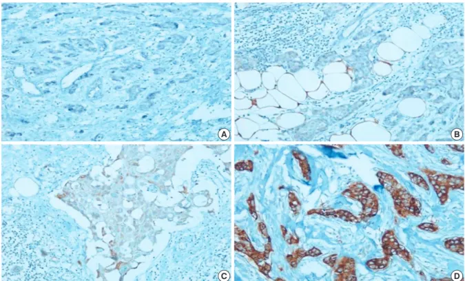
Figure 1. αB-crystallin expression scoring. Staining was graded as follows: 0, negative staining; 1, weakly positive staining; 2, moderately positive staining; 3, highly positive staining (Immunohistochemical staining for αB-crystallin, ×100).
(A) Score=0, (B) Score=1, (C) Score=2, (D) Score=3.
A
C
B
D
to αB-crystallin (1:200 in antibody diluents, SPA-222; Stress- gen Biotechnologies, Victoria, Canada).
Scoring of αB-crystallin staining
Cytoplasmic expression of αB-crystallin was scored using a four-tiered system. Staining was graded as follows: 0, negative staining; 1, weakly positive staining; 2, moderately positive staining; 3, highly positive staining in cytoplasm (Figure 1).
αB-crystallin expression was analyzed according to various clinical and biological characteristics such as tumor size, lymph node metastasis, lymphovascular invasion, histological grade, tumor markers such as ER, PR, HER2, and time to recurrence.
Statistical analysis
DBSTAT software version 4.1 (DBSTAT Co., Seoul, Korea) was used. Correlations between αB-crystallin and clinico- pathological characteristics were assessed using chi-square and Fisher’s exact tests. Time to recurrence between the αB- crystallin positive and negative groups was analyzed by the Kaplan-Meier method. A p<0.05 was considered significant.
RESULTS
Patient age ranged from 28-76 years with a median age of 53
years. The mean follow-up period was 50 months. All of the path- ological types were IDC. Nine cases of distant metastasis to bone, lung, liver, adrenal gland and the leptomenix were found.
Immunohistochemical staining for αB-crystallin
Twenty nine tumors (35.4%) had no cytoplasmic αB-crystallin staining (score 0), 23 (28.0%) had weakly positive staining (score 1), 15 (18.3%) had moderate staining (score 2), and 15 tumors (18.3%) had strong cytoplasmic staining (score 3). We defined scores of 0 and 1 as being indicative of “negative or low expression” and scores of 2 and 3 as being indicative of “posi- tive or high expression.” As a result, 52 tumors (63.4%) had low expression, and 30 tumors (36.6%) had high expression.
Correlation between αB-crystallin expression and clinicopathological features (Table 1)
αB-crystallin was not correlated with tumor size (p=0.602), lymph node status (p=0.403), or distant metastasis (p=0.064).
However, it was correlated with histological grade (p=0.004) and lymphovascular invasion (p=0.022). No statistical corre- lation was found between αB-crystallin score and lymph node metastasis (Figure 2). Six of nine cases who developed distant metastasis positively expressed αB-crystallin. Although the patient population was small and the p value was >0.05, αB- crystallin expression tended to have a marginal association with distant metastasis. Furthermore, when lymph node sta- tus was classified into pN stages by the number of metastatic lymph nodes, αB-crystallin was expressed more strongly in pN2 than in pN0 or pN1 (p=0.020) (Figure 3).
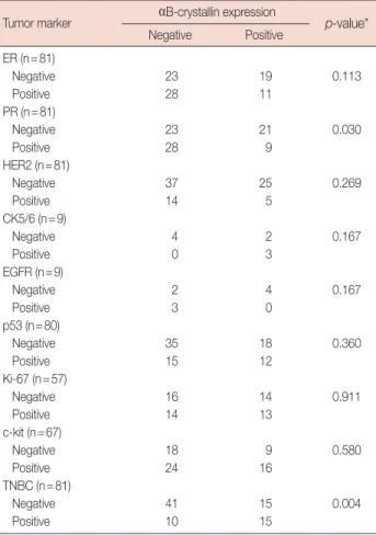
Correlation between αB-crystallin expression and breast tumor markers
αB-crystallin expression was associated with PR (p=0.030) Table 1. Correlation between αB-crystallin expression and patient’s
clinicopathological features Clinicopathologic feature (n=82)
αB-crystallin expression
p-value*
Negative Positive
Tumor size (cm) 0.602
≤2 24 20
>2, ≤5 24 8
≥5 4 2
Lymph node metastasis 0.403
Negative 24 11
Positive 28 19
pN 0.020
pN0 (LN=0) 24 11
pN1 (LN=1-3) 15 3
pN2 (LN ≥4) 13 16
Histologic grades 0.004
1 15 1
2 27 15
3 10 14
Lymphovascular invasion 0.022
Negative 31 10
Positive 21 20
Distant metastasis (n=74) 0.064
Negative 43 22
Positive 3 6
LN=number of lymph node metastases.
*Statistical analysis was performed with the chi-square test. p<0.05 was con- sidered significant.
No. of patients
αB-crystallin score
0 1 2 3
20
15
10
5
0
Lymph node negative Lymph node positive p>0.05
Figure 2. αB-crystallin score distributions among patients with lymph node negative and lymph node positive breast cancer. No statistical correlation was found between αB-crystallin score and each group.
and triple negative breast cancer (TNBC) (p=0.004). A total of 60% of the TNBCs were αB-crystallin positive, whereas 26.8% of non-TNBCs were αB-crystallin positive. Other fac-
tors such as ER, HER2, cytokeratin 5/6 (CK 5/6), and epider- mal growth factor receptor (EGFR) had no association with αB-crystallin expression (Table 2).
Time to distant metastasis according to αB-crystallin expression
The difference in the time to distant metastasis between pa- tients who were αB-crystallin negative and positive was as- sessed by the Kaplan-Meier method using the log-lank test.
αB-crystallin expression significantly decreased time to dis- tant metastasis. A statistical significance was found between any type of distant metastasis and αB-crystallin expression (p=0.039) (Figure 4).
DISCUSSION
Clinical indices such as tumor size, grade and axillary lymph node metastasis are useful prognostic factors in breast cancer.
Among these factors, axillary lymph node metastasis is the most important prognostic factor for patients with breast can- cer [9]. Many other studies have been conducted to identify pre- dictors related to axillary lymph node status. However, no fac- tor accurately predicts axillary lymph node metastasis.
Recently, Chelouche-Lev et al. [5] found significantly more αB-crystallin-positive tumors among patients with lymph node- positive disease than patients with lymph node-negative dis- ease (p<0.001). They reported that constitutive αB-crystallin expression in human breast cancer cells in vitro was associated with the ability to metastasize in nude mice, and that the high- est expression levels were observed in cell lines established from metastatic cells. Similarly, αB-crystallin staining was signifi- cantly associated with lymph node metastasis in our study.
No. of patients
The number of metastatic lymph node (pN)
pN0 (0) pN1 (1-3) pN2 (more than 3)
25
20
15
10
5
0
αB-crystallin negative p=0.020 αB-crystallin positive
Figure 3. αB-crystallin expression according to the number of metasta- tic lymph nodes (pN stage). αB-crystallin expression in breast cancers was expressed more strongly in pN2 than in pN0 and pN1 (p=0.006).
Figure 4. Time to distant metastasis based on αB-crystallin expression.
αB-crystallin expression significantly decreased time to distant metasta- sis. Statistical significance was found between any type of distant me- tastasis and αB-crystallin expression (p=0.039).
Log survival
Time to distant metastasis (mo)
0 20 40 60 80 100
0.00
-0.05
-0.10
-0.15
-0.20
-0.25
-0.30
αB-crystallin negative
αB-crystallin positive p=0.039
Table 2. Correlation between αB-crystallin expression and tumor markers Tumor marker αB-crystallin expression
p-value*
Negative Positive ER (n=81)
Negative 23 19 0.113
Positive 28 11
PR (n=81)
Negative 23 21 0.030
Positive 28 9
HER2 (n=81)
Negative 37 25 0.269
Positive 14 5
CK5/6 (n=9)
Negative 4 2 0.167
Positive 0 3
EGFR (n=9)
Negative 2 4 0.167
Positive 3 0
p53 (n=80)
Negative 35 18 0.360
Positive 15 12
Ki-67 (n=57)
Negative 16 14 0.911
Positive 14 13
c-kit (n=67)
Negative 18 9 0.580
Positive 24 16
TNBC (n=81)
Negative 41 15 0.004
Positive 10 15
ER=estrogen receptor; PR=progesterone receptor; CK5/6=cytokeratin5/6;
EGFR=epidermal growth factor receptor; TNBC=triple negative breast can cer.
*Statistical analysis was performed with the chi-square test. p<0.05 was con- sidered significant.
However, in this study, αB-crystallin was expressed significant- ly more in the pN2 breast cancer group than in pN0 or pN1 groups, which was different from Chelouche-Lev’s study. We thought that the differences in the patient cohorts made it dif- ficult to compare the results of Chelouche-Lev et al. with ours.
Although αB-crystallin seemed to be associated with lymph node metastasis, we do not think αB-crystallin is an accurate enough factor to eliminate axillary lymph node dissection. We thought that other factors co-expressed with αB-crystallin such as stromal cell-derived factor-1 and its receptor, CXC chemo- kine receptor 4 [10] need to be studied to accurately predict prognosis and survival.
Additionally, 60% of TNBCs were αB-crystallin positive, whereas 26.8% of non-TNBCs were αB-crystallin positive. αB- crystallin may be associated with TNBC. Patients with a TNBC had significantly shorter survival following the first metastatic event than those with a non-TNBC [11,12]. As most BLBCs are ER-negative and HER2-negative, the term TNBC has pre- viously been substituted for BLBCs [13]. Although there is over- lap between TNBC and BLBC, 76% of BLBCs expressed either EGFR or CK5/6, and these markers define BLBCs [14]. Both gene expression data and a recent immunohistochemistry anal- ysis of breast cancer tissue have suggested an association be- tween αB-crystallin and BLBCs [15]. The gene expression data revealed that αB-crystallin is included in the basal-like gene cluster [5,16,17]. αB-crystallin is commonly expressed in BLBCs and is thought to be a sensitive (81%) and specific (100%) mark- er for BLBCs [18]. These studies provide additional indepen- dent validation linking αB-crystallin to BLBCs [18]. We did not find a relationship between BLBC and αB-crystallin because of insufficient immunohistochemistry staining for CK5/6 and EGFR.
It seems that αB-crystallin is resistant to neoadjuvant chemo- therapy. Ivanov et al. [19] reported that αB-crystallin-positive tumors had poorer overall response rates than αB-crystallin- negative tumors (clinical overall response rate, 21% vs. 59%, respectively, p=0.005; overall pathological response rate, 16%
vs. 70%, respectively, p<0.001).
Despite the pathogenic significance of αB-crystallin, the reg- ulatory mechanism of its expression related to aggressiveness is poorly understood. Heat-shock proteins such as αB-crystallin play a major role in the ability of in vitro tumor cells to over- come stress caused by external stimuli, and enhance resistance to apoptosis [20,21]. Such resistance may result from the par- tial binding of αB-crystallin to caspase-3 and the resulting in- hibition of the autoproteolytic maturation of caspase-3, a key effector molecule in the apoptotic cascades [22]. These findings suggest that the anti-apoptotic effect of αB-crystallin may be related to the aggressiveness of breast cancer. Furthermore,
recent research shows that Ets1, an oncogenic transcription factor, binds to the αB-crystallin promoter and regulates its expression through an ETS-binding site dependent mechanism.
Ets1 overexpression in breast cancer cells increases αB-crystallin protein level, whereas silencing Ets1 reduces αB-crystallin lev- els. Moreover, Ets1 is expressed in BLBC and is associated with poor survival [8]. The rapid time to distant metastasis in our study may be linked to these in vitro findings.
Consistent with earlier studies, we demonstrated that αB- crystallin expression was associated with poor prognosis such as axillary lymph node metastasis in pN2, TNBC, and rapid time to recurrence. We think that αB-crystallin could be used as an oncoprotein to predict poor clinical outcomes. However, further studies are needed to prospectively elucidate the role of this novel tumor marker as a clinical prognostic factor in breast cancer.
REFERENCES
1. Yehiely F, Moyano JV, Evans JR, Nielsen TO, Cryns VL. Deconstructing the molecular portrait of basal-like breast cancer. Trends Mol Med 2006;
12:537-44.
2. Pinder SE, Balsitis M, Ellis IO, Landon M, Mayer RJ, Lowe J. The expres- sion of alpha B-crystallin in epithelial tumours: a useful tumour marker?
J Pathol 1994;174:209-15.
3. Mehlen P, Mehlen A, Guillet D, Preville X, Arrigo AP. Tumor necrosis factor-alpha induces changes in the phosphorylation, cellular localiza- tion, and oligomerization of human hsp27, a stress protein that confers cellular resistance to this cytokine. J Cell Biochem 1995;58:248-59.
4. Sørlie T, Perou CM, Tibshirani R, Aas T, Geisler S, Johnsen H, et al. Gene expression patterns of breast carcinomas distinguish tumor subclasses with clinical implications. Proc Natl Acad Sci U S A 2001;98:10869-74.
5. Chelouche-Lev D, Kluger HM, Berger AJ, Rimm DL, Price JE. AlphaB- crystallin as a marker of lymph node involvement in breast carcinoma.
Cancer 2004;100:2543-8.
6. Chin D, Boyle GM, Williams RM, Ferguson K, Pandeya N, Pedley J, et al. Alpha B-crystallin, a new independent marker for poor prognosis in head and neck cancer. Laryngoscope 2005;115:1239-42.
7. Moyano JV, Evans JR, Chen F, Lu M, Werner ME, Yehiely F, et al. AlphaB- crystallin is a novel oncoprotein that predicts poor clinical outcome in breast cancer. J Clin Invest 2006;116:261-70.
8. Bosman JD, Yehiely F, Evans JR, Cryns VL. Regulation of alphaB-crys- tallin gene expression by the transcription factor Ets1 in breast cancer.
Breast Cancer Res Treat 2010;119:63-70.
9. Fisher ER, Anderson S, Redmond C, Fisher B. Pathologic findings from the National Surgical Adjuvant Breast Project protocol B-06. 10-year patho- logic and clinical prognostic discriminants. Cancer 1993;71:2507-14.
10. Kim JO, Suh KS, Lee DH, Sul HJ, Lee JU, Song KS. Expression of CXCR4 and SDF-1alpha in primary breast cancers and metastatic lymph nodes.
J Breast Cancer 2009;12:249-56.
11. Dent R, Trudeau M, Pritchard KI, Hanna WM, Kahn HK, Sawka CA, et al. Triple-negative breast cancer: clinical features and patterns of re-
currence. Clin Cancer Res 2007;13(15 Pt 1):4429-34.
12. Tischkowitz M, Brunet JS, Bégin LR, Huntsman DG, Cheang MC, Akslen LA, et al. Use of immunohistochemical markers can refine prognosis in triple negative breast cancer. BMC Cancer 2007;7:134.
13. Carey LA, Dees EC, Sawyer L, Gatti L, Moore DT, Collichio F, et al. The triple negative paradox: primary tumor chemosensitivity of breast cancer subtypes. Clin Cancer Res 2007;13:2329-34.
14. Rakha EA, Tan DS, Foulkes WD, Ellis IO, Tutt A, Nielsen TO, et al. Are triple-negative tumours and basal-like breast cancer synonymous? Breast Cancer Res 2007;9:404.
15. Nielsen TO, Hsu FD, Jensen K, Cheang M, Karaca G, Hu Z, et al. Im- munohistochemical and clinical characterization of the basal-like sub- type of invasive breast carcinoma. Clin Cancer Res 2004;10:5367-74.
16. Perou CM, Sørlie T, Eisen MB, van de Rijn M, Jeffrey SS, Rees CA, et al.
Molecular portraits of human breast tumours. Nature 2000;406:747-52.
17. Sorlie T, Tibshirani R, Parker J, Hastie T, Marron JS, Nobel A, et al. Re- peated observation of breast tumor subtypes in independent gene ex-
pression data sets. Proc Natl Acad Sci U S A 2003;100:8418-23.
18. Sitterding SM, Wiseman WR, Schiller CL, Luan C, Chen F, Moyano JV, et al. AlphaB-crystallin: a novel marker of invasive basal-like and meta- plastic breast carcinomas. Ann Diagn Pathol 2008;12:33-40.
19. Ivanov O, Chen F, Wiley EL, Keswani A, Diaz LK, Memmel HC, et al.
AlphaB-crystallin is a novel predictor of resistance to neoadjuvant che- motherapy in breast cancer. Breast Cancer Res Treat 2008;111:411-7.
20. Parcellier A, Gurbuxani S, Schmitt E, Solary E, Garrido C. Heat shock proteins, cellular chaperones that modulate mitochondrial cell death pathways. Biochem Biophys Res Commun 2003;304:505-12.
21. Beere HM. Stressed to death: regulation of apoptotic signaling pathways by the heat shock proteins. Sci STKE 2001;2001:re1.
22. Kamradt MC, Chen F, Cryns VL. The small heat shock protein alpha B- crystallin negatively regulates cytochrome c- and caspase-8-dependent activation of caspase-3 by inhibiting its autoproteolytic maturation. J Biol Chem 2001;276:16059-63.