http://dx.doi.org/10.4167/jbv.2018.48.1.23
pISSN 1598-2467, eISSN 2093-0429
JBV
LPS-stimulated Macrophage Activation Affects Endothelial Dysfunction
Naehwan Baek, Sohyun Sim and Kyung-Sun Heo*
Department of Pharmacology, Chungnam National University, College of Pharmacy, Daejeon, Korea
Corresponding Kyung-Sun Heo
Department of Pharmacology, Chungnam National University, College of Pharmacy, 99 Daehak-ro (St.), Yuseong-gu, Daejeon 34134, Korea
Phone : +82-42-821-5927 Fax : +82-42-821-8925 E-mail : [email protected]
Received : March 12, 2018 Revised : March 21, 2018 Accepted : March 22, 2018
No potential conflict of interest relevant to this article was reported.
Copyright © 2018 Journal of Bacteriology and Virology
©This is an Open Access article distributed under the terms of the Creative Commons Attribution Non-Commercial License (http://creativecommons.org/
license/by-nc/3.0/).
Intestinal microbiota is involved in the atherosclerotic process by development of an atheromatous core with foam cells in carotid arteries. It has reported that lipopolysaccharide (LPS) from Escherichia coli localizes in human atherosclerotic plaque and causes inflammation via interaction with toll like receptor 4. However, there is no evidence that whether LPS-activated macrophages regulate endothelial cell (EC) function. We evaluated whether LPS-activated macrophage acts as one of the stimulants activating EC and its underlying signaling pathways. Using Western blotting and quantitative reverse transcription-polymerase chain reaction (qRT- PCR), we confirmed that intraperitoneal injection with LPS increases iNOS protein and inflammatory cytokine, TNF-α and IL-6 mRNA expressions. To determine whether LPS-mediated macrophage inflammatory condition affects EC activation and inflammation, human umbilical vein endothelial cells (HUVECs) were incubated with isolated peritoneal macrophages from LPS-injected mice. Interestingly, p90RSK Serine 380 phosphorylation and protein expression were significantly increased by macrophage treatment in EC. Messenger RNA levels of vascular cell adhesion molecule 1 and p90RSK was increased, but endothelial nitric oxide synthase was decreased. In addition, NF-κB promoter activity, which plays an important role in the pathogenesis of inflammation, was strongly enhanced by the macrophage treatment in EC. We further evaluated the effects of LPS on EC function in the mouse aorta using en face staining. In agreement with in vitro result, p90RSK expression was strongly increased in the steady laminar flow region of the mouse aorta in mice injected with LPS. Together, our study demonstrates that p90RSK might be a one of the major therapeutic candidates for the prevention of vascular diseases mediated by LPS.
Key Words: Atherosclerosis, EC inflammation, Lipopolysaccharide, p90RSK
INTRODUCTION
The endothelium modulates a lot of biological processes within the blood vessel to regulate vascular tone and blood pressure through stimulation of nitric oxide and endothelin (1, 2) and to stimulate cell proliferation and angiogenesis through secretion of various growth factors and vasoactive substances. Therefore, endothelium plays a key role in the initiation and development of inflammatory atherosclerosis (3).
Endothelial dysfunction is characterized by atherosclerosis with early events including vasoregulation, activation of inflammatory processes, and damaged barrier function due to endothelial cell (EC) death (4, 5). Hemodynamic shear stress affects vascular
JBV
pathophysiologic conditions such as progression of atherosclerosis, vascular dilation, and aneurysms (4, 5).
p90 ribosomal S6 kinase (p90RSK) is one of the downstream regulators of ERK1/2 (6). p90RSK is involved in activation of nuclear factor-κB by phosphorylation of Iκ-B (7) or phosphorylation of transcription factors such as c-Fos (8), Nur77 (9), and cAMP-response element-binding protein (10). Furthermore, we recently reported that p90RSK was highly expressed in atherosclerotic lesion and was associated with EC inflammation and apoptosis via regulating ERK5 and p53-mediated signaling pathway (5, 11).
There is a report that intestinal microbiota is involved in the atherosclerotic process (12). The patients with atheroma showed higher levels of circulating LPS and the presence of an atheromatous core with foam cells in carotid arteries compared with the control patients (12). Mechanistically, it demonstrated that lipopolysaccharide (LPS) from Escherichia coli (E. coli) localizes in human atherosclerotic plaque and causes inflammation via interaction with toll like receptor 4. However, there is no evidence that weather LPS-activated macrophages affect EC function.
Here, we have attempted to verify whether LPS-activated macrophage acts as one of the agonists activating EC and its underlying signaling pathways. We found that LPS-induced macrophages increase inducible nitric oxide synthase (iNOS) and inflammatory cytokines, including TNF-α and IL-6. When these macrophages cocultured with EC, they up-regulates p90RSK, VCAM-1, and NF-κB, whereas down-regulates endothelial nitric oxide synthase (eNOS), resulting in EC activation and inflammation.
Finally, EC dysfunction was assessed after exposure of LPS in mouse aorta. Consistent with in vitro experiment data, p90RSK expression was up-regulated in the LPS-exposed aorta in mice. Together, our finding demonstrates that p90RSK might be a one of the key candidates for treating vascular diseases mediated by LPS.
MATERIALS AND METHODS
Cell cultures
Human umbilical vein endothelial cells (HUVECs) were purchased from Lonza (Walkersville, MD, USA) and cultured in M200 medium supplemented with low serum growth supplement (LSGS; Cascade biologic, Portland, OR, USA) and 5% fetal bovine serum (FBS, Gibco, Grand Island, NY, USA). HUVECs were cultured on 0.2% gelatin pre-coated dishes. Peritoneal neutrophils and macrophages were obtained after intraperitoneal (i.p.) injection of 2 ml autoclaved 3% (w/v) thioglycolate in H2O (Sigma-Aldrich, St. Louis, MO, USA). After 3 days, peritoneal cells were harvested by injecting 10 ml DMEM containing 10%
FBS into the peritoneal cavity, the abdomen gently massaged to dislodge cells, and cell suspension was collected. The mouse monocyte/macrophage cell line, RAW264.7 was obtained from American Type Culture Collection (ATCC, Manassas, VA, USA;
#TIB-71) and was propagated in DMEM containing 10% FBS at 37°C and 5% CO2 in air.
Plasmids and luciferase assay
The NF-κB-luc reporter plasmid was obtained from JD Li (13) lab and the construct was verified by sequencing. Peritoneal macrophages were plated on 12-well plates at 5 x 10⁴ cells/well. For a promoter assay, cells were transfected in Opti-MEM (#31985-088; Gibco) with lipofectamine (#15338100, Invitrogen, Waltham, MA) mixture containing the NF-κB-luc reporter plasmid along with pRL-tk for 4 hrs. Opti-MEM was replaced by fresh complete culture medium 4 hrs post transfection. The cells were collected 36 hrs after transfection, and the luciferase activity was assayed with the dual luciferase kit (#E1960, Promega, Madison, WI, USA) using a TD-20/20 luminometer (Turner Designs, San Jose, CA, USA). Since pRL-tk contains the Renilla luciferase gene, the expression and transfection efficiencies were normalized with the Renilla luciferase activity.
Transfections were performed in triplicate, and each experiment was repeated at least three times.
Rabbit anti-p90RSK S380 (#11989), rabbit anti-p90RSK1 (#9333), eNOS (#32027) were purchased from Cell Signaling Technology (Danvers, MA, USA). Rabbit anti-VCAM-1 (#sc-8304) was purchased from Santa Cruz Biotechnology (Santa Cruz, CA, USA). TNF-α (#KMC3011) and IL-6 (#KMC0061) mouse ELISA kit were purchased from Invitrogen. LPS from E. coli 0127:B8 was purchased from Sigma-Aldrich (#L-3137).
Animals
C57BL/6 mice were purchased from Samtako Animals (Osan, Korea). All mice were maintained under pathogen-free conditions at the Chungnam National University. All animal procedures were performed with the approval from the University Committee on Animal Resources at the Chungnam National University and approved protocol number was CNU-00826.
Western blot analysis
Cell extracts were prepared in modified radioimmunoprecipitation assay (RIPA) buffer including 50 mM Tris-HCl [pH 7.4], 150 mM NaCl, 1 mM EDTA, 1% Nonidet P-40, 0.1% SDS, 1 mM dithiothreitol, 1:200-diluted protease inhibitor cocktail (Sigma), 1 mM PMSF and 10 mM NEM, and 0.1 mM iodoacetamide. The protein extracts were resolved by SDS-PAGE, electro transferred onto a Hybond enhanced chemiluminescence nitrocellulose membrane, and visualized by using the enhanced chemiluminescence detection reagents (Amersham Pharmacia Biotech, Piscataway, NJ, USA) according to the manufacturer's instructions. Each protein expression was detected by Western blotting with each corresponding antibody.
Quantitative real-time polymerase chain reaction (qRT-PCR) assay
Total RNA was extracted using the TRIzol reagent according to manufacturer's instructions. The cDNA synthesis was performed in 20 μl mixtures containing 1 μg of total RNA using an iScript cDNA synthesis kit according to the manufacture's protocol (#170-8890, Bio-Rad, Hercules, CA, USA). For qRT-PCR, specific mouse primers including TNF-α and IL-6, and human VCAM-1, eNOS, and p90RSK were designed using Primer Express 3.0 software. The primer sequences used were: (Mouse) β-actin forward 5'-GGCGCTTTTGACTCAGGATT, β-actin reverse 3'-GGGATGTTTGCTCCAACCAA; TNF-α forward 5'-ATCCGCGAC- GTGGAACTG, TNF-α reverse 3'-ACCGCCTGGAGTTCTGGAA. IL-6 forward 5'-CCTCGGTCTTCTGGAGTACC, IL-6 reverse 3'- ACTCCTTCTGTGACTCCAGC; (Human) GAPDH forward 5'-CCTGCACCACCAACTGCTTA, GAPDH reverse 3'-GGCCATCCAC- AGTCTTCTGAG, VCAM-1 forward 5'-GAACCACTATTTTCTCATCACGACA, VCAM-1 reverse 3'-TTTAAAAGCTTGAGAAGCT- GCAAAC, eNOS forward 5'-TGGCTTTCCCTTCCAGTTC, eNOS reverse 3'-AGAGGCGTTTTGCTCCTTC.
En Face immunostaining
We performed en face staining as described in our previous report (14, 15). Briefly, animals of 6~8 weeks of age were euthanized by CO2 inhalation. The arterial tree was perfused via the left ventricle first with saline containing heparin (40 USPU /ml) and then with 4% paraformaldehyde in PBS for 10 min. Fixed aorta was dissected out and the adipose tissue around the vessel was removed. It was cut open longitudinally and permeabilized with PBS containing 0.1% Triton X-100 and blocked by TBS containing 10% goat serum and 2.5% Tween-20 for 30 min. Aortas were incubated with serum-free protein blocking buffer (X0909, DAKO, Carpinteria, CA, USA) for 30 min followed by incubation with rabbit anti-p90RSK and anti-rabbit IgG as a control (10 μg/ml) in the presence of rat anti-VE-cadherin (7 μg/ml, an endothelial cell marker) in the antibody diluent buffer (#S0809, DAKO) for overnight. After a PBS rinse, anti-rabbit IgG and anti-rat IgG antibodies (1:1,000 dilution, Alexa Fluor 546 and 488, respectively, Molecular Probes, Eugene, OR, USA) were applied for 1 h at room temperature. The samples were analyzed using a K1-Fluo laser scanning confocal microscope equipped with a PLAPON 60X (NA 1.42) oil objective lens (Nanoscope Systems, Daejeon, Korea).
JBV
Statistical Analysis
Data are presented as mean ± SEM. Comparison between 2 independent groups were subjected to the Wilcoxon rank sum test. For multiple comparisons of >2 groups, the Kruskal-Wallis test followed by Dunn post hoc analysis was performed with the use of GraphPad Prism (GraphPad Software, San Diego, CA, USA). A p value <0.05 was considered statistically significant.
RESULTS
LPS treatment induces inflammatory environment in vivo
To determine whether LPS treatment induces inflammation in mice, various concentrations of LPS (1, 5, and 10 mg/kg) were i.p. injected in mice. Six hours later, peritoneal macrophage was isolated and evaluated iNOS expression, which produces toxic oxygen species, nitric oxide (NO). As shown in Fig. 1A, iNOS expression was significantly increased at 1, 5, and 10 mg/
kg compared with control (PBS treatment). Since induction of nitric oxide is associated with increased inflammation (16), key inflammatory cytokines such as TNFα and IL-6 were evaluated in isolated peritoneal macrophages using qRT-PCR (Fig. 1B). As expected, LPS treatment significantly increases both TNFα and IL-6 mRNA levels in a concentration-dependent manner. Based on this data on LPS-induced inflammatory conditions, we set up a 5 mg/kg LPS treatment for 6 h for further experiments.
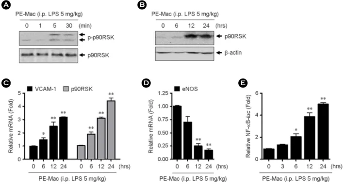
Co-culture of HUVECs with LPS-treated peritoneal macrophages leads to EC inflammation
To evaluate whether LPS treatment induces macrophage activation-mediated EC inflammation, macrophages were prepared from peritoneum of 5 mg/kg LPS i.p. injected mice. Then, HUVECs were co-cultured with isolated macrophages for various Figure 1. The expression of iNOS and inflammatory cytokines in LPS-activated peritoneal macrophages. LPS was i.p. injected with indicated doses in C57BL/6 mice. After 6 hrs, peritoneal macrophages were isolated and then assessed iNOS protein expression by western blotting using specific antibody (A) and TNF-α and IL-6 mRNAs by qRT-PCR using specific primers (B).
Bar graph represents the quantification of relative fold changes of iNOS protein expression compared to the control in three independent experiments (0, A, lower panel). Data present mean ± SEM. *p < 0.05 and **p < 0.01, compared with control (0, PBS treated) by 1-way ANOVA followed by Bonferroni's post hoc test.
regulates p90RSK phosphorylation. Interestingly, LPS-activated macrophages increase p90RSK S380 phosphorylation and p90RSK expression at time-dependent manner (Fig. 2A, B). We further evaluated whether these macrophages regulate inflammatory molecule such as vascular adhesion molecule (VCAM)-1. VCAM-1 mRNA expression was significantly increased as well as p90RSK mRNA levels in RNA of co-cultured cells (Fig. 2C). eNOS is a key regulator of cellular function that is important to maintain EC homeostasis (17). When eNOS mRNA level was determined under the same conditions, eNOS mRNA levels significantly reduced in a LPS-activated macrophage treatment time-dependent manner (Fig. 2D). Since nuclear factor κB (NF-κB) transcription factor is involved in the activation of many genes in response to inflammatory stimuli (18), NF-κB transcriptional activity was determined in co-cultured cells. Consistent with other inflammatory molecule expression, NF-κB transcriptional activities were increased by HUVECs co-cultured with LPS-activated macrophages after 6 hrs incubation (Fig. 2E).
LPS treatment induces inflammatory environment in vivo
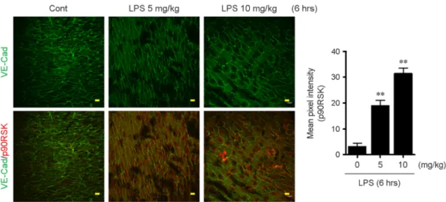
To confirm the previous result in vivo, male C57BL/6 mice (7-week old) were i.p. injected with 0 (PBS), 5, and 10 mg/kg LPS and the aortas were isolated from the mice. After perfusion with Saline including heparin, it was fixed with 4% paraformaldehyde, and then en face preparations were made. The fixed aorta was double-stained with anti-VE-Cad (as an EC marker) or anti-p90RSK. Consistent with the in vitro data, increased level of p90RSK was appeared in regions where steady laminar flow area of the aorta of 5 and 10 mg/kg LPS-treated mice compared with control (0, PBS-treated mice) (Fig. 3).
Figure 2. The effect of LPS-activated macrophages on EC functions. The HUVECs were co-cultured with macrophages isolated from 5 mg/kg LPS-injected mice for indicated times. (A-B) Phospho-p90RSK and total-p90RSK were determined by western blotting using specific antibody, respectively. (C-D) Total RNAs were isolated and qPT-PCR for VCAM-1, p90RSK, and eNOS mRNA levels were performed. (E) HUVECs were transfected with NF-κB-luc and pRT-tk using Lipofectamine 2000. The cells were co-cultured with the LPS-activated macrophages for indicated times and then promoter assay was performed. Data present mean ± SEM. *p < 0.05 and **p < 0.01, compared with control (0, PBS treated) by 1-way ANOVA followed by Bonferroni's post hoc test.
JBV
DISCUSSION
ECs are major determinant of vascular tone, leukocyte adhesion, and SMC proliferation (19). In its intact state, the endothelium maintains vasodilator, anti-thrombotic and anti-inflammatory status (20). However, when inflammatory conditions occur, ECs are a major target of inflammatory molecules such as cytokine signals from various immune cells and can cause EC dysfunction and inflammation (20). Thus, endothelial dysfunction is an early event in atherosclerotic disease prior to clinical manifestations and complications. A recent report indicates that intestinal microorganisms are involved in the atherosclerotic process, but there is still lacking informational weather the microorganism infection affects EC inflammation and its underlying signaling pathway. Here, we clearly demonstrate that LPS-induced macrophage activation leads to EC inflammation via p90RSK activation.
To determine the role of LPS-induced macrophage activation, we first assessed the effect of LPS on iNOS and cytokine expressions in peritoneal macrophages by treatment with LPS in mice. The enzyme responsible for the NO production by macrophages is the iNOS that catalyzes arginine to form NO and citrulline (21). Compared with the neuronal and endothelial isoforms of NOS (nNOS and eNOS, respectively), iNOS has drawn considerable attention for its important functions in inflammatory diseases (22). Fig. 1 showed that iNOS expressions were dramatically increased by treatment with low dose of LPS for 6 hrs compared to the control (PBS treated) in mice. Consistent with this result, cytokine expression was also greatly increased by LPS treatment in mice (Fig. 1B).
To prove our hypothesis that LPS-activated macrophages affect EC function, peritoneal macrophages was isolated from mice after LPS injection, and then HUVECs were co-cultured various times with these activated macrophages. Interestingly, EC co-cultured with LPS-activated macrophages increases p90RSK S380 phosphorylation at 5 mins as well as p90RSK protein expression after 12 hrs of stimulation (Figs. 2A and B). Previously, we reported that EC transduced with adenovirus containing dominant negative p90RSK (K94A/K447A, Ad-DN-RSK), but in those transduced with Ad-LacZ, abolished atherogenic flow- induced proinflammatory adhesion molecule expression (5). It suggests that p90RSK kinase activation plays a crucial role in regulating EC inflammation. In particular, mRNA levels of VCAM-1 and p90RSK and NF-κB promoter activity were increased by LPS-activated macrophage treatment in HUVECs, whereas mRNA level if eNOS was decreased (Fig. 2).
Figure 3. LPS stimulation increases p90RSK expression in aortic arch. After stimulation with 0 (PBS), 5, and 10 mg/kg LPS for 6 hrs, en face preparations of the aortic arch of 7-week C57BL/6 mice were processed. The samples were double-stained with anti-VE-Cad (as an EC marker) and anti-p90RSK. Images were recorded using a confocal microscope equipped with a Planapo x60 1.42 NA oil objective lens. Scale bars: 10 μm. Bar graph indicates quantification of percentage of p90RSK fluorescence intensity. Data present mean ± SEM. **p < 0.01, compared with control (PBS treated) by 1-way ANOVA followed by Bonferroni's post hoc test.
inflammatory responses that contribute to the production of atheroma (14). The endothelium in the aortic arch is susceptible to two different flows, which are steady laminar flow (anti-atherogenic flow) and disturbed flow (atherogenic flow) (15).
Especially laminar shear stress plays a protective role by inhibiting activation of adhesion molecule and inflammatory cytokines (23). It has been reported that p90RSK overexpression was observed in athero-prone area, where is disturbed flow area and p90RSK expression was colocalized with VCAM-1 expression in endothelium of mouse aorta suggesting that p90RSK plays an inflammatory regulator (5, 11, 24). To investigate the effect of LPS on endothelial cells-exposed by laminar shear stress, LPS- injected mouse aorta was co-stained with VE-Cad as EC marker and p90RSK in Fig. 3. Interestingly, LPS stimulation disrupted the laminar shear stress vascular protective function by increasing expression of p90RSK in laminar shear stress-exposed legion. It suggests that LPS-mediated inflammatory environment affects p90RSK expression in EC and is thought be critical for the regulation of atherosclerosis. Therefore, targeting the p90RSK in LPS-induced inflammatory condition is a one of the key therapeutic approaches for the prevention of vascular diseases.
ACKNOWLEDGEMENT
This work is supported by grants from the Chungnam National University to Kyung-Sun Heo (2016-1766-01).
REFERENCES
1) Li Q, Park K, Li C, Rask-Madsen C, Mima A, Qi W, et al. Induction of vascular insulin resistance and endothelin-1 expression and acceleration of atherosclerosis by the overexpression of protein kinase C-beta isoform in the endothelium. Circ Res 2013;113:418-27.
2)Pucci ML, Miller KB, Dick LB, Guan H, Lin L, Nasjletti A. Vascular responsiveness to nitric oxide synthesis inhibition in hypertensive rats. Hypertension 1994;23:744-51.
3) Ziche M, Morbidelli L, Masini E, Amerini S, Granger HJ, Maggi CA, et al. Nitric oxide mediates angiogenesis in vivo and endothelial cell growth and migration in vitro promoted by substance P. J Clin Invest 1994;94:2036-44.
4)Heo KS, Chang E, Le NT, Cushman H, Yeh ET, Fujiwara K, et al. De-SUMOylation enzyme of sentrin/SUMO-specific protease 2 regulates disturbed flow-induced SUMOylation of ERK5 and p53 that leads to endothelial dysfunction and atherosclerosis. Circ Res 2013;112:911-23.
5) Heo KS, Le NT, Cushman HJ, Giancursio CJ, Chang E, Woo CH, et al. Disturbed flow-activated p90RSK kinase accelerates atherosclerosis by inhibiting SENP2 function. J Clin Invest 2015;125:1299-310.
6) Blenis J. Signal transduction via the MAP kinases: proceed at your own RSK. Proc Natl Acad Sci U S A 1993;90:5889-92.
7) Ghoda L, Lin X, Greene WC. The 90-kDa ribosomal S6 kinase (pp90rsk) phosphorylates the N-terminal regulatory domain of IkappaBalpha and stimulates its degradation in vitro. J Biol Chem 1997;272:21281-8.
8) Chen RH, Chung J, Blenis J. Regulation of pp90rsk phosphorylation and S6 phosphotransferase activity in Swiss 3T3 cells by growth factor-, phorbol ester-, and cyclic AMP-mediated signal transduction. Mol Cell Biol 1991;11:1861-7.
9) Fisher TL, Blenis J. Evidence for two catalytically active kinase domains in pp90rsk. Mol Cell Biol 1996;16:1212-9.
JBV
10) Xing J, Ginty DD, Greenberg ME. Coupling of the RAS-MAPK pathway to gene activation by RSK2, a growth factor- regulated CREB kinase. Science 1996;273:959-63.
11) Le NT, Heo KS, Takei Y, Lee H, Woo CH, Chang E, et al. A crucial role for p90RSK-mediated reduction of ERK5 transcriptional activity in endothelial dysfunction and atherosclerosis. Circulation 2013;127:486-99.
12)Sanduzzi Zamparelli M, Compare D, Coccoli P, Rocco A, Nardone OM, Marrone G, et al. The Metabolic Role of Gut Microbiota in the Development of Nonalcoholic Fatty Liver Disease and Cardiovascular Disease. Int J Mol Sci 2016;17.
13) Lim JH, Jono H, Komatsu K, Woo CH, Lee J, Miyata M, et al. CYLD negatively regulates transforming growth factor- beta-signalling via deubiquitinating Akt. Nat Commun 2012;3:771.
14) Heo KS, Fujiwara K, Abe J. Disturbed-flow-mediated vascular reactive oxygen species induce endothelial dysfunction. Circ J 2011;75:2722-30.
15) Heo KS, Lee H, Nigro P, Thomas T, Le NT, Chang E, et al. PKCzeta mediates disturbed flow-induced endothelial apoptosis via p53 SUMOylation. J Cell Biol 2011;193:867-84.
16) McNeill E, Crabtree MJ, Sahgal N, Patel J, Chuaiphichai S, Iqbal AJ, et al. Regulation of iNOS function and cellular redox state by macrophage Gch1 reveals specific requirements for tetrahydrobiopterin in NRF2 activation. Free Radic Biol Med 2015;79:206-16.
17) Heiss C, Rodriguez-Mateos A, Kelm M. Central role of eNOS in the maintenance of endothelial homeostasis. Antioxid Redox Signal 2015;22:1230-42.
18)Kempe S, Kestler H, Lasar A, Wirth T. NF-kappaB controls the global pro-inflammatory response in endothelial cells:
evidence for the regulation of a pro-atherogenic program. Nucleic Acids Res 2005;33:5308-19.
19) Sprague AH, Khalil RA. Inflammatory cytokines in vascular dysfunction and vascular disease. Biochem Pharmacol 2009;
78:539-52.
20) Goveia J, Stapor P, Carmeliet P. Principles of targeting endothelial cell metabolism to treat angiogenesis and endothelial cell dysfunction in disease. EMBO Mol Med 2014;6:1105-20.
21) Jacobs AT, Ignarro LJ. Lipopolysaccharide-induced expression of interferon-beta mediates the timing of inducible nitric- oxide synthase induction in RAW 264.7 macrophages. J Biol Chem 2001;276:47950-7.
22) Cedergren J, Forslund T, Sundqvist T, Skogh T. Inducible nitric oxide synthase is expressed in synovial fluid granulocytes.
Clin Exp Immunol 2002;130:150-5.
23)van Thienen JV, Fledderus JO, Dekker RJ, Rohlena J, van Ijzendoorn GA, Kootstra NA, et al. Shear stress sustains atheroprotective endothelial KLF2 expression more potently than statins through mRNA stabilization. Cardiovasc Res 2006;72:231-40.
24) Heo KS, Berk BC, Abe J. Disturbed Flow-Induced Endothelial Proatherogenic Signaling Via Regulating Post-Translational Modifications and Epigenetic Events. Antioxid Redox Signal 2016;25:435-50.