J Clinical Otolaryngol 2010;21:118-122 □ 증 례□
외상성 경부 림프관 파열로 인해 발생한 유미종 치험 1예
건양대학교 의과대학 이비인후과학교실
이 호 진·박 병 건
A Case of Post-traumatic Cervical Chyloma
Ho-Jin Lee, MD and Byung Kuhn Park, MD
Department of Otorhinolaryngology-Head and Neck Surgery, College of Medicine, Konyang University, Daejeon, Korea
- ABSTRACT -
Chyloma is a cyst or a pseudocyst with chyle collection due to disruption of the thoracic duct and its tributaries.
Congenital malformation of lymphatic channels and various acquired lesions can cause chyloma. Acquired chyloma can result from thoracic surgery, radical neck dissection, blunt or penetrating trauma, and various pro- cedures such as subclavian vein line placement. Complications caused by chyle leakage include malnutrition, hypovolemia, hyponatremia, hypocalcemia, metabolic acidosis, electrolyte abnormalities, and immune com- promise. Chyloma can be diagnosed with sonography, neck CT or MR, chemical analysis of cystic fluid with fine needle aspiration. Chyle leakage site can be identified with lymphangiography. Treatment consists of me- dical management of chyle leakage with nutritional modification and surgical management of mass and ligation of lymphatics. We present a case report of left supraclavicular chyloma secondary to blunt trauma. (J Clinical Otolaryngol 2010;21:118-122)
KEY WORDS:Chyles·Neck·Cysts.
서 론
유미종은 흉관 및 분지에서 기원하는 낭종 또는 가성 낭종을 일컬으며, 림프계의 선천성 기형이나 여러 가지 후천성 원인에 의해 발생할 수 있다. 후천성 유미종은 흉부수술, 경부절제술, 경부 둔상 및 관통성 외상, 종양 에 의한 미란 또는 쇄골하정맥관삽관 등에 의해 흉관
또는 경부나 상흉부 림프관의 손상으로 발생한다.1,2) 유 미 유출로 인해 환자는 호흡곤란, 피로감 등을 호소할 수 있고, 지속적 유미 유출은 주변 조직의 감염을 일으 킬 수 있으며, 저단백혈증, 저나트륨혈증, 영양 흡수 장 애와 수액 손실, 유미흉, 면역저하 등의 합병증을 유발 하기도 한다.3)
유미종의 진단을 위해서는 초음파, 경부 컴퓨터 단층 촬영 및 자기공명촬영을 통해 낭종의 범위, 위치를 확인 하고 세침흡입검사를 통한 성분 분석이 필요하다. 중성 지방 수치의 증가와 병리조직검사 및 CD31, CD34, factor VIII, keratin 등 내피막 세포 염색 표지자 확인 으로 흉관 낭종, 낭림프관종, 새열낭, 갑상혀관낭, 가성낭 종 등을 감별할 수 있다. 치료는 식이 조절을 이용한 보 존적 치료, 낭종의 절제와 유미 유출 부위를 직접 결찰 논문접수일:2010년 2월 17일
논문수정일:2010년 3월 05일 심사완료일:2010년 4월 06일
교신저자:박병건, 302-718 대전광역시 서구 가수원동 685 건양대학교 의과대학 이비인후과학교실
전화:(042) 600-9215·전송:(042) 543-8959 E-mail:[email protected]
하는 수술적 치료를 시행할 수 있다.4,5)
저자들은 61세 남환에서 교통사고 후 경부 둔상에 의 해 발생한 좌측 쇄골 상부의 유미종 1예를 치험하였기에 문헌 고찰과 함께 보고하는 바이다.
증 례
61세 남환이 자동차를 타고 가다 발생한 교통사고로 타 병원 입원 치료 중 우측 무릎 슬개골 골절 진단하에 본원 정형외과로 전원되어 수술을 받았다. 사고 당시 안 전벨트 매었던 부위에 발생한 좌측 경부의 낭성 종물에
대해 절개 배액술을 시행 받았던 분으로, 하루 9장 정도 의 외과용 패드가 젖을 정도로 우윳빛 액체가 배액된다 며 절개 배액술 시행 3일째 이비인후과로 진료 의뢰되 었다.
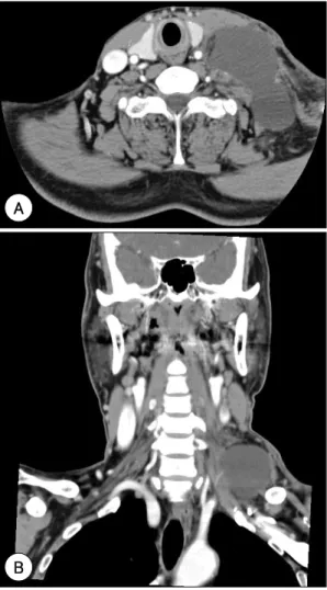
병력 청취상 사고 후 좌측 경부에 부드럽고 가동성 있 는 낭성 종물이 발생하였다고 하며 압박감 외에 다른 증 상은 없었다고 하였다. 타 병원에서 시행한 경부 컴퓨터 단층촬영에서 좌측 쇄골 상부에 9×4 cm 크기로 액체 가 고여 있었으며(Fig. 1), 이학적 검사상 좌측 쇄골 상 부 종창이 관찰되었고 펜로즈 배액관(penrose drain) 을 통해 우유 빛 액체가 배액되고 있었으나, 압통이나 염증 소견은 없었다(Fig. 2). 우윳빛 액체에 대한 성분 분 석과 경부 자기공명촬영을 계획하였으며, 환자에게 저지 방 식이를 하도록 하고 국소압박을 유지하며 경과 관찰하 였다.
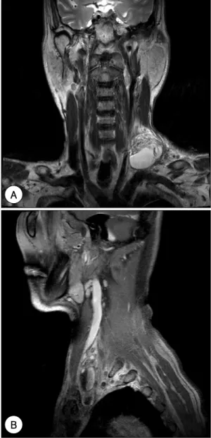
성분 분석 결과 중성지방(triglyceride) 653 mg/dL, 총 콜레스테롤(total cholesterol) 63 mg/dL였고, 5일 간의 국소압박 및 저지방 식이에도 불구하고 배액량이 감소되지 않았다. 경부 자기공명촬영에서 액체-액체 층 (fluid-fluid level)으로 구성된 6×2 cm 크기의 낭성 종 물이 내경정맥을 압박하고 있어(Fig. 3) 경부 림프관 손상 에 의한 유미종 진단하에 종물 절제 및 림프관 결찰술을 시행하였다(Fig. 4). 조직 검사상 섬유성 막으로 이루어진 가성낭종 이었으며(Fig. 5), 수술 1주일 후 배액량이 거 의 없어 배액관을 제거하였고, 2일 후 환자는 퇴원하였 다. 수술 2개월까지 유미 유출의 재발은 보이지 않았다.
Fig. 1. Neck computed tomography shows 9×4 cm sized low density fluid collection at the left supraclavicular area (A:Axial view. B:Coronal view).
A
B
Fig. 2. Yellowish milky fluid was drained through penrose drain.
고 찰
유미종은 드물게 발생하는 질환으로 흉관 및 분지의 손 상으로 유미가 유출되어 형성되는 낭종 및 가성낭종을 일 컫는다. 유미종은 림프계의 선천성 기형이나 여러 가지
후천성 원인에 의해 발생할 수 있다. 후천성 유미종은 흉 부 수술, 경부 절제술, 경부 둔상 및 관통성 외상, 종양 에 의한 미란 또는 쇄골하정맥관삽관 등의 처치에 의해 림프관이 손상되어 발생한다.1,2)
경부 흉관 낭종을 포함한 경부 유미종 환자의 대부분 은 무증상이나 약간의 압박감 및 드물게 호흡곤란을 호 소하기도 한다.6,7) Jang 등의 보고에 따르면 16명의 경 부 흉관 낭종 환자에서 11명이 무증상, 5명이 경부 압 박감, 1명이 호흡곤란을 호소하였다고 하였다.4) 본 증례 에서도 좌측 경부 종창 및 압박감 외에 통증이나 호흡 곤란 등의 특이 증상은 없었다. 그러나 지속적 유미 유출 은 주변 조직의 감염을 일으킬 수 있으며, 저단백혈증, 저나트륨혈증, 저칼슘혈증, 대사성 산증, 영양 흡수 장애 와 수액 손실, 유미흉, T-세포와 세포매개 면역 감소에 따른 면역저하 등의 합병증을 유발하기도 한다.3)
유미는 세포사이액의 림프와 장 유미관(intestinal lac- teals)에서 나온 유화 지방으로 된 액체이다. 탁한 우윳 빛의 유미는 중성지방, 단백질, 포도당, 전해질, 림프구, 적혈구, 항트롬빈인자, 글로불린, 프로트롬빈, 피브리노 겐 등으로 구성되며 매일 2~4L가 흉관을 통해 흐른다.1,3) 유미종의 진단을 위해서는 초음파, 경부 컴퓨터 단층촬 영 및 자기공명촬영을 통해 낭종의 범위, 위치를 확인하 고 세침흡입검사를 통한 성분 분석이 필요하다. 흉관 낭
Fig. 3. Neck magnetic resonance image shows 6×2 cm sized fluid-fluid leveled cystic lesion at the left supracla- vicular area and collapsed left internal jugular vein due to large fluid collection (A:T2 coronal view. B:T1 sagit- tal view).
B A
Fig. 4. Lymphatic channel (arrow) was ligated after cystic mass excision.
종의 경우 낭종액의 성분분석과 병리조직검사로 진단할 수 있다. 흉관 낭종은 중성 지방의 수치가 800~2,300 mg/dL로 높아져 있고, 상피세포없이 단층의 세포로 구성 된 내막과 섬유결합조직이 관찰되며 CD31, CD34, factor VIII, keratin 등 내피막 세포 염색 표지자 양성이므로 낭 림프관종, 새성낭, 갑상혀관낭, 가성낭종 등과 감별할 수 있다.4,5) 림프관 조영술을 이용하면 흉관을 포함한 림프 관의 윤곽을 알 수 있어 유출 부위를 확인할 수 있다.8,9)
유미 유출의 치료는 식이 조절을 통한 보존적 치료 와 수술적 치료로 크게 나뉘며, 보존적 치료가 우선된다.
보존적 치료로는 금식이나 중쇄중성지방식이, 총경정맥 영양법 등이 사용된다. 중쇄중성지방은 장쇄중성지방과 는 달리 림프계를 거치지 않고 문맥순환으로 바로 들어
가므로 흉관 림프의 유량을 줄여준다. 수술적 치료는 보 존적 치료가 실패했을 때 하는데, 경부절제술 후 유미 유출의 경우에는 하루 500 mL 이상의 유출이 5~7일 이 상 지속되는 경우 시행한다. 본 증례에서는 펜로즈 배액 관이 삽입되어 유출되는 양을 정확히 확인할 수는 없었지 만 5일간의 저지방식 및 국소압박에도 불구하고 유출액 이 감소되지 않아 수술을 결정하였다. 수술은 낭종의 절 제와 유미 유출 부위를 직접 결찰하는 방법이 주로 사용 된다. 드물지만 Picibanil 등을 이용한 경화요법으로 유 미종과 유미 유출을 치료하기도 한다.3,5,10) 하지만 본 증 례에서는 이미 절개배농술이 시행되어 있어 경화요법은 고려하지 않았다.
외상 후 발생한 좌측 쇄골 상부 낭성 종물에 대해서는 유미종의 가능성을 항상 고려해야 하고, 치료 전 진단을 위해 경부 전산화 컴퓨터촬영, 세침흡인검사를 통한 성분 분석이 필요하며, 보존적 치료 및 유미 유출 부위의 결찰 과 종물 절제를 통한 수술적 처치를 시행할 수 있겠다.
외상성 경부 유미종 치험 1예를 문헌 고찰과 함께 보고 함으로써 향후 유사한 질환의 치료에 도움이 되고자 한다.
중심 단어:유미·경부·낭종.
REFERENCES
1) Gomez J, Palacios E, Gupta JD. Post-traumatic cervical ch- yloma. Ear Nose Throat J 2008;87(1):12-4.
2) Sinclair D, Woods E, Saibil EA, Taylor GA. ‘Chyloma’: a persistent post-traumatic collection in the left supraclavicu- lar resion. J Trauma 1987;27(5):567-9.
3) Smoke A, Delegge MH. Chyle leaks: consensus on manage- ment? Nutr Clin Pract 2008;23(5):529-32.
4) Jang HU, Youn SJ, Park JH, Soha JH. A case of cervical tho- racic duct cyst. Korean J Otorhinolaryngol-Head Neck Surg 2006;49(11):1123-5.
5) Park SH, Han JK, Lee CK, Jo SS, Kim HH, Bae WK, et al.
Cervical thoracic duct cyst: a case report. J Korean Radiol Soc 2007;56(6):541-4.
6) Mattila PS, Tarkkanen J, Mattila S. Thoracic duct cyst: a case report and review of 29 cases. Ann Otol Rhinol Laryngol 1999;108(5):505-8.
7) Gottwald F, Iro H, Finke C, Zenk J. Thoracic duct cysts: a rare differential diagnosis. Otolaryngol Head Neck Surg 2005;132(2):330-3.
8) Matsumoto T, Yamagami T, Kato T, Hirota T, Yoshimatsu R, Masunami T, et al. The effectiveness of lymphangiography as a treatment method for various chyle leakages. Br J Ra- diol 2009;82(976): 286-90.
9) Kolbenstvedt A, Aanesen J. Cystic dilatation of the thoracic Fig. 5. Histologic section of specimen shows thick wall
composed of inflammatory cells, fibrinoid material and granulation tissue, but epithelial lining is not seen (H & E, A:×40. B:×200).
A
B
duct presenting as a supraclavicular mass. Br J Radiol 1986;
59(708):1228-9.
10) Chang H, Ahn YJ, Sung MW, Kim KH. Picibanil sclerothe-
rapy for intractable chylous leakage after neck dissection.
Korean J Otorhinolaryngol-Head Neck Surg 2008;51(9): 846-9.