Korean J Gastroenterol Vol. 74 No. 2, 119-122 https://doi.org/10.4166/kjg.2019.74.2.119 pISSN 1598-9992 eISSN 2233-6869
IMAGE OF THE MONTH
Korean J Gastroenterol, Vol. 74 No. 2, August 2019 www.kjg.or.kr
담낭 파열에 의한 간농양
조성호, 송상현
단국대학교 의과대학 단국대학교병원 외과
Intrahepatic Abscess due to Gallbladder Perforation
Sungho Jo and Sanghyun Song
Department of Surgery, Dankook University Hospital, Dankook University College of Medicine, Cheonan, Korea
CC This is an open access article distributed under the terms of the Creative Commons Attribution Non-Commercial License (http://creativecommons.org/licenses/
by-nc/4.0) which permits unrestricted non-commercial use, distribution, and reproduction in any medium, provided the original work is properly cited.
Copyright © 2019. Korean Society of Gastroenterology.
교신저자: 송상현, 31116, 천안시 동남구 망향로 201, 단국대학교 의과대학 단국대학교병원 외과
Correspondence to: Sanghyun Song, Department of Surgery, Dankook University Hospital, Dankook University College of Medicine, 201 Manghyang-ro, Dongnam-gu, Cheonan 31116, Korea. Tel: +82-41-550-3087, Fax: +82-41-550-6034, E-mail: [email protected], ORCID: https://orcid.org/0000-0002-9208-9537 Financial support: None. Conflict of interest: None.
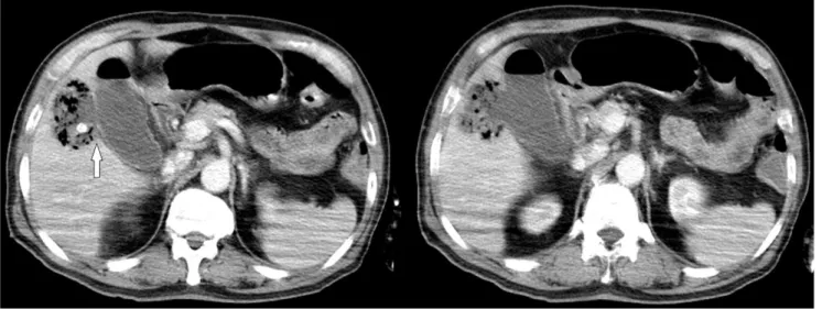
Fig. 1. Initial computed tomography scan showed acute calculous cholecystitis accompanied by focal perforation (arrow) and a 6×3.8 cm sized liver abscess and gallstone outside gallbladder.
Case: A 69-year-old male patient presented with right upper quadrant pain for 10 days. He was febrile (39℃), but the other vital signs were normal. A physical examination revealed mild tenderness on the right upper quadrant, and no rebound tenderness. The laboratory results revealed the following: a
white blood cell count 244,000/mm3 (neutrophil 88.1%), AST 188 U/L, ALT 245 U/L, ALP 78 U/L, GGT 26 U/L, and total bilirubin 8.32 mg/dL. A CT scan revealed acute calculous chol- ecystitis accompanied by focal perforation along the gall- bladder bed of segment V and a 6×3.8 cm sized abscess
120 조성호, 송상현. 담낭 파열에 의한 간농양
The Korean Journal of Gastroenterology Fig. 2. Percutaneous drainage catheter was inserted into the gallbladder.
Fig. 3. Endoscopic retrograde cholangiopancreatography presented a filling defect in common bile duct, which was removed by balloon sweeping.
(Fig. 1). Percutaneous drainage into the gallbladder was per- formed and cefotaxime and metronidazole were administered (Fig. 2). ERCP was conducted and a large amount of sludge was extracted from the common bile duct using a balloon (Fig. 3). Enterococcus faecalis and Staphylococcus aureus were cultured in pus. The laparoscopic cholecystectomy and drainage of the hepatic abscess were performed eight days after admission (Fig. 4). The post-operative course was un- eventful and the patient was discharged 14 days after surgery.
The follow-up CT scan revealed a decrease in the size of the hepatic abscess.
Diagnosis: intrahepatic abscess due to gallbladder perfo- ration
Gallbladder perforation is a rare but very serious complication. The frequency of this complication in patients with acute cholecystitis has decreased over time; the in- cidence was reported to be 2.9-15.4% before the 1990s, but
Jo S and Song S. Intrahepatic Abscess due to Gallbladder Perforation 121
Vol. 74 No. 2, August 2019 Fig. 4. Necrotic tissue area in the gallbladder bed was observed
after cholecystectomy in operation field image.
this recently fell to approximately 0.8%.1 The reason for this decrease in incidence is that recently, more cholecystectomy of symptomatic gallbladder stones has been carried out than in the past, and there is a tendency to conduct surgical treat- ment for acute cholecystitis at an early stage. Niemeier classi- fied a gallbladder perforation into three types: type 1, chronic perforation with the presence of a fistulous communication between the gallbladder and some other viscus; type 2, sub- acute perforation where the perforated gallbladder is sur- rounded by an abscess walled off by adhesions from the gen- eral peritoneal cavity; and type 3, acute perforation of the gallbladder into the free peritoneal cavity without protective adhesions.2 A gallbladder perforation occurs mainly in the fun- dus beause the blood supply is weakest in it. The morbidity and mortality due to a gallbladder perforation have been re- ported to be 37% and 7%, respectively. The risk factors for gallbladder perforation are old age, male gender, and car- diovascular comorbidity.1 Most of the gallbladder perforations are acute cholecystitis due to gallstones. On the other hand, acute acalculous cholecystitis may cause gallbladder perfo- ration because acute acalculous cholecystitis is more com- mon in sepsis and other co-morbidities than in other patients.
A gallbladder perforation usually begins with cystic duct ob- struction by gallstones. This phenomenon causes bile stasis and gallbladder distension and increases the pressure in the gallbladder, interfering with venous and lymphatic drainage, eventually damaging the blood vessels causing gallbladder necrosis and perforation. An intrahepatic gallbladder is buried within the liver tissue, and a perforation wound results in a
hepatic abscess.3
A gallbladder perforation has no specific clinical manifestations. The symptoms may occur acutely or slowly, and pain may decrease suddenly at the time of the perforation.
General weakness and weight loss may occur over a long period and may be considered to be a malignant tumor.4 Interestingly, this patient did not have Murphy’s sign because the peritoneal layers did not appear to be affected. An elevation of liver enzymes, particularly alkaline phosphatase, is common in blood tests. A ‘hole sign’, indicating that a gallbladder wall defect on ultrasonography, is useful for diagnosing gallbladder perforation. But, it may be difficult to be visualized by ultra- sonography due to bowel gas and pain.5 For this reason, it is better to perform a CT scan. According to Pedrosa et al.,6 the direct indicators of perforation on a CT scan include a demonstration of either calculi outside the gallbladder or a ruptured segment of the gallbladder wall. Indirect indicators include a demonstration of an abscess outside the gallbladder, presence of gallstones, and thickening of gallbladder wall. In the present case, a ‘hole’ can be found between the gallbladder wall and liver abscess (Fig. 1).
Because this complication is rare, it is difficult to stand- ardize the treatment. On the other hand, it is reasonable to perform interval open cholecystectomy after percutaneous drainage in addition to antibiotics to control a pyogenic liver abscess.4,7-9 If percutaneous transhepatic drainage or aspira- tion is contraindicated or anatomically impossible, recently in- troduced techniques, such as ultrasound-guided trans- duodenal (or transgastric) gallbladder drainage with stenting and endoscopic transpapillary gallbladder stenting, can be applied.10 Although laparoscopic cholecystectomy may be troublesome due to adhesion and severe inflammation, it could be performed successfully in selected cases, as in this patient.
REFERENCES
1. Stefanidis D, Sirinek KR, Bingener J. Gallbladder perforation: risk factors and outcome. J Surg Res 2006;131:204-208.
2. Niemeier OW. Acute free perforation of the gall-bladder. Ann Surg 1934;99:922-924.
3. Peer A, Witz E, Manor H, Strauss S. Intrahepatic abscess due to gallbladder perforation. Abdom Imaging 1995;20:452-455.
4. Donati M, Biondi A, Basile F, Gruttadauria S. An atypical pre- sentation of intrahepatic perforated cholecystitis: a modern in- dication to open cholecystectomy. Report of a case. BMC Surg
122 조성호, 송상현. 담낭 파열에 의한 간농양
The Korean Journal of Gastroenterology 2014;14:6.
5. Kochar K, Vallance K, Mathew G, Jadhav V. Intrahepatic perfo- ration of the gall bladder presenting as liver abscess: case report, review of literature and Niemeier's classification. Eur J Gastroenterol Hepatol 2008;20:240-244.
6. Pedrosa CS, Casanova R, Rodríguez R. CT findings in subacute perforation of the gallbladder: report on 5 cases. Eur J Radiol 1981;1:137-142.
7. Singh K, Singh A, Vidyarthi SH, Jindal S, Thounaojam CK.
Spontaneous intrahepatic type II gallbladder perforation: a rare cause of liver abscess - case report. J Clin Diagn Res 2013;7:
2012-2014.
8. Liao CY, Tsai CC, Kuo WH, et al. Emphysematous cholecystitis pre-
senting as gas-forming liver abscess and pneumoperitoneum in a dialysis patient: a case report and review of the literature. BMC Nephrol 2016;17:23.
9. Hussain T, Adams M, Ahmed M, Arshad N, Solkar M. Intrahepatic perforation of the gallbladder causing liver abscesses: case studies and literature review of a rare complication. Ann R Coll Surg Engl 2016;98:e88-e91.
10. Jang JW, Lee SS, Park DH, Seo DW, Lee SK, Kim MH. Feasibility and safety of EUS-guided transgastric/transduodenal gall- bladder drainage with single-step placement of a modified cov- ered self-expandable metal stent in patients unsuitable for cholecystectomy. Gastrointest Endosc 2011;74:176-181.