598
대한안과학회지 2015년 제 56 권 제 4 호 J Korean Ophthalmol Soc 2015;56(4):598-601 ISSN 0378-6471 (Print)⋅ISSN 2092-9374 (Online)
http://dx.doi.org/10.3341/jkos.2015.56.4.598
Case Report
일차 수술에서 발견된 다형성 샘암종 1예
A Case of Carcinoma Ex Pleomorphic Adenoma at 1st Operation
전수지⋅김수아⋅백지선⋅양석우
Soo Ji Jeon, MD, Su Ah Kim, MD, Ji Sun Paik, MD, PhD, Suk Woo Yang, MD, PhD
가톨릭대학교 의과대학 안과 및 시과학교실
Department of Ophthalmology and Visual Science, The Catholic University of Korea College of Medicine, Seoul, Korea
Purpose: To report a case of carcinoma ex pleomorphic adenoma observed during the patient’s first operation.
Case summary: A 63-year-old female presented with proptosis and ptosis that was aggravated 1 year prior. On preoperative CT image, a 32 x 20 x 21 mm-sized well demarcated mass (suspected as pleomorphic adenoma) was observed and was removed entirely by anterolateral orbitotomy. The excised mass surface was uneven but the capsule appeared intact on gross examination. Hard, yellow-colored and soft, dark-colored materials were found concurrently on cross section. The histological examination showed malignant cells as part of the soft material and was diagnosed as carcinoma ex pleomorphic adenoma.
Conclusions: We report a case of carcinoma ex pleomorphic adenoma of the lacrimal gland that presented with malignant change during the patient’s first operation. Supposedly, during the process of mass growth, minimal rupture occurred causing malignant transformation. Clinically, although a mass is believed benign based on imaging, the possibility of malignant trans- formation of a tumor increasing rapidly or enlargement causing development of rapid proptosis should be considered.
J Korean Ophthalmol Soc 2015;56(4):598-601
Key Words: Carcinoma ex pleomorphic adenoma, Lacrimal gland tumor, Malignant mixed tumor, Pleomorphic adenoma
■Received: 2014. 11. 28. ■ Revised: 2014. 12. 30.
■Accepted: 2015. 3. 7.
■Address reprint requests to Suk Woo Yang, MD, PhD Department of Ophthalmology, College of Medicine, The Catholic University of Korea, Seoul St. Mary’s Hospital,
#222 Banpo-daero, Seocho-gu, Seoul 137-701, Korea Tel: 82-2-2258-1200, Fax: 82-2-533-6718
E-mail: [email protected]
ⓒ2015 The Korean Ophthalmological Society
This is an Open Access article distributed under the terms of the Creative Commons Attribution Non-Commercial License (http://creativecommons.org/licenses/by-nc/3.0/) which permits unrestricted non-commercial use, distribution, and reproduction in any medium, provided the original work is properly cited.
눈물샘의 종양은 전체 안와 종양 중 10% 미만 정도를 차 지하는 드문 종양이며, 절반 정도는 상피성 기원으로 알려 졌다.1,2 상피성 기원의 눈물샘 종양 중에서 반수 정도가 다 형성 샘종이며 이는 완전히 절제된다면 예후는 좋은 편이 다. 일반적으로 양성 다형성 샘종의 제거 수술에서 종양이 모두 제거되지 않은 경우나 종괴 피막이 터지는 경우 악성 변화를 일으킬 수 있으므로 조직검사 등의 조작은 금기로 되어 수술로 완전 절제하는 것이 원칙으로 알려졌다.3 악성
변화한 다형성 샘종은 다형성 샘암종이라고 부르며, 임상 적으로 악성 혼합종(malignant mixed tumor)이라는 용어와 혼용된다. 다형성 샘암종은 전체 눈물샘 악성 종양 중 10%
정도를 차지하며 예후는 좋지 않다.4
본 증례에서는 수술 전 전산화단층촬영과 자기공명검 사에서 경계가 명확하고 주변 조직으로의 침윤과 골 파괴 소견이 없어 양성 다형성 샘종 의심하에 조직검사 없이 완전 절제 수술을 계획하였고 수술 중 파열이 없이 완전 절제하였으나, 술 후 조직학적으로 다형성 샘암종으로 최 종 진단되었다. 눈물샘 종양은 양성과 악성 간에 그 예후 와 치료 방침이 상이하므로 수술 전 검사에서 흔히 알려 진 것처럼 안와 벽을 침범하거나 경계가 불분명한 악성 양상의 종괴가 아닐 경우에도 항상 악성 변이의 가능성을 고려하고 접근해야 한다는 임상적 의의가 있기에 고찰해 보고자 한다.
599 - 전수지 외 : 다형성 샘암종 증례보고 -
Figure 1. The patient shows ptosis of right eye at primary posi-
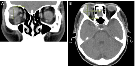
tion in preoperative frontal view.Figure 2. On CT image,
32 × 20 × 21 mm sized well dermacated mass is seen (A: coronal view;B: axial view).
증례보고
기저질환과 안과 과거력이 없는 63세 여자 환자가 1년 전부터 시작된 우안의 안구 돌출과 눈꺼풀 처짐을 주소로 내원하였다. 양안 시력 0.8, 세극등 검사에서 전안부에 특이 소견은 없었으나, 안구돌출계 검사에서 우안 18 mm, 좌안 14 mm로 심한 안구돌출이 보였으며, 환자의 눈꺼풀각막반 사 거리는 우안은 2 mm, 좌안은 3 mm로 우안의 눈꺼풀 처 짐을 보이면서 안구가 아래쪽으로 편위되어 있었다(Fig. 1).
우안 상안검 이측으로 단단한 종괴가 촉지되었고, 상방 주 시 시 우측 안구의 외안근 운동 제한이 관찰되었다.
안와 전산화단층촬영 및 자기공명영상에서 32×20×21 mm 크기의 경계가 명확한 고음영 종괴가 우측 안구를 누르고 있는 모습이 확인되었다(Fig. 2).
전신마취하 앞가측 안와절개술을 통한 종양 제거술 시행 하였으며 수술 과정에서 특이 사항 없었고 절개부위는 깨 끗하였으며, 육안상 종괴는 울퉁불퉁한 모양으로 피막은 매끈하게 유지되는, 경계가 명확한 종괴가 주변 조직과의 유착이나 파열 없이 제거되었다.
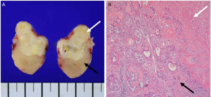
31×21×16 mm 크기의 경계가 명확한 종괴 내의 단면을
절개하여 흰색과 옅은 노란색으로 구성된 단단한 부분과 좀 더 어두운 색의 점액질 성상이 섞여 있는 부분이 동시에 존재함을 관찰할 수 있었다(Fig. 3A). 병리조직검사상 단단 한 부분에서 유상피세포와 결합조직으로 구성된 눈물샘 샘 종 소견을 보였으며, 점액질 성상이 섞여 있는 부분에서는 분열이 진행 중이거나 선명한 핵소체가 관찰되었고 이는 눈물샘 샘암종 소견과 일치하였다(Fig. 3B). 종괴 내에서 양 성과 악성이 공존하는 양상을 보여 병리학적으로 최종 다 형성 샘암종으로 진단되었다.
술 후 안와 자기공명촬영과 전신 양전자방출단층촬영 시 행하였으며 남아 있는 병변이나 전신 전이는 보이지 않았 고 눈꺼풀 처짐은 술 전보다 호전되었다(Fig. 4).
고 찰
눈물샘의 종양 중 가장 흔한 것은 상피성 종양의 50% 정 도에 해당하는 양성 다형성 샘종이며 나머지 상피성 종양 은 샘낭암종(adenoid cystic carcinoma)을 포함한 악성 종양 으로서 환자군이 다른 종양에 비해 젊은 편으로 예후는 치 명적일 수 있다.5,6 눈물샘의 다형성 샘암종은 전체 안와 종 양에서도 0.3% 정도에서 나타나며 병기 설정이나 치료 방 침에 대하여 여러 가지 의견이 논의되어 왔지만 조직학적 으로는 침샘의 모습과 동일하여 최근에는 침샘 종양의 병 기 설정과 치료 방침을 따르는 것이 일반적으로 받아들여 지고 있다.5,7 증례의 수가 많지 않아 임상적으로나 병리학 적으로 알려져 있는 것이 적으므로, 양성 종양인 다형성 샘 종에서 악성 변화를 겪게 되는 기전이나 예후 등에 대해서 도 거의 알려져 있는 바가 없다. 그에 따라 눈물샘의 다형 성 샘암종도 침샘의 다형성 샘암종에 준하여 임상적 판단 을 하게 되는데, 보통 침샘의 다형성 샘암종에서도 재발된
A B
600
- 대한안과학회지 2015년 제 56 권 제 4 호 -
Figure 3. On cross sectional photo of excised mass (A), the pathologic finding of upper- nodular, translucent lesion has benign char-
acteristics (white arrow), and lower- tan-yellow, firm mass with ill-defined lesion has malignant characterisctics (black arrow). The malignant component is distinguished by its collagenous stromal hyalinization. On microscopic examination (B), morphologic diver- sity with both epithelioid and connective tissue components is seen, which is consistent with pleomorphic adenoma (white arrow).This component is partially replaced by the malignant component (black arrow) – duct formation with nucleus containing chromatin condensation and prominent nucleoli is seen on high power, which is consistent with adenocarcinoma.
Figure 4. After operation, ptosis is improved in frontal view.
다형성 샘종 또는 다형성 샘종 수술 중 파열이 일어난 경우 에 악성 변화를 일으키는 것으로 알려졌다.7,8 또한 오래된 다형성 샘종 역시 다형성 샘암종으로 변화 가능한 것으로 알려져 있는데 병리학적으로 광범위한 유리질화는 오래된 다형성 샘종을 나타내는 소견으로,7 본 증례에서도 광범위 한 유리질화 소견을 보여주고 있다. 따라서 증례의 환자에 서는 오래된 다형성 샘종에서 종괴의 크기 증가나 외부의 충격에 의해 미세한 파열이 일어나 악성 변화를 일으켰고, 안구돌출이 1년 동안 급격하게 진행된 것으로 보아 1년 동안 악성화가 진행되었을 것으로 추정 가능하다. 또는 최근 발생 한 양성 종괴에서 크기가 급격히 커지면서 미세 파열이 일어 나 악성 변화를 일으킨 가능성도 생각해 볼 수 있다.
종괴의 성장속도가 매우 빠르거나9 종괴의 크기가 커서 안구돌출이 빠른 속도로 진행되는 경우에는 악성 종양의
가능성을 염두에 두어야 하며,10 수술 전 임상적으로는 다 형성 샘종의 양성과 악성의 구별이 쉽지 않으므로 경험적 으로 양성 종양에 준한 환자 진료를 경계해야 할 것이다. 침샘의 다형성 샘암종이 수술적 절제에 이어 국소 방사선 치료를 시행하여 생존율을 올리려는 것이 치료의 근간인 것처럼, 눈물샘의 다형성 샘암종도 이와 같은 치료 방침을 가져야 할 것으로 생각한다.11
REFERENCES
1) Shields JA, Shields CL, Scartozzi R. Survey of 1264 patients with orbital tumors and simulating lesions: The 2002 Montgomery Lecture, part 1. Ophthalmology 2004;111:997-1008.
2) Jung BJ, Cho YK, La TY. A giant pleomorphic adenoma of lac- rimal gland involving the palpebral lobe causing severe mechan- ical ptosis. J Korean Ophthalmol Soc 2011;52:241-5.
3) Shields JA, Shields CL, Eagle RC, Rizzo J. Pleomorphic adenoma ('benign mixed tumor') of the lacrimal gland. Arch Ophthalmol 1987;105:560-1.
4) von Holstein SL, Fehr A, Persson M, et al. Lacrimal gland pleomor- phic adenoma and carcinoma ex pleomorphic adenoma: genomic profiles, gene fusions, and clinical characteristics. Ophthalmology 2014;121:1125-33.
5) von Holstein SL. Tumours of the lacrimal gland. Epidemiological, clinical and genetic characteristics. Acta Ophthalmol 2013;91 Thesis 6:1-28.
6) Shields CL, Shields JA, Eagle RC, Rathmell JP. Clinicopathologic review of 142 cases of lacrimal gland lesions. Ophthalmology
A B
601
= 국문초록 =
일차 수술에서 발견된 다형성 샘암종 1예
목적: 눈물샘에 발생한 다형성 샘암종(Carcinoma ex pleomorphic adenoma)의 1예를 경험하고 이를 보고하고자 한다.
증례요약: 63세 여자 환자가 1년 전부터 시작된 우안 안구 돌출과 안검하수를 주소로 내원하였다. 수술 전 검사한 전산화단층촬영에서 다형성 샘종으로 의심되는 32×20×21 mm 크기의 경계가 명확한 종괴가 보여 앞가측 안와절개를 통하여 완전 적출술을 시행하였다.
육안상 종괴는 울퉁불퉁한 모양으로 피막은 매끈하게 유지되는 것으로 보였다. 종괴를 잘랐을 때 단면에서는 흰색과 옅은 노란색을 띈 단단한 부분과 더 어두운 색의 점액질 성상이 섞여 있는 부분이 동시에 존재하였고, 병리조직검사상 점액질 성상이 섞여 있는 부분에서 악성 세포가 관찰되어 다형성 샘암종으로 진단되었다.
결론: 본 증례에서는 일차 수술에서 이미 악성 변화가 진행된 다형성 샘종이 발견되었는데 이는 오래된 양성 다형성 샘종에서 종괴의 크기가 커지면서 미세한 파열이 일어나 악성 변화를 일으킨 것으로 추측해 볼 수 있다. 수술 전 영상 검사에서 양성 종양으로 의심되 었으나, 임상적으로 종괴의 성장속도가 빠르거나 종괴의 크기가 커지면서 안구돌출이 빠른 속도로 진행되는 경우에는 악성 종양의 가능성을 염두에 두는 것이 좋겠다.
<대한안과학회지 2015;56(4):598-601>
- 전수지 외 : 다형성 샘암종 증례보고 -
1989;96:431-5.
7) Sakuma T, Ohashi H, Yamamoto K, Kawano K. Carcinoma ex pleomorphic adenoma of the lacrimal gland: a case report. Jpn J Ophthalmol 2008;52:67-8.
8) Takahira M, Minato H, Takahashi M, et al. Cystic carcinoma ex pleomorphic adenoma of the lacrimal gland. Ophthal Plast Reconstr Surg 2007;23:407-9.
9) Won JY, Jung SK, Paik JS, Yang SW. Clinical analysis of epithelial
tumors of the lacrimal gland. J Korean Ophthalmol Soc 2014;
55:795-800.
10) Jung CK, Kim SM, Lee JY, Chung SK. Malignant change of pleo- morphic adenoma. J Korean Ophthalmol Soc 1997;38:2251.
11) Zhao J, Wang J, Yu C, et al. Prognostic factors affecting the clinical outcome of carcinoma ex pleomorphic adenoma in the major sali- vary gland. World J Surg Oncol 2013;11:180.