been considered essential to the success of a TKA2) and release of the contracted medial soft tissues from the tibial attachment site has been widely performed to balance the medial-lateral soft tissue tension1,3,4). However, this technique may result in an intraoperative rupture in the presence of soft tissue contracture or adhesion or medial instability of the knee5-7). Minimal soft tissue release followed by resection of the proximal medial tibia along the longitudinal axis of the tibia can be considered as an alternative. This technique is expected to be a safer option that would reduce the incidence of complications, such as medial collateral ligament (MCL) rupture, by allowing minimal release of the superficial layer of the MCL from the tibial attachment site8). In spite of this, there has been no domestic study on this technique.
The purpose of this study was to compare the results of TKA using the conventional medial soft tissue release and longitudinal bony resection of the proximal medial tibia with minimal soft tissue release in severe varus knees.
Comparative Study of Two Techniques for Ligament Balancing in Total Knee Arthroplasty for Severe Varus Knee: Medial Soft Tissue Release vs. Bony Resection of Proximal Medial Tibia
Ji Hyun Ahn, MD, and Young Woong Back, MD
Department of Orthopaedic Surgery, Dongguk University Ilsan Hospital, Goyang, Korea
This is an Open Access article distributed under the terms of the Creative Commons Attribution Non-Commercial License (http://creativecommons.org/licenses/by-nc/3.0/) which permits unrestricted non-commercial use, distribution, and reproduction in any medium, provided the original work is properly cited.
pISSN 2234-0726 · eISSN 2234-2451
Knee Surgery & Related Research
Purpose: Bony resection of the proximal medial tibia, an alternative technique for soft tissue balancing in total knee arthroplasty (TKA), was compared to the conventional medial soft tissue release technique.
Materials and Methods: From June 2005 to June 2007, we performed 40 TKA in 27 patients with ≥10o tibio-femoral varus deformity. The conventional, medial soft tissue release technique was applied in 20 cases and bony resection of proximal medial tibia in the other 20 cases (vertical osteotomy group). Total operation time, knee range of motion (ROM), hospital for special surgery (HSS) scores, and tibio-femoral medial-lateral gap ratio in 0o, 90o, and 130o flexion at postoperative 6 months were compared between the groups.
Results: The total operation time was shorter in the vertical osteotomy group. Tibio-femoral medial-lateral gap ratio in 130o flexion was closer to 1 in the vertical osteotomy group (p=0.000). There was no significant difference in the ROM, HSS score, or tibio-femoral medial-lateral gap ratio in 0o and 90o flexion at postoperative 6 months.
Conclusions: In severe varus knees, bony resection of proximal medial tibia can be considered as an alternative technique, in order to decrease total operation time and to obtain medial-lateral, soft-tissue balance in deep flexion.
Keywords: Knee, Varus deformity, Total knee arthroplasty, Bony resection, Proximal medial tibia
Received May 3, 2011; Revised (1st) September 20, 2011;
(2nd) April 4, 2012; (3rd) October 27, 2012; Accepted October 30, 2012 Correspondence to: Ji Hyun Ahn, MD
Department of Orthopaedic Surgery, Dongguk University Ilsan Hospital, 27 Dongguk-ro, Ilsandong-gu, Goyang 410-773, Korea
Tel: +82-31-961-7317, Fax: +82-31-961-7290 E-mail: [email protected]
Introduction
Total knee arthroplasty (TKA) has been frequently performed for severe degenerative arthritis in senior patients. Unfortunately, TKA can be technically challenging in knees with profound varus deformity when it is combined with medial soft tissue contracture and lateral soft tissue laxity1). Ligament balancing has
13
Copyright © 2013. THE KOREAN KNEE SOCIETY www.jksrr.org
Materials and Methods
Of the patients who underwent TKA performed by the same surgeon at our institution between June 2005 and June 2007, 27 patients (40 cases) with ≥10o tibio-femoral varus deformity on the preoperative whole leg standing anteroposterior (AP) view (Fig. 1) were included in this prospective randomized controlled study. All study participants were female with a mean age of 71.5 years (range, 62 to 83 years). The mean preoperative, anatomical tibio-femoral axis was varus 13.3o (range, 11o to 19o) (Table 1).
TKA was performed using a posterior-stabilized type prosthesis in all cases. For ligament balancing, the conventional medial soft tissue release was performed in 20 cases (medial release group).
In the remaining 20 cases (vertical osteotomy group), a vertical osteotomy for one size smaller tibial component was performed using an osteotome in the proximal medial tibia for ligament balancing (Fig. 2).
There was no statistically significant difference in age and preoperative varus deformity angle, range of motion (ROM), and hospital for special surgery (HSS) score between the groups.
In the medial release group, the knee joint was exposed using a midvastus approach and medial soft tissue was released up to 1 cm distally from the subperiosteal layer of the joint capsular insertion site, on the proximal tibia and then posteromedially.
Progressive soft tissue release was carried out until symmetrical, medial-lateral balance was confirmed using a trial prosthesis after femoral and tibial articular surface resection. The initial medial release was extended ≥1 cm distally in all of the medial
release group (n=20). Medial-lateral tension was assessed with the knee in extension and 90o flexion. If medial soft tissue contracture was noted in extension, the deep layer of the MCL and the posteromedial joint capsule were released; whereas the superficial layer of the MCL and the anteromedial joint capsule were released, if contracture was noted in 90o flexion to obtain a perfect ligament balance.
In the vertical osteotomy group, a midvastus approach was used for joint exposure. Soft tissue release was started in the subperiosteal layer of the joint capsular insertion site on the proximal tibia and extended 1 cm distally and posteromedially.
Subsequently, without further release, femoral and tibial arti- cular surface resection was carried out and ligament balance was assessed using a trial prosthesis. Taking care to achieve symmetrical medial-lateral tension with the use of a prosthesis that is one size smaller than the trial one, an osteotome was inserted perpendicular to the resected articular surface for bone tissue removal in the proximal medial tibia. The bone resection was performed in 2 mm increments according to the level of the medial soft tissue contracture until a perfect ligament balance was confirmed with the insertion of the trial prosthesis.
Comparisons between the groups were based on the total operation (OP) time, ROM, HSS score, tibio-femoral medial- lateral gap ratio examined with the knee in 0o, 90o, and 130o flexion under an image intensifier at 6 months postoperatively.
Total OP time starting from the initial skin incision to skin closure was measured. On the assessment of ROM at postoperative 6 months, the difference in the angle created by the femoral longitudinal axis and the tibial longitudinal axis with the knee in maximum flexion and extension on lateral radiographs was recorded.
To assess the tibio-femoral medial-lateral gap ratio at post- operative 6 months, the patient was placed in the supine position with the knee in 0o, 90o, and 130o flexion under an image magnifier that was set up to produce a cross-sectional image of the tibial articular surface on the AP view. The medial and lateral tibio-femoral gaps were measured with the knee in 0o, 90o, and 130o flexion on the picture archiving and communication system (PACS) and the lateral value was divided by the medial value to obtain the ratio at each flexion angle (Fig. 3).
Statistical analysis was performed using SPSS ver. 15.0 (SPSS Inc., Chicago, IL, USA) in order to analyze total OP time, ROM, HSS score, and tibio-femoral medial-lateral gap ratio at postoperative 6 months. The Mann-Whitney test was conducted to compare differences between the groups with a 95% confi- dence interval.
Fig. 1. Anteroposterior radiograph of both knees showing arthritic change of the medial, tibio-femoral joint with severe varus deformity.
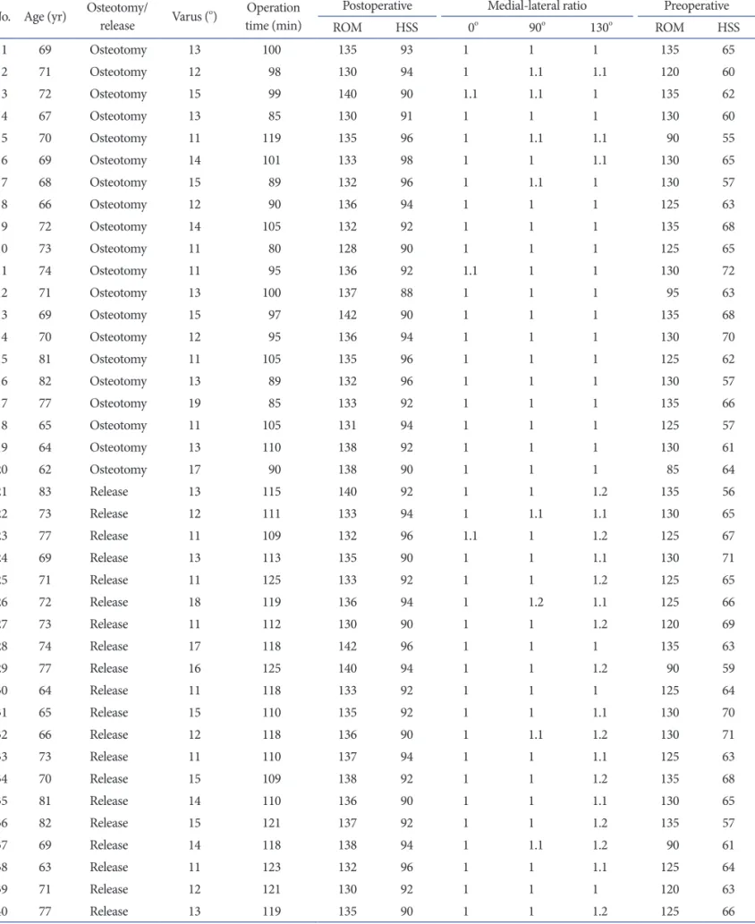
Table 1. Clinical Raw Data for Total Knee Arthroplasty Cases with Mechanical Varus Deformity More Than 10o No. Age (yr) Osteotomy/
release Varus (o) Operation time (min)
Postoperative Medial-lateral ratio Preoperative
ROM HSS 0o 90o 130o ROM HSS
1 69 Osteotomy 13 100 135 93 1 1 1 135 65
2 71 Osteotomy 12 98 130 94 1 1.1 1.1 120 60
3 72 Osteotomy 15 99 140 90 1.1 1.1 1 135 62
4 67 Osteotomy 13 85 130 91 1 1 1 130 60
5 70 Osteotomy 11 119 135 96 1 1.1 1.1 90 55
6 69 Osteotomy 14 101 133 98 1 1 1.1 130 65
7 68 Osteotomy 15 89 132 96 1 1.1 1 130 57
8 66 Osteotomy 12 90 136 94 1 1 1 125 63
9 72 Osteotomy 14 105 132 92 1 1 1 135 68
10 73 Osteotomy 11 80 128 90 1 1 1 125 65
11 74 Osteotomy 11 95 136 92 1.1 1 1 130 72
12 71 Osteotomy 13 100 137 88 1 1 1 95 63
13 69 Osteotomy 15 97 142 90 1 1 1 135 68
14 70 Osteotomy 12 95 136 94 1 1 1 130 70
15 81 Osteotomy 11 105 135 96 1 1 1 125 62
16 82 Osteotomy 13 89 132 96 1 1 1 130 57
17 77 Osteotomy 19 85 133 92 1 1 1 135 66
18 65 Osteotomy 11 105 131 94 1 1 1 125 57
19 64 Osteotomy 13 110 138 92 1 1 1 130 61
20 62 Osteotomy 17 90 138 90 1 1 1 85 64
21 83 Release 13 115 140 92 1 1 1.2 135 56
22 73 Release 12 111 133 94 1 1.1 1.1 130 65
23 77 Release 11 109 132 96 1.1 1 1.2 125 67
24 69 Release 13 113 135 90 1 1 1.1 130 71
25 71 Release 11 125 133 92 1 1 1.2 125 65
26 72 Release 18 119 136 94 1 1.2 1.1 125 66
27 73 Release 11 112 130 90 1 1 1.2 120 69
28 74 Release 17 118 142 96 1 1 1 135 63
29 77 Release 16 125 140 94 1 1 1.2 90 59
30 64 Release 11 118 133 92 1 1 1 125 64
31 65 Release 15 110 135 92 1 1 1.1 130 70
32 66 Release 12 118 136 90 1 1.1 1.2 130 71
33 73 Release 11 110 137 94 1 1 1.1 125 63
34 70 Release 15 109 138 92 1 1 1.2 135 68
35 81 Release 14 110 136 90 1 1 1.1 130 65
36 82 Release 15 121 137 92 1 1 1.2 135 57
37 69 Release 14 118 138 94 1 1.1 1.2 90 61
38 63 Release 11 123 132 96 1 1 1.1 125 64
39 71 Release 12 121 130 92 1 1 1 120 63
40 77 Release 13 119 135 90 1 1 1.2 125 66
ROM: range of motion, HSS: hospital for special surgery.
Results
Statistically significant intergroup differences were found in the total OP time and the tibio-femoral medial-lateral gap ratio in 130o flexion. There was no significant difference in the preoperative HSS score and tibio-femoral angle on the whole leg standing AP view and postoperative ROM, HSS score, and tibio- femoral medial-lateral gap ratio in 0o and 90o flexion. The mean total OP time was remarkably short in the vertical osteotomy group (mean, 96.9 minutes; range, 80 to 119 minutes) compared to that in the medial release group (mean, 116.2 minutes; range, 109 to 125 minutes) (p=0.000). The mean tibio-femoral medial- lateral gap ratio in 130o flexion at postoperative 6 months was notably smaller in the vertical osteotomy group (1.02) than in the medial release group (1.14; p=0.000). However, the ratio was not significantly different between the groups in 0o and 90o flexion.
No significant intergroup difference was found in the ROM and HSS score at postoperative 6 months (Table 2).
In the vertical osteotomy group, the tibial component size was determined after bone resection by assessing the medial-lateral tension with a trial prosthesis. In this group, the final prosthesis of choice was one size smaller than the one selected before bone resection, which did not result in a size mismatch with the femoral component. In the medial release group, there were 2 cases of intraoperative partial MCL tear, which did not lead to any clinical problems after staple fixation (Fig. 4).
Discussion
There is a variety of surgical techniques for the treatment of degenerative arthritis of the knee according to the severity and extent of a lesion. Of these, TKA is the most common surgical intervention for profound degenerative arthritis in senior patients. Ligament balance is essential to the success of TKA and imbalance has been recognized as one of the major causes of early failure of TKA as well as pain during ambulation5,6,9). Sequential medial soft tissue release has been widely performed in varus arthritic knees10), however, profound varus deformity often necessitates additional soft tissue release in the MCL and joint capsule11,12). An extensive medial soft tissue release performed in severe varus knees may result in overcorrection, for which a thick polyethylene insert or a constrained prosthesis should be used when a patient undergoes TKA13). The conventional ligament balancing procedure involves removal of degenerative osteophytes and release of the contracted medial soft tissue at the tibial attachment site to obtain symmetrical medial-lateral tension in extension and 90o flexion of the knee1,3,14). This technique is advantageous in that various medial soft tissues, including the superficial and deep layers of the MCL, can be selectively released
Fig. 3. Anteroposterior radiographs by an X-ray image intensifier showing tibio- femoral medial-lateral gap ratio changes according to different flexion angles at postoperative 6 months. (A) 0o flexion. (B) 90o flexion. (C) 130o flexion.
Fig. 2. Intraoperative photograph showing bony resection of proximal medial tibia using an osteotome for ligament balancing.
to adjust medial tension in extension and 90o flexion of the knee5,7). However, it is difficult to avoid medial soft tissue rupture during the procedure. To overcome this disadvantage, Dixon et al.8) suggested longitudinal resection of the proximal medial tibia.
Their method minimizes soft tissue release to prevent medial soft tissue rupture and uses vertical resection of the proximal medial tibia for ligament balance. Hence, it is expected to be more effective and safer than the conventional procedure, allowing minimal release of the superficial layer of the MCL in the tibial
attachment site8,15). In this study, we compared the 2 ligament balancing techniques based on the total OP time, preoperative and 6-month postoperative ROM and HSS score, and tibio- femoral medial-lateral gap ratio in 0o, 90o, and 130o flexion. There were statistically significant differences in the total OP time and the ratio in 130o knee flexion. The mean total OP time was longer in the medial release group than in the vertical osteotomy group. We attributed this to the use of a sequential release procedure to prevent excessive soft tissue release according to recommendations and technical difficulties caused by adhesion and contracture of medial soft tissues in knees with degenerative arthritis4,15). The mean total OP time in the medial release group could have been lengthened due to staple fixation in 2 cases with a partial rupture of the superficial layer of the MCL. The 2 cases were not excluded from the analysis under the assumption that the ruptures represented the risk of medial soft tissue release. The significantly lower tibio-femoral medial-lateral gap ratio in 130o flexion in the vertical osteotomy group could be attributable to the lack of an aggressive medial release to avoid the possibility of medial soft tissue instability16). The difference in the ratio in 130o knee flexion did not result in significant intergroup difference in ROM. In spite of this, the results are worth consideration in TKA for Asian patients, because hyperflexion of the knee is often necessary in Asian culture17,18).
Proximal medial tibial resection can be an effective method for ligament balancing. However, an extensive bone resection may cause tibial component loosening, difficulty in revision surgery, Table 2. Comparison between Vertical Osteotomy Group and Medial
Release Group
Group Mean±SD (range) p-value
Age (yr) 0.222
Osteotomy 70.6±5.12 (62–82)
Release 72.5±5.77 (63–83)
Varus (°) 0.978
Osteotomy 13.25±2.15 (11–19)
Release 13.25±2.17 (11–18)
Operation time (min) 0.000
Osteotomy 96.85±9.44 (80–119)
Release 116.20±5.39 (109–125)
Postoperative ROM 0.376
Osteotomy 134.45±3.58 (128–142)
Release 135.40±3.28 (130–142)
Postoperative HSS 0.718
Osteotomy 92.90±2.65 (88–98)
Release 92.60±2.06 (90–96)
Medial-lateral ratio (0o) 0.553
Osteotomy 1.01±0.03 (1.0–1.1)
Release 1.01±0.02 (1.0–1.1)
Medial-lateral ratio (90o) 0.938
Osteotomy 1.02±0.04 (1.0–1.1)
Release 1.03±0.06 (1.0–1.2)
Medial-lateral ratio (130o) 0.000
Osteotomy 1.02±0.04 (1.0–1.1)
Release 1.14±0.07 (1.0–1.2)
Preoperative ROM 0.567
Osteotomy 123.75±15.21 (85–135)
Release 124.25±12.59 (90–135)
Preoperative HSS 0.212
Osteotomy 63.00±4.58 (55–72)
Release 64.65±4.21 (56–71)
SD: standard derviation, ROM: range of motion, HSS: hospital for special surgery.
Fig. 4. (A) Anteroposterior radiograph of the right knee showing repair by staple for torn medial collateral ligament. (B) Intraoperative photograph showing torn medial collateral ligament during medial release.
and kinematic changes in the knee due to lateral translation of the tibial component. Although extensive medial soft tissue release has been associated with knee instability in some studies, Choi et al.2) suggested that proper postoperative fixation could improve stability in knees with varus deformity even after extensive release of medial soft tissues including the MCL.
One of the limitations of this study is the 6-month short-term follow-up period in comparison to other studies. Although TKAs were bilateral in 13 of the 27 patients, the influence of personal differences on the postoperative ROM and HSS score were not taken into consideration in the analysis. In addition, the medial- lateral gap was measured without weight bearing and thus the results may not reflect the possibility of instability during walking or daily living activities. The difference in the tibio-femoral medial-lateral gap ratio in 130o flexion might have originated from the difference in the axis of knee flexion. Furthermore, the results could have been affected by the surgeon’s preference or skills considering that all the operations were performed by the same surgeon in this study.
Conclusions
The 6-month short-term follow-up results of TKA showed that proximal medial tibial resection in severe varus knees can be effective in reducing operation time and achieving ligament balance in high flexion. We believe possible complications related to the procedure should be investigated in future long-term follow-up studies.
Conflict of Interest
No potential conflict of interest relevant to this article was reported.
References
1. Mullaji AB, Padmanabhan V, Jindal G. Total knee arthro- plasty for profound varus deformity: technique and radio- logical results in 173 knees with varus of more than 20 degrees. J Arthroplasty. 2005;20:550-61.
2. Choi CH, Kim JH, Chung HK, Kim JH. Clinical and radiological evaluation of detached MCL cases in medial release during TKA: MCL detached vs stable CR vs stable PS. J Korean Knee Soc. 2002;14:1-8.
3. Bottros J, Gad B, Krebs V, Barsoum WK. Gap balancing in total knee arthroplasty. J Arthroplasty. 2006;21(4 Suppl 1):11-5.
4. Yagishita K, Muneta T, Ikeda H. Step-by-step measurements of soft tissue balancing during total knee arthroplasty for patients with varus knees. J Arthroplasty. 2003;18:313-20.
5. Kim JH, Choi CH, Kang SK, Lee BK, Shon KH, Chung HK. Assessment of failure modes in total knee replacement arthroplasty. J Korean Knee Soc. 2003;15:177-84.
6. Lee CC, Cho SD, Ko SH, Jang KH, Gwak CY, Jeong JY.
Instability after total knee arthroplasty. J Korean Knee Soc.
2006;18:175-81.
7. Sim JA, Kwak JH, Yang SH, Kim JY, Lee BK. Short-term follow-up results of medial epicondylar osteotomy for the varus knee in TKA. J Korean Knee Soc. 2009;21:197-204.
8. Dixon MC, Parsch D, Brown RR, Scott RD. The correction of severe varus deformity in total knee arthroplasty by tibial component downsizing and resection of uncapped proximal medial bone. J Arthroplasty. 2004;19:19-22.
9. Insall JN, Binazzi R, Soudry M, Mestriner LA. Total knee arthroplasty. Clin Orthop Relat Res. 1985;(192):13-22.
10. Laskin RS, Schob CJ. Medial capsular recession for severe varus deformities. J Arthroplasty. 1987;2:313-6.
11. Teeny SM, Krackow KA, Hungerford DS, Jones M. Primary total knee arthroplasty in patients with severe varus deformity: a comparative study. Clin Orthop Relat Res.
1991;(273):19-31.
12. Vince KG, Abdeen A, Sugimori T. The unstable total knee arthroplasty: causes and cures. J Arthroplasty. 2006;21(4 Suppl 1):44-9.
13. Matsueda M, Gengerke TR, Murphy M, Lew WD, Gustilo RB. Soft tissue release in total knee arthroplasty: cadaver study using knees without deformities. Clin Orthop Relat Res. 1999;(366):264-73.
14. Whiteside LA. Soft tissue balancing: the knee. J Arthroplasty.
2002;17(4 Suppl 1):23-7.
15. McPherson EJ, Portugal D. Revision total knee arthroplasty for excessive ligamentotaxis. J Arthroplasty. 2007;22:1214-6.
16. Stahelin T, Kessler O, Pfirrmann C, Jacob HA, Romero J.
Fluoroscopically assisted stress radiography for varus-valgus stability assessment in flexion after total knee arthroplasty. J Arthroplasty. 2003;18:513-5.
17. Sugama R, Kadoya Y, Kobayashi A, Takaoka K. Preparation of the flexion gap affects the extension gap in total knee arthroplasty. J Arthroplasty. 2005;20:602-7.
18. Yasgur DJ, Scuderi GR, Insall JN. Medial release for fixed varus deformity. In: Scuderi GR, Tria AJ, eds. Surgical technique in total knee arthroplasty. New York: Springer;
2002. p189-96.