The Effects of Closed Kinetic and Open Kinetic Chain Exercises Using Knee Reposition Sense in Chronic Stroke Patients
Purpose: This study aimed to determine the effects of open kinetic chain exercise (OKCE) and closed kinetic chain exercise (CKCE) using knee reposition sensing on balance, strength, and knee joint reposition sense (JPS) in chronic stroke patients.
Methods: Twenty-nine hemiplegic patients participated in this study. Participants were randomly divided into 3 groups, CKCE, OKCE, and controls, with 9, 10, and 10 participants, respectively. The CKCE group completed CKCE using knee reposition sensing, whereas the OKCE group completed OKCE using knee reposition sensing. The control group completed conventional physical therapy.
Results: Significant differences between the CKCE and OKCE groups were apparent for all outcomes except the functional reaching test. The CKCE group displayed significant improvements in knee JPS versus the OKCE and control groups (p<0.01). The OKCE group displayed significant improvements in knee extensor muscle strength versus the CKCE and control groups (p<0.05).
The CKCE and OKCE groups displayed significantly improvements in static balance versus the control group (p<0.05).
Conclusion: CKCE and OKCE improved balance, strength, and knee JPS. Additionally, CKCE might provide a more useful intervention benefit than OKCE for increasing knee JPS, a weight-bearing task. OKCE was sufficient to improve the knee extensor muscle strength.
Key Words:Stroke, Closed-kinetic chain exercise, Open-kinetic chain exercise, Joint position sense Kyu-Young Lee1, Won-Seob Shin2
1Department of Physical Therapy, Graduate School of Daejeon University, 2Department of Physical Therapy, College of Natural Science, Daejeon University
The Journal of Korean Society of Physical Therapy Original articles
I. Introduction
Stroke patients experience difficulties such as reduced balance, weakness, and a loss of proprioception consequent to brain damage.1 Reduced balance limits participation in activities of daily living.2 Weakness is the main stroke symptom that limits movement of the body.3,4 Proprioception refers to information provided by mechanoreceptors about the positions and
Copylight ⓒ 2014 The Korea Society of Physical Therapy
This is an Open Access article distribute under the terms of the Creative Commons Attribution Non-commercial License (Http:// creativecommons.org/license/by-nc/3.0.) which permits unrestricted non-commercial use, distribution,and reproduction in any medium, provided the original work is properly cited.
Received May 16, 2014 Revised Jun 10, 2014 Accepted Jun 13, 2014
Corresponding author Won-Seob Shin, [email protected]
movements of the bones and joint capsules.5 Proprioception is an essential element for participation in activities of daily living such as locomotion, eating, and spontaneous movement.6 Joint position sense (JPS), a type of proprioception, is the specific ability to recognize and reproduce information about the position of a joint and comprises parameters such as velocity, direction, and magnitude.5 JPS also contributes to postural control and involuntary movement.7
Two types of physical exercises are used to strengthen the knee joints, the open kinetic chain exercises (OKCE) and closed kinetic chain exercises (CKCE). In OKCE, the hand or foot can freely move, usually in response to elbow or knee joint movements that induce motion during non-weight- bearing (NWB) support. Weight may be applied to the distal
extremities. OKCE have a limited range of motion and are effective strength-training exercises.8,9 Additionally, OKCE target muscles to maintain the shape and strength of additional training.10 OKCE predominantly target concentric muscle contraction as well as the generation of increased traction and rotational forces and provide stability via external means.11 Additionally, during OKCE, the acceleration increases, resistance decreases, distraction force and torque is lost, and increased strain is placed on the joint and muscle mechanoreceptors, while concentric acceleration and deceleration are associated with an increase in efferent functional features such as the promotion of activity.12 During CKCE, the hand (arm movement) or foot (leg movement) remains fixed while performing an exercise. The hand or foot is placed on the ground and remains in contact with the surface of the apparatus. In closed-chain movement, the dynamic stability of the muscle contraction predominates over the simultaneous eccentric contraction, and the pressing joint shear force reduces the inherent stability, thus sensitizing the capsule mechanoreceptors to pressure changes and promoting proprioception. CKCE also strengthens the antagonist of the damaged target to influence the stability of the joint. The exercises involve typical external weights and body weight- bearing (WB) exercises. Squat exercises are a good example of CKCE.
OKCE and CKCE have both advantages and disadvantages;
however, in recent years CKCE have been more frequently recommended. The reason for this preference is that CKCE include more functional tasks. Additionally, CKCE may provide more sensory feedback and hence improve the sensorimotor functions, including motor control and joint proprioception, to a greater extent than OKCE.13 Although previously published studies have reported using OKCE14-17 and CKCE18 as strength-training exercises to activate muscles via proprioceptive effects or to rehabilitate patients with musculoskeletal injuries, the effects of these exercises have not been reported as extensively in stroke patients. Moreover, the limited available evidence regarding the independent effects of kinetic exercise using knee reposition sensing makes it difficult to specifically describe this effect.
Furthermore, a small study compared WB versus NWB
exercises in the context of chronic stroke. The current study aimed to evaluate the effects of OKCE and CKCE using knee reposition sensing on balance, strength, and JRS in chronic stroke patients.
II. Methods
1. Subjects
The subjects were 29 stroke patients who had been admitted to a clinic. The subjects were randomized into 3 groups by a physical therapist who did not participate in the study: the CKCE group (n=9), OKCE group (n=10), and control group (n=10). This randomization was performed by selecting an opaque closed envelope from among envelopes in which the group assignments were written. The assignments were given to the physical therapist in sealed numbered envelopes. The inclusion criteria were (1) an interval of >6 months post- event; (2) sufficient cognition to participate in the study:
a Mini-Mental State Exam-Korea (MMSE-K) score of
≥24; (3) the ability to remain in an unassisted standing position for >30 s; (4) the ability to perform knee flexion and extension; (5) a higher than fair lower extremity strength grade; and (6) a modified Ashworth scale (MAS) grade <2.
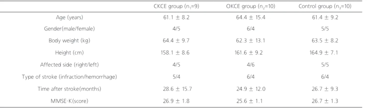
The exclusion criteria were (1) knee injury or disease and (2) any uncontrolled health conditions. Table 1 shows the general characteristics of the subjects. Participation in the study was voluntary and fully understanding the contents of this study.
Written informed consent, after providing an explanation of the study purpose, as well as the experimental method and processes, was obtained from all patients. The study was approved by the Daejeon University institutional review board (1040647-201403-HR-015-03).
2. Instruments and measurement
Static balance was measured using a Wii Balance Board (WBB) (Nintendo, Kyoto, Japan) and Balancia software (Mintosys, Seoul, Korea). The WBB was used as the input instrument for a consumer machine of 50 × 50 cm. A pressure sensor was embedded into a square in a horizontal grid. The pressure center information was collected through the 4 load cells on the square edges and entered into a computer. The sampling
ratio of the collected data was controlled by software linked with the computer. The WBB is a highly reliable, effective, and validated tool for evaluating stance balance. In this study, standing position information from stroke patients was entered into the COP analysis program for analysis using Balancia software ver. 2.0. The WBB measured the COP information and submitted it to Bluetooth-connected computer program for analysis using Balancia. The COP analysis results included the X and Y-axes, sway distance and sway velocity for the left and right weight distributions, 95% slope, and 95% area.
This test has high inter (r=0.93) and intra-observer (r=0.96) reliability for analyzing the sway distance and sway velocity.19 The dynamic balance was measured using the timed up and go test (TUG) and functional reaching test (FRT). The TUG test records the time required to rise from a chair (height: 50 cm), walk 3 meters, turn around a marker, walk back to the chair, and sit. The participants were asked to perform this test 3 times, and the mean round-trip time was recorded.
This test has high inter (r=0.98) and intra-observer (r=0.99) reliability.20 The FRT records the maximal reaching distance in the front, left, and right directions while in a comfortable standing position. If participant could not reach with their arm, the acromion distance was measured. This test also has inter (r=0.92) and intra-observer (r=0.98) reliability.21
Strength was measured using a PowerTrack II Commander hand-held device (PTCH; J Tech Medical, Salt Lake City, UT, USA). Participants held the PTCH for 5 s during maximal isometric contraction. The participants were asked to perform this test 3 times, and the mean result was recorded. This test has high inter-observer reliability (ICC=0.86-0.94).22
Knee JRS (KJRS) was measured using a smartphone application. A free angle measurement software application (Angle; Smudge Apps, Christchurch, New Zealand) was downloaded to the smartphone from the Apple application store. At each step, the smartphone was used to measure the knee extension angle by placing the device on the anterior surface of the thigh and on the anterior surface of the distal tibia; the device then displayed the angle relative to a horizontal line in both positions. The knee extension angle was calculated as the sum of the 2 measurements. The intra- observer and inter-observer reproducibility were good.23
With the subjects blindfolded, the JPS of the affected leg were assessed at knee extension angles of 120° and 150°.
For the NWB joint position test, the subjects were seated in an elevated chair so that their legs could freely move. The affected side was positioned at either 120° or 150° according to the smartphone application, and the subjects were then asked to move their leg towards a 90° angle (relaxed seated position) and then return their leg as closely as possible to the initial angle.24
For the WB joint position sense test, the subjects stood, bent their knees to either 120° or 150° according to the smartphone application, straightened their legs to the full extension (standing upright), and then again bent their knees as closely as possible to the initial angle. The participants were allowed to practice each task and then repeated each task twice in a random order.25
3. Intervention
In this study, all participants, including those in the control group, were subjected to conventional physical therapy.
Conventional physical therapy included neuro-developmental therapy (NDT) and functional electronic stimulation (FES) and was conducted during a 4-week program with 60-min sessions, 5 days per week. The CKCE and OKCE also used knee reposition sensing. Participants in the CKCE and OKCK groups underwent after conventional treatment during a 30-min sessions, 3 days per week, 4 weeks.
For knee reposition sensing in the OKCE group, the subjects were seated in an elevated chair so that their legs could freely move. Before exercise, each subject’s maximum affected side knee extension strength was measured. During weeks 1 and 2, the therapist applied a sandbag weight equivalent to <50% of the maximum strength to the ankle and during weeks 3 and 4, applied a weight >50%.
The knee reposition sense was determined via the subject's ability to reproduce a specific knee angle that had been randomly selected among the possible knee extension angles between 0° and 90°. Each subject moved their knee toward the target extension angle and maintained this position for 5 s. After a resting time of 10 s, the subjects returned to the starting position and reproduced the knee extension at the
same target angle. If the subject failed to reproduce an angle, the therapist would correct a JPS error and reset.
For knee reposition sensing in the CKCE group, the subjects stood on the template with their weight equally distributed over both feet. All subjects’ maximum knee joint angles had been measured while sitting. The therapist subjected the knee to <50% of the maximum angle during weeks 1 and 2 and to
>50% of the maximum angle during weeks 3 and 4. During this test, the subjects moved their knee toward the target extension angle and maintained this position for 5 s. After a resting time of 10 s, the subjects returned to the starting position and reproduced the knee extension at the same target angle.
4. Statistical analyses
All statistical analyses were conducted using the Windows SPSS 18.0 statistical software package (SPSS Inc., Chicago, IL, USA). The paired Student’s t-test was used to analyze changes between the pre and post-test balance, strength, and JRS measurements. A one-way ANOVA was used to compare differences between the pre and post-test balance, strength, and JRS measurements between the groups. A post-hoc Sheffe test was used for statistically significance results. Statistical significance was defined as a P-value
<0.05.
3. Results
1) Comparison of pre and post-intervention balance
The mean changes in the sway distances were -52.21 ± 48.18 mm in the CKCE group and -48.21 ± 49.38 mm in the
CKCE group (n1=9) OKCE group (n2=10) Control group (n3=10)
Age (years) 61.1 ± 8.2 64.4 ± 15.4 61.4 ± 9.2
Gender(male/female) 4/5 6/4 5/5
Body weight (kg) 64.4 ± 9.7 62.3 ± 13.1 63.5 ± 8.2
Height (cm) 158.1 ± 8.6 161.6 ± 9.2 164.9 ± 7.1
Affected side (right/left) 4/5 4/6 5/5
Type of stroke (infraction/hemorrhage) 5/4 6/4 6/4
Time after stroke(months) 28.6 ± 15.7 24.9 ± 12.0 26.7 ± 9.3
MMSE-K(score) 26.9 ± 1.8 25.6 ± 1.1 26.7 ± 1.3
CKCE group (n1=9) OKCE group (n2=10) Control group (n3=10) F
Sway distance (mm)
Pre 137.69 ± 53.57 134.90 ± 47.51 123.07 ± 54.09 0.22
Post 85.48 ± 21.39 86.69 ± 27.31 98.10 ± 42.97 0.46
t 3.25* 3.09* 1.31
Change -52.21 ± 48.18 † -48.21 ± 49.38 † -24.96 ± 43.72 4.41*
Sway velocity (mm/s)
Pre 4.59 ± 1.76 4.50 ± 1.58 4.10 ± 1.80 0.22
Post 2.85 ± 0.71 2.89 ± 0.91 3.27 ± 1.43 0.46
t 3.25* 3.09* 1.31
Change -1.74 ± 1.60 † -1.61 ± 1.65 † -0.13 ± 0.32 4.41*
Table 1. General characteristics of subjects
Table 2. A comparison of static balance changes
CKCE: Closed Kinetic Chain Exercises, OKCE: Open Kinetic Chain Exercises, MMSE-K: Mine-Mental State Examination Korean version
CKCE: Closed Kinetic Chain Exercises, OKCE: Open Kinetic Chain Exercises
*p<0.05
† significant difference compared with the Control group (p<0.05).
OKCE group (p<0.05). In contrast, no significant change was observed in the control group, although this group differed significantly from the other groups (p<0.05). The post hoc analysis indicated that patients in the CKCE and OKCE groups displayed a significant improvement relative to those in the control group. However, there was no difference between the CKCE and OKCE groups. The mean changes in the sway velocity were -1.74 ± 1.60 mm/s in the CKCE group and -1.61±1.65 mm/s in the OKCE group (p<0.05). In contrast, no significant change was observed in the control group;
however, a significant difference was observed between the groups (p<0.05). The post hoc analysis indicated that patients in the CKCE and OKCE groups displayed significant improvement relative to those in the control group. However, there was no difference between the CKCE and OKCE groups (Table 2).
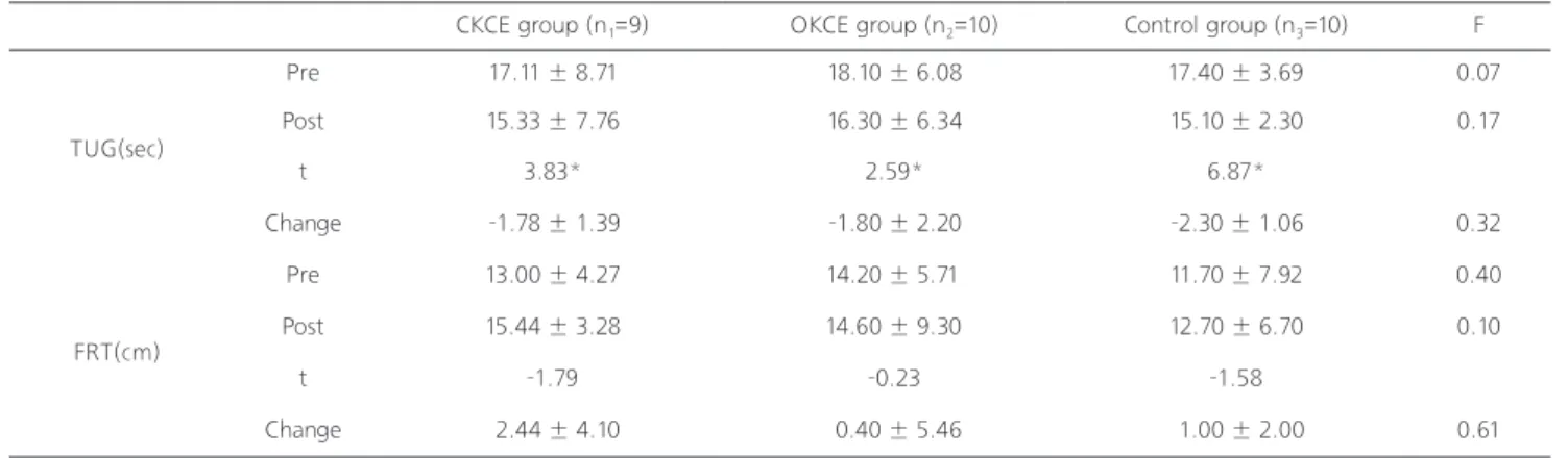
The mean changes in the TUG test results were -1.78
± 1.39 s in the CKCE group, -1.80 ± 2.20 s in the OKCE group, and -2.30 ± 1.06 s in the control group (p<0.01).
There were no significant differences between the groups.
The mean changes in the FRT for dynamic balance did not significantly differ among the CKCE, OKCE, and control groups (Table 3).
2) Comparison of pre and post-intervention knee extensor strength The mean changes in the knee extensor strength were 9.44
± 5.34 Nm in the in CKCE group, 12.70 ± 4.06 Nm in the OKCE group, and 6.30 ± 4.47 Nm in the control group (p<0.01), and these values differed significantly among the groups (p<0.05). The post hoc analysis indicated that patients in the OKCE group displayed significant improvement relative to those in the CKCE and control groups (Table 4).
3) Comparison of pre and post- intervention JPS
CKCE group (n1=9) OKCE group (n2=10) Control group (n3=10) F
TUG(sec)
Pre 17.11 ± 8.71 18.10 ± 6.08 17.40 ± 3.69 0.07
Post 15.33 ± 7.76 16.30 ± 6.34 15.10 ± 2.30 0.17
t 3.83* 2.59* 6.87*
Change -1.78 ± 1.39 -1.80 ± 2.20 -2.30 ± 1.06 0.32
FRT(cm)
Pre 13.00 ± 4.27 14.20 ± 5.71 11.70 ± 7.92 0.40
Post 15.44 ± 3.28 14.60 ± 9.30 12.70 ± 6.70 0.10
t -1.79 -0.23 -1.58
Change 2.44 ± 4.10 0.40 ± 5.46 1.00 ± 2.00 0.61
CKCE group (n1=9) OKCE group (n2=10) Control group (n3=10) F
Knee extension strength
(Nm)
Pre 78.00 ± 12.57 70.70 ± 9.90 76.10 ± 26.16 0.43
Post 87.44 ± 11.69 83.40 ± 11.13 82.40 ± 29.74 0.17
t -5.31* -9.90* -4.45*
Change 9.44 ± 5.34 12.70 ± 4.06 † 6.30 ± 4.47 4.79*
Table 3. A comparison of dynamic balance changes
Table 4. A comparison of knee extension strength changes CKCE: Closed Kinetic Chain Exercises, OKCE: Open Kinetic Chain Exercises TUG: Timed Up and Go Test, FRT: Functional Reaching Test
*p<0.05
CKCE: Closed Kinetic Chain Exercises, OKCE: Open Kinetic Chain Exercises
*p<0.05
† significant difference compared with the Control group (p<0.05).
The mean error degree changed in NWB exercise at 120° were -3.89 ± 1.83° in the CKCE group and -4.10 ± 1.37° in the OKCE group (p<0.01). In contrast, no significant change was observed in the control group, although a significant difference was observed among the groups (p<0.01). The post hoc analysis indicated that patients in the CKCE and OKCE groups displayed significant improvement relative to those in the control group. However, there was no difference between the CKCE and OKCE groups. The mean error degree changes in NWB exercise at 150° were -4.78 ± 1.92° in the CKCE group, -5.20 ± 2.25° in the OKCE group, and -1.40 ± 1.17°
in the control group (p<0.01), with significant differences among the groups (p<0.01). The post hoc analysis indicated that patients in the CKCE and OKCE groups displayed significant improvement relative to those in the control group.
However, there was no difference between the CKCE and OKCE groups. The mean error degree changes in WB exercise at 120° were -5.11 ± 1.76° in the CKCE group, -2.30 ± 1.06° in the OKCE group, and -1.40 ± 1.26° in the control group (p<0.01), with significant differences between the groups (p<0.01). The post hoc analysis indicated that patients in the CKCE group displayed significant improvement relative to those in the OKCE and control groups. The mean error degree changes in WB exercise at 150° were -6.22 ± 3.42°
in the CKCE group and -2.50 ± 0.85° in the OKCE group (p<0.01), In contrast, no significant change was observed in the control group, although significant differences were observed among groups (p<0.01). The post hoc analysis indicated that patients in the CKCE and OKCE groups displayed significant improvement relative to those in the control group. The CKCE group also exhibited significant improvement relative to the OKCE group (Table 5).
4. Discussion
The aim of this study was to determine the effects of OKCE and CKCE using knee reposition sensing on balance, strength, and JPS in chronic stroke patients. The results of the present study indicate that (1) comparing the means of the OKCE and CKCE groups revealed significant differences in balance, strength, and JRS and (2) a significant difference in knee position sensing between WB and NWB tests.
The effects of increased muscle strength on static/dynamic balance should also be considered. In this study, static balance was measured using the sway distance and sway speed and dynamic balance was measured using the TUG and FRT. Regarding static balance, significant changes in the pre and post-intervention sway distance and sway velocity measurements were observed in the CKCE and OKCE groups (p<0.05), and the CKCE and OKCE groups displayed significant increases relative to the control group. Significant changes in the pre and post-intervention TUG measurements were observed in all groups (p<0.01), and the CKCE and OKCE groups exhibited significant increases relative to the control group. A previous study reported a strong correlation between balance and leg strength.26-28 However, these comparisons should be made carefully because the sample baselines can differ substantially. Therefore, it is difficult to assess the efficacy of a training program without addressing this factor;
however, it seems clear that the lower the baseline, the more difficult it will be to achieve significant gains (i.e., a low score is better for this test).27 No significant changes between the pre and post-intervention FRT measurements were observed in any group. These results were evaluated in stroke patients while performing task motions to assess compensation due to motor function impairment.28
This study found that all groups exhibited significant increases in knee extension strength. OKCE might be more useful than CKCE and conventional therapy for increasing the knee extension strength. In a previous study of patients with knee osteoarthritis, a NWB exercise group demonstrated improved knee extensor strength.29,30 This was because NWB exercises mostly affect the isolated knee extensor muscles, as they are performed from 90° of knee flexion to full extension with concentric quadriceps action, followed by flexing of the knee joint to the starting position with eccentric quadriceps action. This differs from normal knee extension with concentric quadriceps, followed by knee flexion with concentric hamstrings. This could explain why greater improvements in knee extensor strength were observed in the NWB exercise group.
This study found that the OKCE and CKCE groups exhibited significantly improved knee JRS relative to the control group.
In the weight bearing test, CKCE might be more effective than OKCE for increasing proprioception. This finding agreed with those of previous studies.31-34 Lim et al.35 studied that CKCE effectively improved knee JPS in normal adults. Jan et al.30 studied that CKCE effectively improved knee JPS in patients with knee osteoarthritis. CKCE is thought to enhance knee joint proprioception by increasing the intra-articular pressure and thereby stimulating the Ruffini nerve endings, which are sensitive to changes in the intracapsular fluid volume.30,36 For this reason, the present study subjects experienced improved knee JPS after CKCE. Weight bearing included hip, knee and ankle motions, more closely paralleling the CKCE than the OKCE pattern. CKCE place less strain on joint and are thus less likely to produce pain and discomfort.11,37 The proprioceptive feedback in CKCE helps protect the joint and decrease ligament strain by approximating the joint and stimulating Golgi ligament endings and muscle spindles. It also allows the athlete to perform rehabilitative exercises with the knee in a more functional position that will simulate activities of sport and daily living.
Our study was conducted to compare the effects of WB and NWB exercise on knee JPS in chronic stroke patients, a finding that has not been reported previously. However, previous studies have measured the knee JPS in the context of aging, muscular skeletal disease, knee injury, or knee ligament injury.38,39 The absolute error means previously reported during WB exercise ranged from 1.8 ± 0.81° to 3.4 ± 2.0°.38 The absolute error mean reported during NWB exercise in a previous study was 4.1 ± 2.6°.40 In our study, the absolute error means during WB exercise ranged from 3.00 ± 0.78° to 6.45 ± 1.44° and the absolute error mean ranges during NWB exercise ranged from 7.09 ± 4.97° to 12.73 ± 2.72°. In our study, the absolute errors observed for knee JPS according to position in chronic stroke patients appeared to be higher than those reported in previous studies.
For this reason, we thought that the neurologic disorder might have affected knee position sensing. However, the WB exercise position yielded lower absolute error means than did the NWB exercise position. This finding agreed with those of previous studies.25,41 In a study by Ghiasi and Akbari,25 the authors investigated differences in the knee JPS
reproducibility in healthy subjects performing WB and NWB exercises and reported more accurate reproduction of knee position sensing during the WB protocol. According to Hopper et al., WB exercise is more important than NWB exercise for performance and injury prevention. WB tasks are more functional and can make the subject more easily aware of information around the knee joint via summation.41
The clinical implications of our findings are that both CKCE and OKCE improved knee joint position sense and balance in chronic stroke patients. OKCE might be more useful for increasing the strength than CKCE. Especially, such a proprioceptive training in weight bearing to improve neuromuscular control may also be of benefit to individuals with various balance deficits. This study included only 29 subjects; overall, there are small stroke patients and it is therefore difficult to generalize the results of this study.
Additionally, the relatively short 4-week treatment period and lack of a long-term follow-up conducted in accordance with the training methods makes it difficult to predict the effects of the differences in the methods. Although the selected subjects were not classified according to the degree of disability or increased strength and speed of movement during the intervention process, the use of quantitative tools to measure proprioception balanced the limitations of this study. These limitations were further complemented by the usefulness of developing treatment intervention programs for stroke patients.
References
1. Cauraugh JH, Kim S. Two coupled motor recovery protocols are better than one electromyogram-triggered neuromuscular stimulation and bilateral movements. Stroke. 2002;33(6):1589- 94.
2. Flansbjer UB, Miller M, Downham D et al. Progressive resistance training after stroke: Effects on muscle strength, muscle tone, gait performance and perceived participation. J Rehabil Med.
2008;40(1):42-8.
3. Williams DA, Farrell MJ, Cunningham J et al. Knee pain and radiographic osteoarthritis interact in the prediction of levels of self-reported disability. Arthritis Rheum. 2004;51(4):558-61.
4. Walsh LD, Hesse CW, Morgan DL et al. Human forearm position sense after fatigue of elbow flexor muscles. J Physiol.
2004;558(Pt 2):705-15.
5. Johnson EO, Babis GC, Soultanis KC et al. Functional neuroanatomy of proprioception. J Surg Orthop Adv. 2007;17 (3):159-64.
6. Carey LM. Somatosensory loss after stroke. Crit Rev Phys RehabilMed. 1995;7(1).
7. Partridge C, Edwards S. The bases of practice-neurological physiotherapy. Physiother Res Int. 1996;1(3):205-8.
8. Kim YJ. The effects of closed kinetic chain exercises of unstable floor on the stability of the knee joints of patients with anterior cruciate ligament reconstruction. Graduate School of Daegu University. 2007.
9. Jang JW. The change of muscle activation in quadriceps femoris muscle during taking open kinetic chain exercise and closed kinetic chain exercise and closed kinetic chain exercise : on the subject of soccer players. Graduate School of Bio-Medical Science, Korea University, 2003
10. Escamilla RF, Fleisig GS, Zheng N et al. Biomechanics of the knee during closed kinetic chain and open kinetic chain exercises. Med Sci Sports Exerc. 1998;30(4):556-69.
11. Kwon SB, Lee HO. Effect of closed and open kinetic chain exercise after cruciate ligament reconstruction. J Korean Soc Phys Ther. 2005;17(3):297-310.
12. Kwon YJ, Park SJ, Kim K, The effect of open and closed chain exercise on lower extremity muscle activity in adults. J Korean soc phys med. 2012;7(2):173-82.
13. Lee NK, Kwon JW, Son SM et al. Changes of plantar pressure distributions following open and closed kinetic chain exercise in patients with stroke. NeuroRehabilitation. 2013;32(2):385-90.
14. Skinner HB, Barrack RL, Cook SD et al. Joint position sense in total knee arthroplasty. J Orthop Res. 1984;1(3):276-83.
15. Rogol IM, Ernst G, Perrin DH. Open and closed kinetic chain exercises improve shoulder joint reposition sense equally in healthy subjects. J Athl Train. 1998;33(4):315-8.
16. Neufeld SD. Reproducing movement in the lower extremity using kinesthetic cues of distance and location. Phys Ther.
1981;61(8):1147-51.
17. Barrett DS, Cobb AG, Bentley G. Joint proprioception in normal, osteoarthritic and replaced knees. J Bone Joint Surg Br.
1991;73(1):53-6.
18. Magalhaes T, Ribeiro F, Pinheiro A et al. Warming-up before sporting activity improves knee position sense. Phys Ther Sport.
2010;11(3):86-90.
19. Park DS, Lee DY, Choi SJ et al. Reliability and validity of the balancia using wii balance board for assessment of balance with stroke patients. Journal of the Korea Academia-Industrial cooperation Society. 2013;14(6):2767-72.
20. Mehrholz J, Wagner K, Rutte K et al. Predictive validity and responsiveness of the functional ambulation category in
hemiparetic patients after stroke. Arch Phys Med Rehabil.
2007;88(10):1314-9.
21. Duncan PW, Weiner DK, Chandler J et al. Functional reach: A new clinical measure of balance. J Gerontol. 1990;45(6):192-7.
22. Knols RH, Aufdemkampe G, De Bruin ED et al. Hand-held dynamometry in patients with haematological malignancies:
Measurement error in the clinical assessment of knee extension strength. BMC musculoskeletal disorders. 2009;10(1):1-11.
23. Jenny J-Y. Measurement of the knee flexion angle with a smartphone-application is precise and accurate. J Arthroplasty.
2013;28(5):784-7.
24. Bennell KL, Hinman RS, Metcalf BR et al. Relationship of knee joint proprioception to pain and disability in individuals with knee osteoarthritis. J Orthop Res. 2003;21(5):792-7.
25. Ghiasi F, Akbari A. Comparison of the effects of open and closed kinematic chain and different target position on the knee joint position sense. Journal of Medical Sciences. 2007;7(6):969-76.
26. Lee HJ, Yi CH, Yoo EY. Correlations among the berg balance scale, gait parameters, and falling in the elderly. Physical Therapy Korea. 2002;9(3):47-65.
27. Sousa N, Sampaio J. Effects of progressive strength training on the performance of the functional reach test and the timed get‐up‐and‐go test in an elderly population from the rural north of portugal. Am J Hum Biol. 2005;17(6):746-51.
28. Roerdink M, Geurts AC, de Haart M et al. On the relative contribution of the paretic leg to the control of posture after stroke. Neurorehabil Neural Repair. 2009;23(3):267-74.
29. Tagesson S, Öberg B, Good L et al. A comprehensive rehabilitation program with quadriceps strengthening in closed versus open kinetic chain exercise in patients with anterior cruciate ligament deficiency a randomized clinical trial evaluating dynamic tibial translation and muscle function. J Orthop Sports Phys Ther. 2008;36(2):298-307.
30. Jan MH, Lin CH, Lin YF et al. Effects of weight-bearing versus nonweight-bearing exercise on function, walking speed, and position sense in participants with knee osteoarthritis:
A randomized controlled trial. Arch Phys Med Rehabil.
2009;90(6):897-904.
31. Palmitier RA, An K-N, Scott SG et al. Kinetic chain exercise in knee rehabilitation. Sports Medicine. 1991;11(6):402-13.
32. Lutz G, Palmitier R, An K et al. Comparison of tibiofemoral joint forces during open-kinetic-chain and closed-kinetic-chain exercises. J Bone Joint Surg Am. 1993;75(5):732-9.
33. Hurd WJ, Chmielewski TL, Snyder-Mackler L. Perturbation- enhanced neuromuscular training alters muscle activity in female athletes. Knee Surgery, Sports Traumatology, Arthroscopy. 2006;14(1):60-9.
34. Hilberg T, Herbsleb M, Puta C et al. Physical training increases isometric muscular strength and proprioceptive performance in
haemophilic subjects. Haemophilia. 2003;9(1):86-93.
35. Lim GR, Kim DS, Park J et al. The effects of closed kinetic chain exercise and open kinetic chain exercise on the knee position sense in the normal adults. Journal of International Academy of Physical Therapy Research. 2010;1(2):126-7.
36. Ferrell WR, Tennant N, Sturrock RD et al. Amelioration of symptoms by enhancement of proprioception in patients with joint hypermobility syndrome. Arthritis Rheumatol.
2004;50(10):3323-8.
37. Naseri N, Pourkazemi F. Difference in knee joint position sense in athletes with and without patellofemoral pain syndrome. Knee Surgery, Sports Traumatology, Arthroscopy. 2012;20(10):2071-6.
38. Bullock-Saxton JE, Wong WJ, Hogan N. The influence of age
on weight-bearing joint reposition sense of the knee. Exp Brain Res. 2001;136(3):400-6.
39. Givoni NJ, Pham T, Allen TJ et al. The effect of quadriceps muscle fatigue on position matching at the knee. J Physiol.
2007;584(1):111-9.
40. Drouin JM, Houglum PA, Perrin DH et al. Weight-bearing and non-weight-bearing knee-joint reposition sense and functional performance. J Sport Rehabil. 2003;12(1):54-66.
41. Hopper DM, Creagh MJ, Formby PA et al. Functional measurement of knee joint position sense after anterior cruciate ligament reconstruction. Arch Phys Med Rehabil.
2003;84(6):868-72.