ISSN: 2233-601X (Print) ISSN: 2093-6516 (Online)
Received: January 20, 2016, Revised: April 26, 2016, Accepted: April 26, 2016, Published online: October 5, 2016
Corresponding author: Kyung Bok Lee, Department of Neurology, Soonchunhyang University Hospital, Soonchunhyang University School of Medicine, 59 Daesagwan-ro, Yongsan-gu, Seoul 04401, Korea
(Tel) 82-2-709-9026 (Fax) 82-2-709-9226 (E-mail) [email protected]
© The Korean Society for Thoracic and Cardiovascular Surgery. 2016. All right reserved.
This is an open access article distributed under the terms of the Creative Commons Attribution Non-Commercial License (http://creativecommons.org/
licenses/by-nc/4.0) which permits unrestricted non-commercial use, distribution, and reproduction in any medium, provided the original work is properly cited.
Delayed Surgery for Aortic Dissection after Intravenous Thrombolysis in Acute Ischemic Stroke
Nari Choi, M.D. 1 , Jee-Eun Yoon, M.D. 1 , Byoung-Won Park, M.D. 2 , Won-Ho Chang, M.D. 3 , Hyun-Jo Kim, M.D. 3 , Kyung Bok Lee, M.D. 1
Departments of
1Neurology,
2Cardiology, and
3Thoracic Surgery, Soonchunhyang University Hospital, Soonchunhyang University School of Medicine
We report a case of aortic dissection masquerading as acute ischemic stroke followed by intravenous throm- bolysis. A 59-year-old man presented with dizziness. After examination, the patient had a seizure with bilateral Babinski signs. Soon after identifying multiple acute infarctions in both hemispheres on diffusion-weighted brain magnetic resonance (MR) imaging, tissue plasminogen activator (t-PA) was administered. Both common carotid arteries were invisible on MR angiography, and subsequent chest computed tomography revealed an aortic dissection. The emergency operation was delayed for 13 hours due to t-PA administration. The patient died of massive bleeding.
Key words: 1. Aortic dissection 2. Stroke
3. Thrombolysis
Case report
A 59-year-old male patient visited emergency room of Soonchunhyang University Seoul Hospital for slight uneasiness in the chest and vertigo. He was fully con- scious and no neurological findings were observed.
The patient’s blood pressure was 77/66 mmHg, which was quite low; however, it recovered to 102/65 mmHg w ith a heart rate of 40 beats per minute w ith fluid therapy. Second-degree atrioventricular block was observed in the electrocardiogram. No abnormal- ities were observed in myocardial enzyme levels or in a standard blood examination. The department of emergency medicine collaborated with the department of cardiology to treat this patient. The patient was scheduled to be hospitalized in the cardiology ward for coronary angiography because Mobitz type II at-
rioventricular block and unstable angina were sus- pected.
The patient had a sudden convulsive seizure while waiting to be hospitalized. The seizure did not stop for more than 5 minutes, during which lorazepam was administered. The patient did not have a history of seizure, heavy drinking, or trauma. After the seizure, while in a semicoma, quadriplegia and the Babinski sign were observed. In order to determine whether the convulsive seizure was accompanied by an acute stroke, diffusion-weighted magnetic resonance imag- ing was performed. Multiple cerebral infarctions were found in both the frontal and temporal lobes (Fig.
1A, B). Tissue plasminogen activator (t-PA) was ad- ministered promptly (within 90 minutes after symp- tom occurrence), and magnetic resonance angiography (MRA) was conducted while intravenous thrombol-
http://dx.doi.org/10.5090/kjtcs.2016.49.5.392
Fig. 1. (A, B) Diffusion-weighted imaging revealed multiple acute infarctions in the bilateral frontal and temporal lobes, especially in the territory of the in whole territory of the right middle cerebral artery. (C) On magnetic resonance angiography, both common carotid ar- teries were invisible.
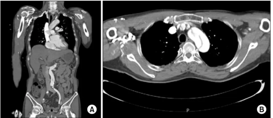
Fig. 2. A Stanford type A aortic dis- section documented on (A) a coro- nal and (B) an axial chest com- puted tomography scan.
ysis was performed. Forty minutes after t-PA admin- istration, the common carotid arteries on bilateral sides showed no contrast on the MRA (i.e., the ar- teries were invisible on the scan), and aortic dis- section was suspected (Fig. 1C).
Stanford type A aortic dissection, which requires an immediate emergency operation, was observed on chest computed tomography (CT) (Fig. 2). However, the operation was postponed due to the high bleed- ing risk caused by the thrombolytic agent; the oper- ation was finally conducted 13 hours later. The pa- tient expired 4 hours after the termination of the op- eration due to excessive bleeding.
Discussion
Aortic dissection generally presents with sudden and severe pain. In the case w here the aortic dis- section invades the ascending aorta, mortality may be as high as 40%–50% if appropriate emergency medical treatment is not obtained promptly [1]. In an aortic dissection, various abnormal neurological findings can occur, and ischemic cerebral infarction is the most frequently observed such finding. However, 10%–55% of all aortic dissection cases are painless.
When a patient seeks care for mental deterioration or language disturbance, the diagnosis of an aortic dissection can be quite difficult.
Thrombolysis, conducted to treat acute cerebral in-
farction, may sometimes be applied without under- standing the secondary cause of cerebral infarction due to limited time. The effects of thrombolysis on aortic dissection patients have not been reported widely [2].
Carotid or vertebral dissection is not an absolute contraindication for intravenous thrombolysis. Intra- cranial hemorrhage and recurrent infarction in pa- tients with carotid dissection were found to occur approximately as frequently as in patients without carotid dissection [3]. The Safe Implementation of Thrombolysis in Stroke-Monitoring Study also re- ported that the occurrence of adverse effects was similar with or without intravenous thrombolysis in patients with cerebral infarction caused by carotid or vertebral dissection [4].
Myocardial infarction in patients with an aortic dissection is a contraindication for intravenous throm- bolysis. In previous studies, it was reported that thrombolysis could cause hemopericardium or car- diac tamponade by aortic rupture [5]. Thrombolysis could reduce the patient’s possibility of survival by leading to the postponement of an emergency oper- ation to treat a Stanford Type A aortic dissection [4].
Antiplatelet or anticoagulant agents, which have lower bleeding risks than t-PA, can have higher bleeding risks during aortic surgery. In the case of cardiac procedures such as coronary artery bypass grafting, the amount of bleeding and transfusion vol- ume were higher in patients who continued anti- platelet medication than in those who had dis- continued medication 7 days prior to the procedure [3]. The amount of bleeding during aortic surgery can be massive; usage of t-PA before surgery is likely to significantly increase hemorrhagic complications and mortality.
However, few reports have been published con- cerning cerebral infarction caused by aortic dissection.
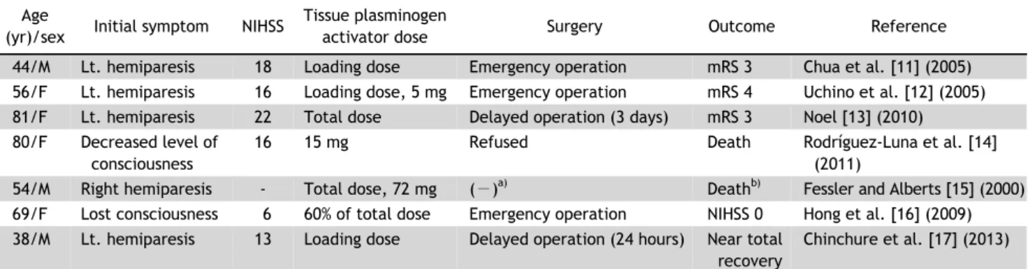
Cases in which intravenous thrombolysis were con- ducted w ithout know ing that aortic dissection w as present in patients with acute cerebral infarction are summarized in Table 1. Although many patients ex- hibit a tendency for increased bleeding, no consensus exists regarding the optimal timing for major cardiac surgery after the administration of t-PA. T-PA was administrated to a total of 7 patients prior to diag- nosis with an aortic dissection. Complete doses of t-PA were infused in 2 patients and administration
ceased in 5 patients because they were diagnosed w ith an aortic dissection. Of the 2 patients w ho re- ceived full dosages of t-PA, 1 of them underwent sur- gery within 3 days and recovered to independent ambulation (modified Rankin Score [mRS], 3 points).
However, the other patient died without undergoing surgery. Among the 5 patients w ho received partial doses of t-PA, emergency operations were conducted for 3 patients, while 1 patient underwent surgery af- ter 24 hours. The other patient did not undergo sur- gery due to the caregiver’s refusal. The 3 patients who underwent surgery had a mRS of 0, 3, and 4 points, respectively, and the patient who underwent surgery after 24 hours recovered to a nearly normal status. Therefore, unlike previous cases that had a relatively positive prognosis, this case represents the first case in which a patient expired after aortic sur- gery and t-PA administration. Since a total dose of 63 mg of t-PA w as applied, the operation w as de- layed for 13 hours, and excessive hemorrhage follow- ing the operation w as the cause of death. Despite the fact that the half-life of t-PA in blood is short, it is possible that a tendency for increased bleeding per- sists for an additional 1–2 days. In this case, how- ever, the increased size of the hemopericardium and the enlarged extent of the cerebral infarction made an emergency operation inevitable. All doses of t-PA were administrated to this patient, and it is thought that the reduced time delay before the operation in- creased the patient’s bleeding risk. Henceforth, t-PA usage in patients with cerebral infarction accom- panied with an aortic dissection will not be recom- mended in treatment guidelines.
In an aortic dissection, cerebral infarctions can be
caused by mechanical occlusion of the common car-
otid artery and vertebral artery, or by relatively low
cerebral perfusion. Approximately 1 out of 3 patients
who have neurologic symptoms from an aortic dis-
section do not feel chest pain [6]. A cerebral in-
farction caused by an aortic dissection usually occurs
in the right hemisphere through infiltration into the
right common carotid artery; however, it can also oc-
cur in both sides (as in this case) [7]. In order to di-
agnose an aortic dissection without symptoms in pa-
tients with acute cerebral infarctions, the pulse dif-
ference between both radial arteries should be con-
firmed, physical examinations including heart mur-
mur auscultation should be performed, and observa-
Table 1. Summary of cases where intravenous thrombolysis without knowledge that an aortic dissection was present Age
(yr)/sex Initial symptom NIHSS Tissue plasminogen
activator dose Surgery Outcome Reference
44/M Lt. hemiparesis 18 Loading dose Emergency operation mRS 3 Chua et al. [11] (2005) 56/F Lt. hemiparesis 16 Loading dose, 5 mg Emergency operation mRS 4 Uchino et al. [12] (2005) 81/F Lt. hemiparesis 22 Total dose Delayed operation (3 days) mRS 3 Noel [13] (2010) 80/F Decreased level of
consciousness
16 15 mg Refused Death Rodríguez-Luna et al. [14]
(2011)
54/M Right hemiparesis - Total dose, 72 mg ( −)
a)Death
b)Fessler and Alberts [15] (2000) 69/F Lost consciousness 6 60% of total dose Emergency operation NIHSS 0 Hong et al. [16] (2009) 38/M Lt. hemiparesis 13 Loading dose Delayed operation (24 hours) Near total
recovery
Chinchure et al. [17] (2013)
NIHSS, National Institute of Health Stroke Scale; M, male; Lt., left; mRS, modified Rankin Score; F, female.
a)